(a)
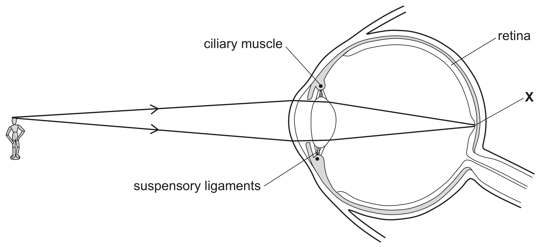
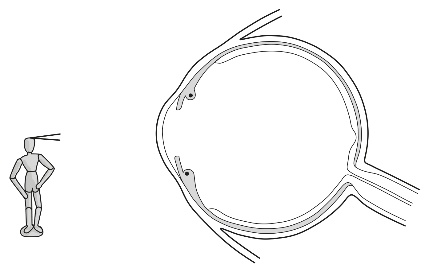
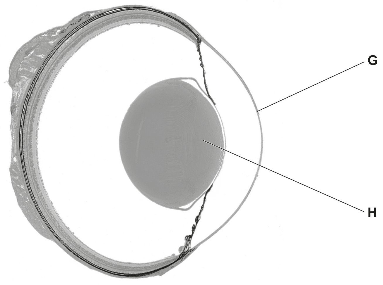
Fig. 1.1 shows part of a human eye and three neurones that conduct electrical impulses between the eye and the brain. These neurones are involved in the pupil reflex.
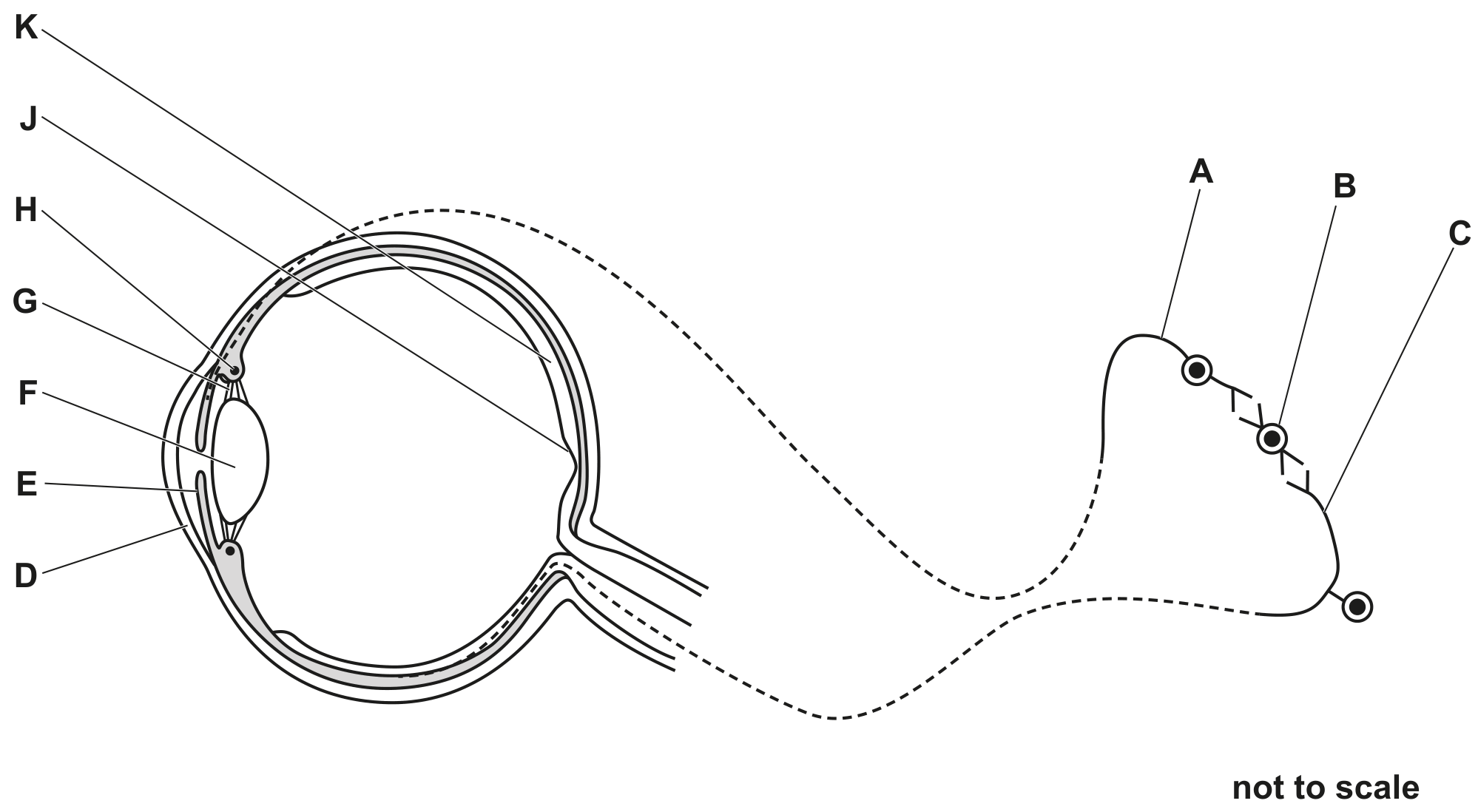
Fig. 1.1
[ 4 ]
(i)
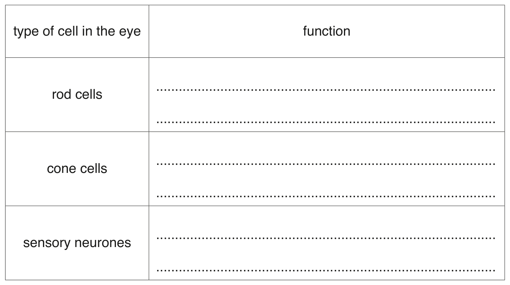
Table 1.1 shows the names of some parts of the eye, their functions and the letters in Fig. 1.1 that identify the parts of the eye.
Complete Table 1.1.
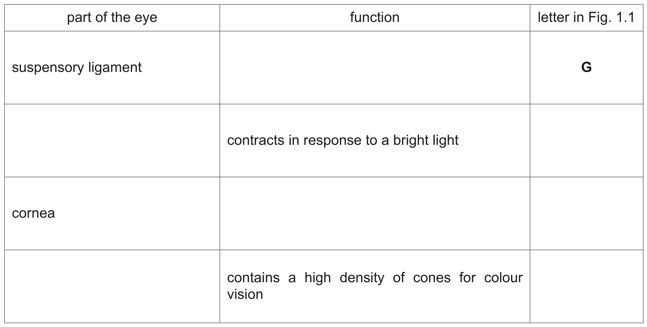
Table 1.1
[ 4 ]
(b)
(i)
The eye can adjust how light is refracted through it in order to focus on a near object.
State one process that uses energy when focusing on a near object.
[ 1 ]