[Maximum number: 1]
What are the features of the cell walls in a xylem vessel?
end wall
side wall
absent
thick
absent
thin
present
thick
present
thin
 EduNinja
What are the features of the cell walls in a xylem vessel?
end wall
side wall
absent
thick
absent
thin
present
thick
present
thin
Phloem is used to transport sucrose and amino acids in plants. Sucrose is a carbohydrate.
Fig. 2.1 shows a diagram of a plant. The arrows point to circles containing magnified cross-sections of those parts of the plant.
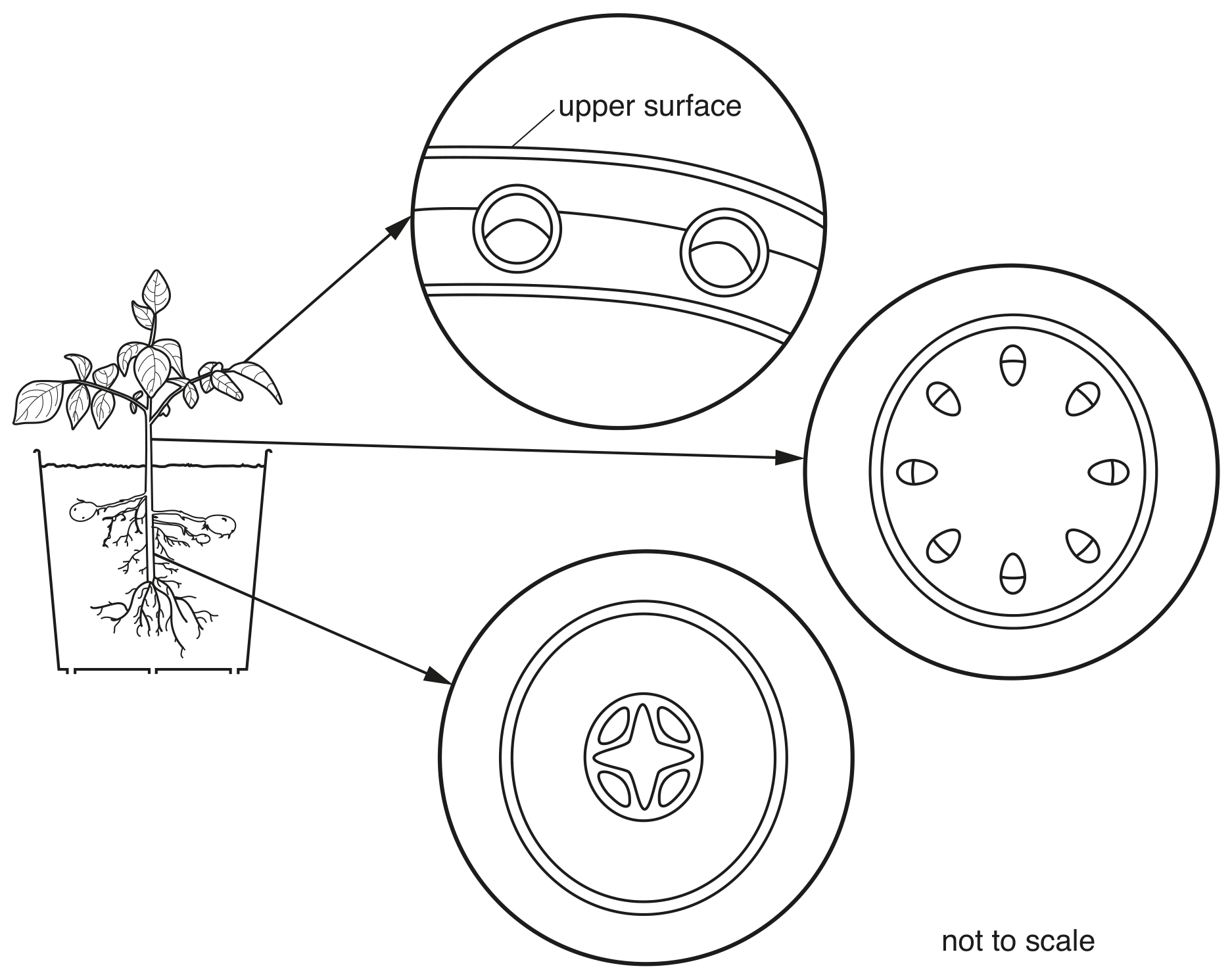
Fig. 2.1
Label the position of the phloem in each of the three magnified sections in Fig. 2.1.
Use a label line and the letter P for each section.
Which sentence explains the importance of vascular bundles for photosynthesis?
Vascular bundles transport carbon dioxide to the leaf.
Vascular bundles transport oxygen to the leaf.
Vascular bundles transport starch to the leaf.
Vascular bundles transport water to the leaf.
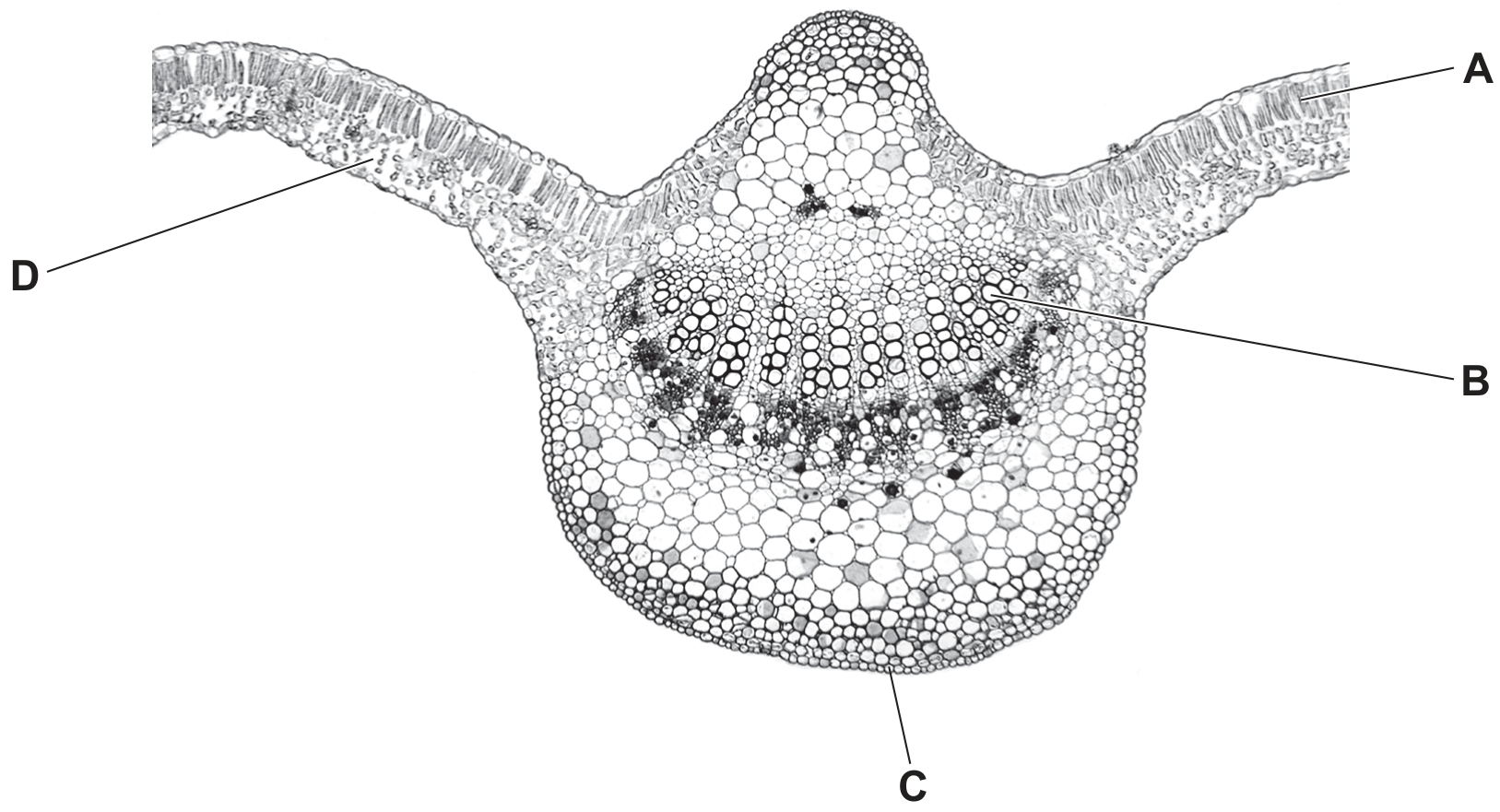
Fig. 2.1 shows part of a cross-section of the stem of a young sunflower plant.
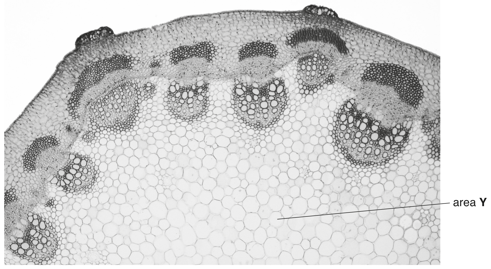
Fig. 2.1
Draw a circle around one vascular bundle on Fig. 2.1.
Label the xylem in the vascular bundle with the letter X.
Explain how the cells in area Y are able to support the stem so that it stays upright.
An experiment was carried out using the apparatus shown.
The carbon dioxide content of the water in each test-tube was measured at the start and again three hours later.
In which test-tube would there be a decrease in carbon dioxide content?
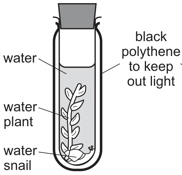
A
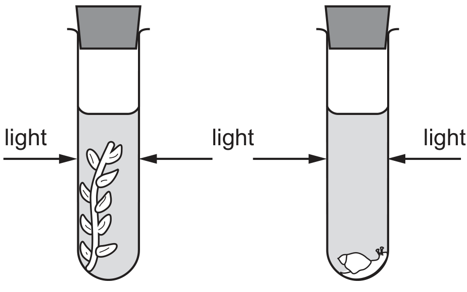
C
Plants produce glucose in leaves and convert some of it to sucrose.
The movement of sucrose in plants can be modelled using laboratory apparatus.
Fig. 2.1 shows the apparatus used to model the movement of sucrose in a plant:
- Partially permeable bags were attached tightly to the ends of tube Q.
- The bag representing a source was filled with a coloured sucrose solution.
- The bag representing a sink was filled with water.
- The containers and tube Q and tube S were filled with water.
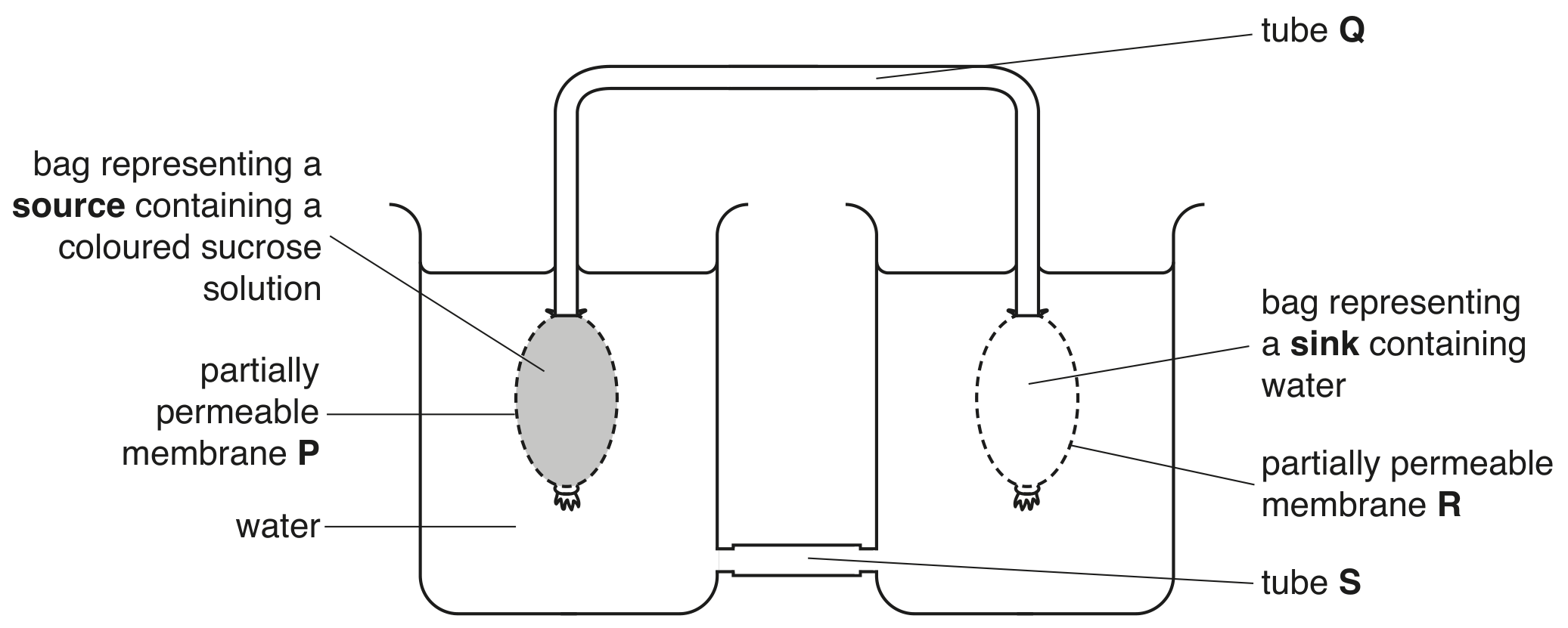
Fig. 2.1
Fig. 2.2 shows the position of the coloured sucrose solution 30 minutes after the apparatus was set up.
The arrows show the direction of the movement of the liquids.
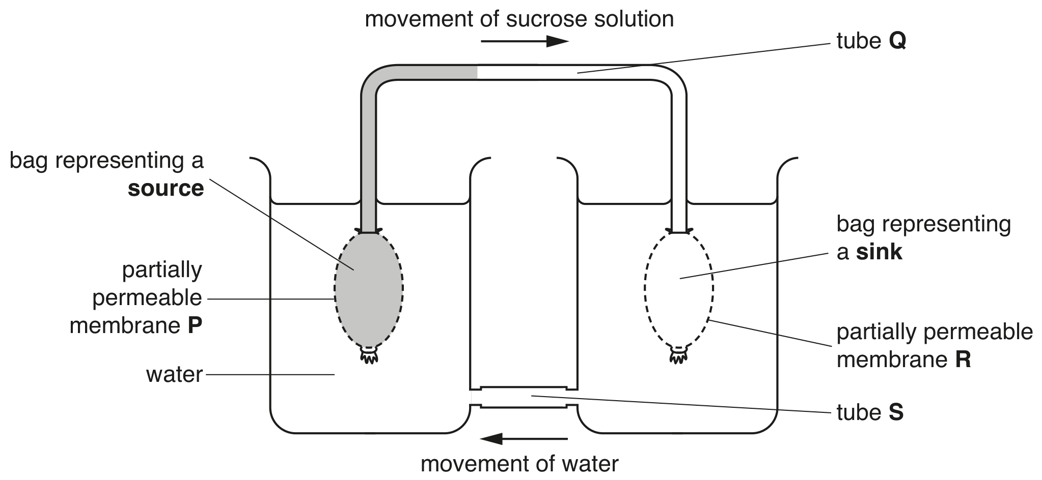
Fig. 2.2
State the name of the tissue represented by tube Q and the name of the tissue represented by tube S in Fig. 2.2.
Q
S
The diagram shows the activity of salivary amylase, pancreatic lipase and stomach protease at different pH levels.
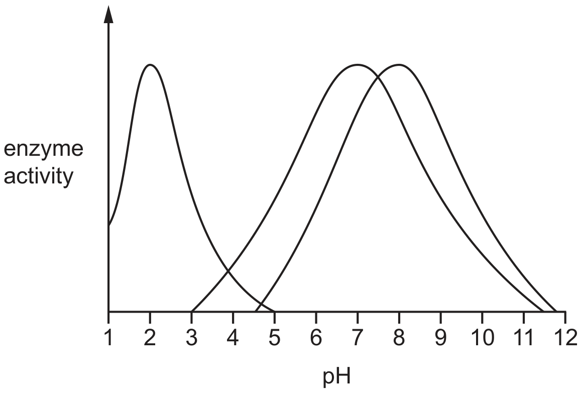
From the graph, what is the optimum pH for the protease enzyme?
2.0
3.5
7.0
8.0
The image shows a cross-section of part of a leaf.
Which labelled structure is the xylem?
A celery stalk was placed in a beaker which contained a red stain. After 24 hours, the red stain appeared at the top of the celery stalk.
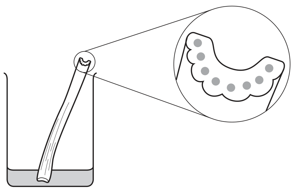
Which structures stained red?
cortex cells
mesophyll cells
phloem
xylem
Fig. 3.1 is a photomicrograph of some xylem vessels.
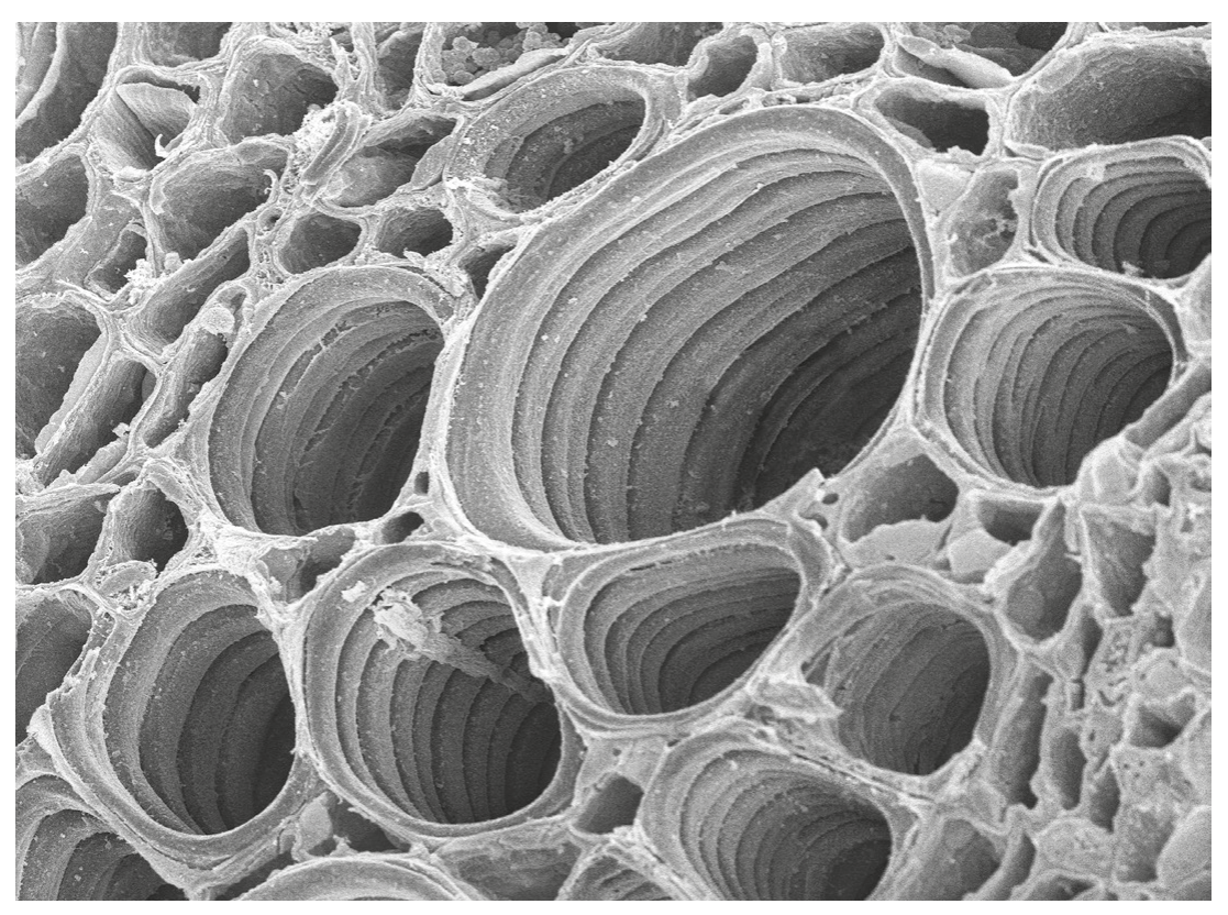
Fig. 3.1
State one structural feature of xylem vessels and explain how this is related to the function of water transport.
feature
explanation
State one role of xylem vessels other than transport.