EduNinja
[Maximum number: 5]

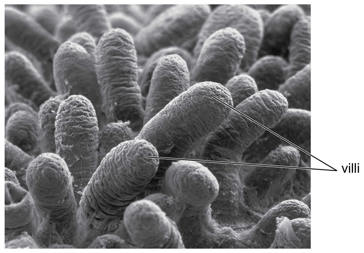
Fig. 1.1 shows several villi from the ileum, which is part of the small intestine.

(a)

State the name of one other part of the small intestine.
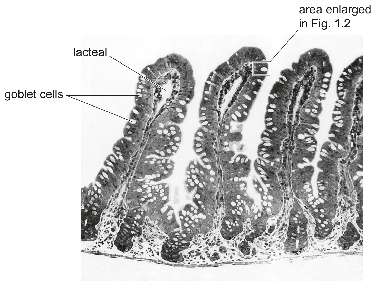
Fig. 1.1

Fig. 1.1

[ 1 ]
(b)

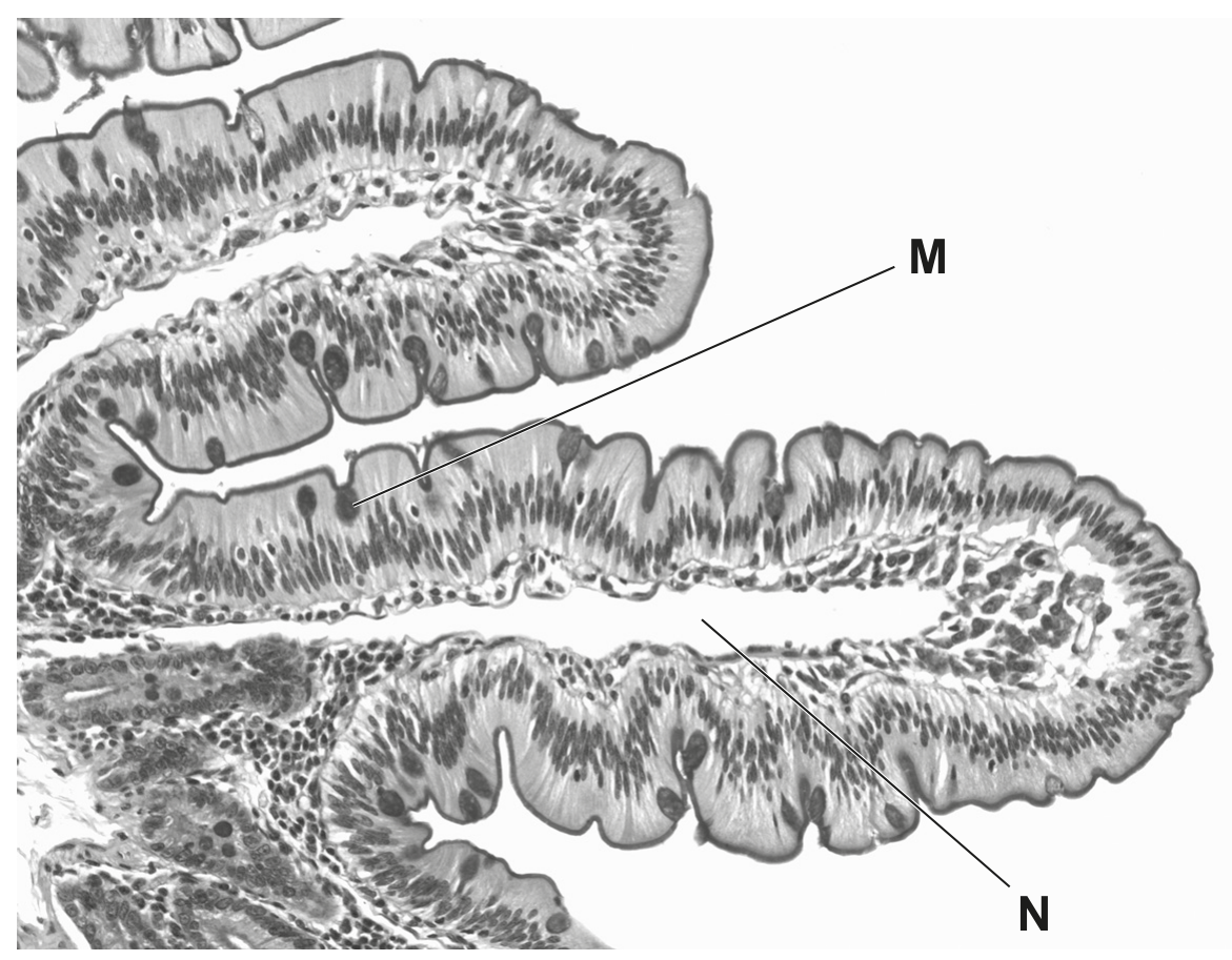
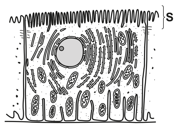
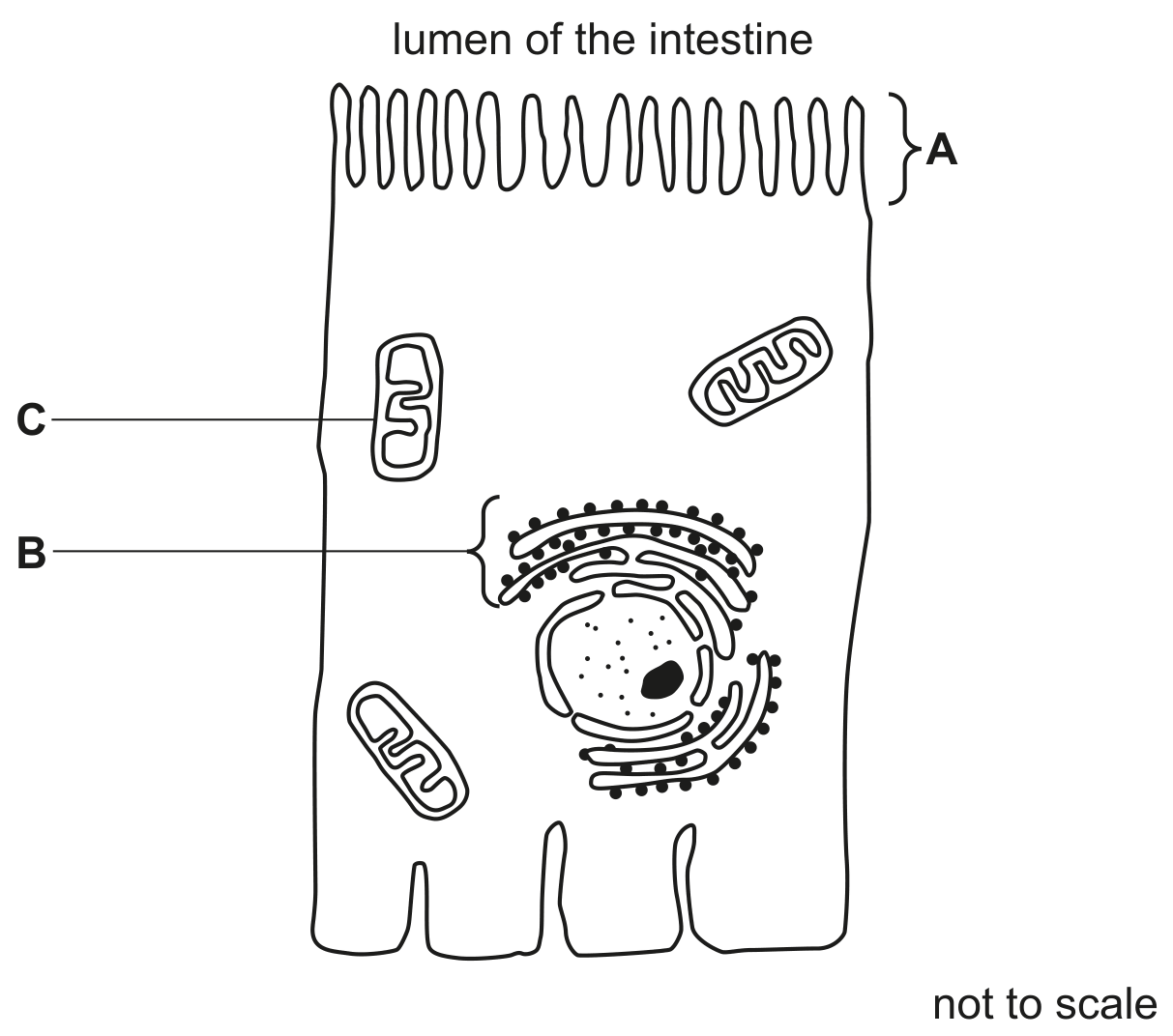
Fig. 1.2 shows the tip of a villus in more detail.
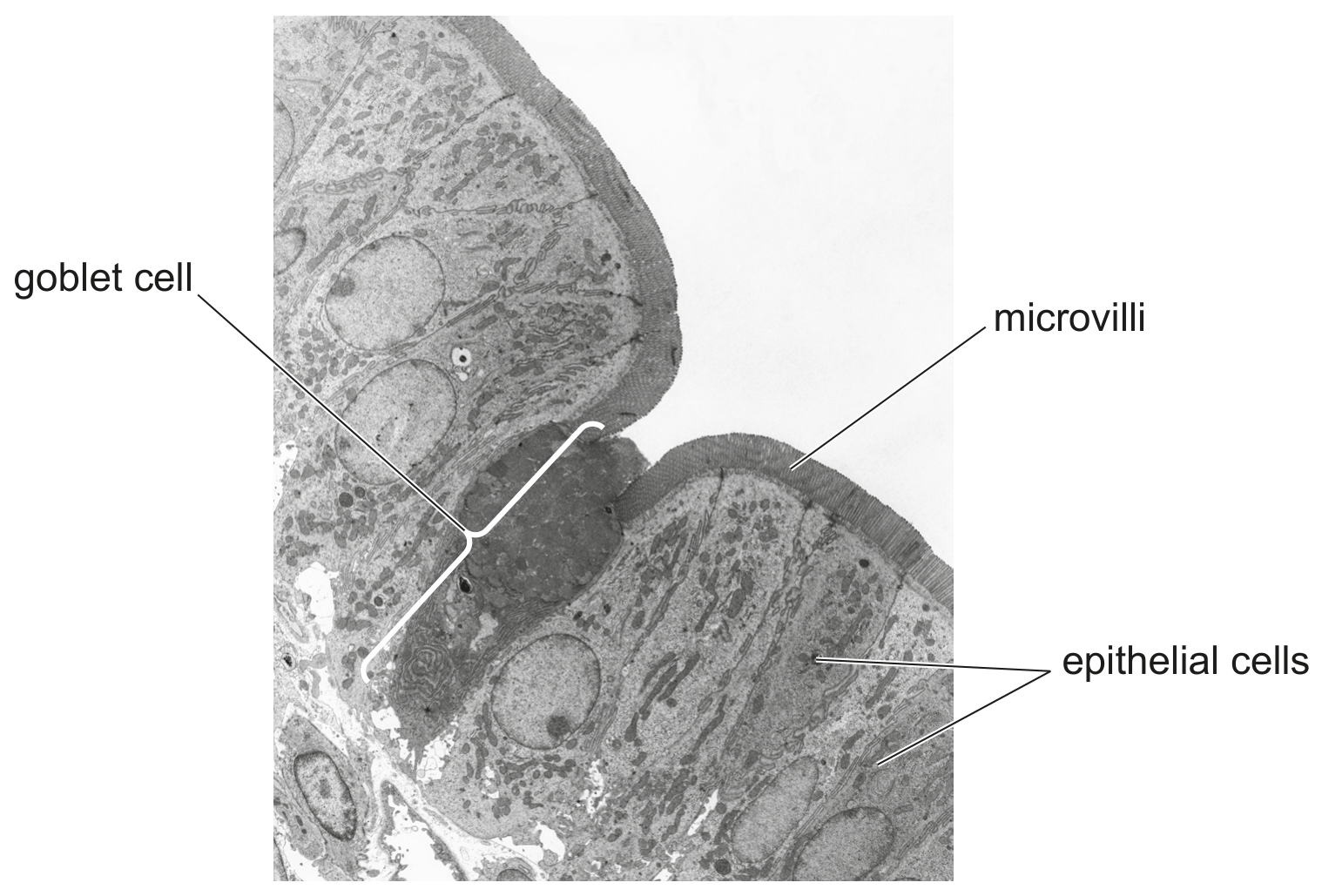
Fig. 1.2

Fig. 1.2

The epithelial cells of the villi absorb nutrients by diffusion and active transport.

[ 2 ]
(i)

Explain the importance of the microvilli shown in Fig. 1.2.

[ 2 ]
(c)

Fig. 1.1 shows a lacteal in the centre of each villus.

Describe the roles of lacteals.

[ 2 ]
[Maximum number: 3]

Fat is a necessary component of the human diet.

(a)

Explain how the products of fat digestion are transported from Q to the rest of the body.

One possible effect of too much fat in the diet is coronary heart disease.

[ 3 ]
(a)

Fig. 1.1 is a photomicrograph showing a surface view of many villi in the small intestine.

Fig. 1.1

Fig. 1.1

[ 4 ]
(i)

State the function of villi.

[ 1 ]
(ii)

Describe the structure of a villus.

[ 3 ]
[Maximum number: 1]

Water is a very important molecule for all living organisms.

(a)
(i)

State the name of the organ in humans where most water is absorbed.

[ 1 ]
(a)

The small intestine is lined with many villi and is adapted to absorb glucose.
Fig. 2.1 shows two villi from the lining of the small intestine.

Fig. 2.1

Fig. 2.1

[ 4 ]
(i)

Describe and explain how the structure of a villus is adapted for its function.

[ 4 ]
(a)

Lacteals are part of the lymphatic system.

[ 2 ]
(i)

State where in the body lacteals are found.

[ 1 ]
(ii)

Describe the role of lacteals.

[ 1 ]
[Maximum number: 1]

The diagram shows the digestive system.
Which part absorbs the most water?

Question image
(a)

Emulsification of fats occurs in the alimentary canal.

[ 1 ]
(i)

State the name of the structure in the villi where digested fats are absorbed.

[ 1 ]
[Maximum number: 2]

The kidneys filter blood, separate useful molecules from excretory wastes and control the water content of the blood.

Fig. 2.1 is a diagram of a kidney tubule and associated blood vessels. The arrows show the direction of blood flow.

Fig. 2.2 is a drawing of a vertical section through a cell from the lining of region 2 of the tubule.

Fig. 2.1

Fig. 2.1

Fig. 2.2

Fig. 2.2

(a)
(i)

The structures labelled S on Fig. 2.2 are microvilli.

Explain the importance of the microvilli on the surface of these cells.

[ 2 ]
(a)

Fig. 3.2 shows a drawing of a cell from the lining of the small intestine. The lumen is the space inside the intestine where food is digested.

Fig. 3.2

Fig. 3.2

State the names of the three labelled structures in Fig. 3.2 and describe the role of each structure in the intestinal cell.

[ 6 ]
0