[Maximum number: 4]
Fig. 1.1 is a diagram of an animal cell.
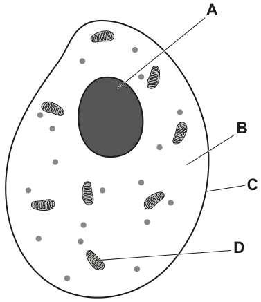
Fig. 1.1
(a)
Complete Table 1.1 by writing the name and function of each of the labelled parts of the cell shown in Fig. 1.1.
| label in Fig. 1.1 | name | function |
|---|---|---|
| A | ||
| B | ||
| C | ||
| D |
Table 1.1
[ 4 ]
 EduNinja
Fig. 1.1 is a diagram of an animal cell.
Fig. 1.1
Complete Table 1.1 by writing the name and function of each of the labelled parts of the cell shown in Fig. 1.1.
| label in Fig. 1.1 | name | function |
|---|---|---|
| A | ||
| B | ||
| C | ||
| D |
Table 1.1
Fig. 1.1 is a diagram of part of a cross-section of a leaf.
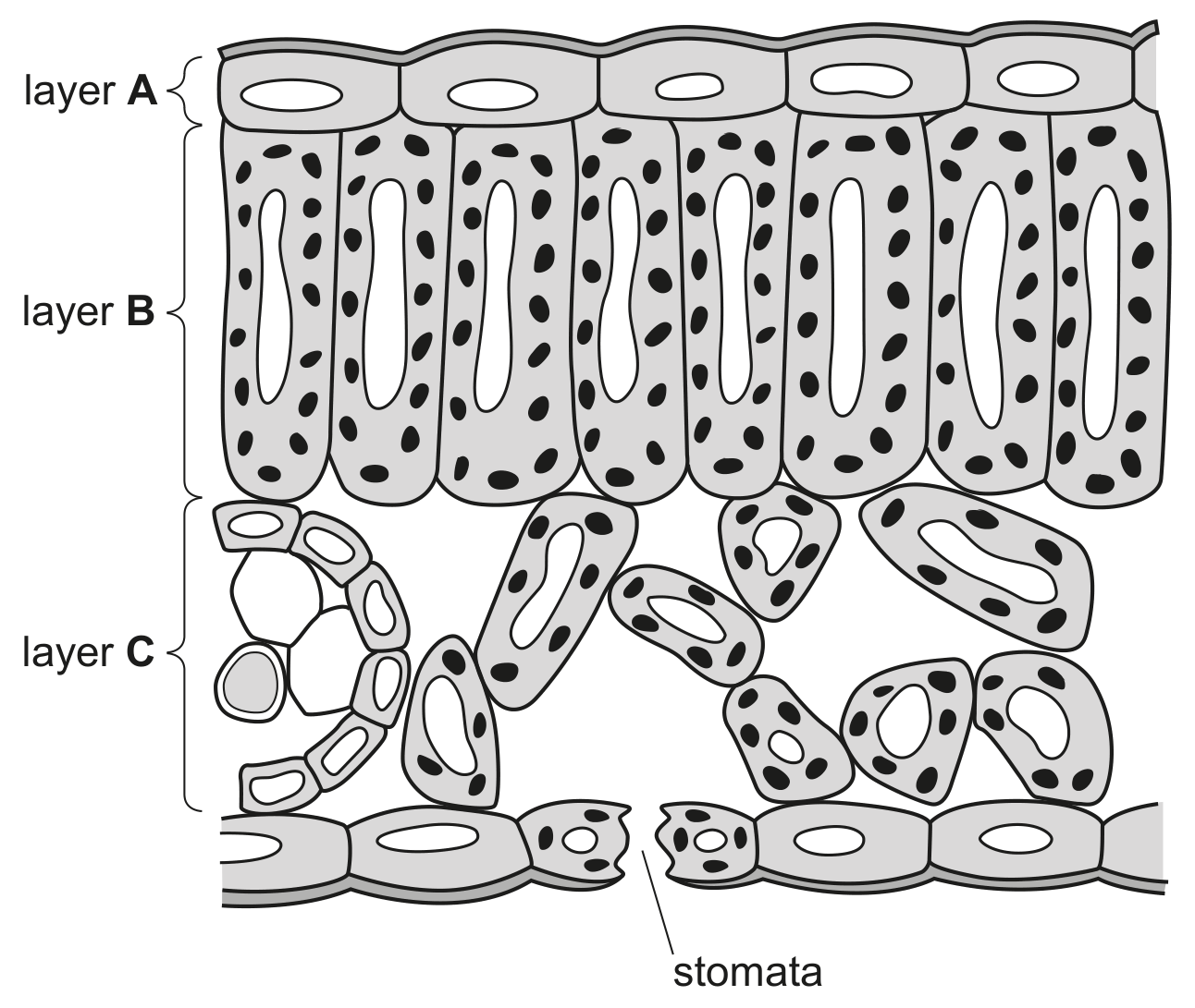
Fig. 1.1
Explain why a leaf is considered to be an organ.
Fig. 1.1 shows a spongy mesophyll cell from the leaf of a plant. The arrows show the net direction of movement of carbon dioxide molecules during daylight.
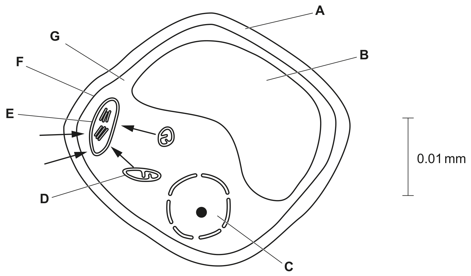
Fig. 1.1
Table 1.1 shows:
- the functions of some of the structures in plant cells
- some of the names of the structures where these functions occur
- some of the letters that label these structures in Fig. 1.1.
Complete Table 1.1.
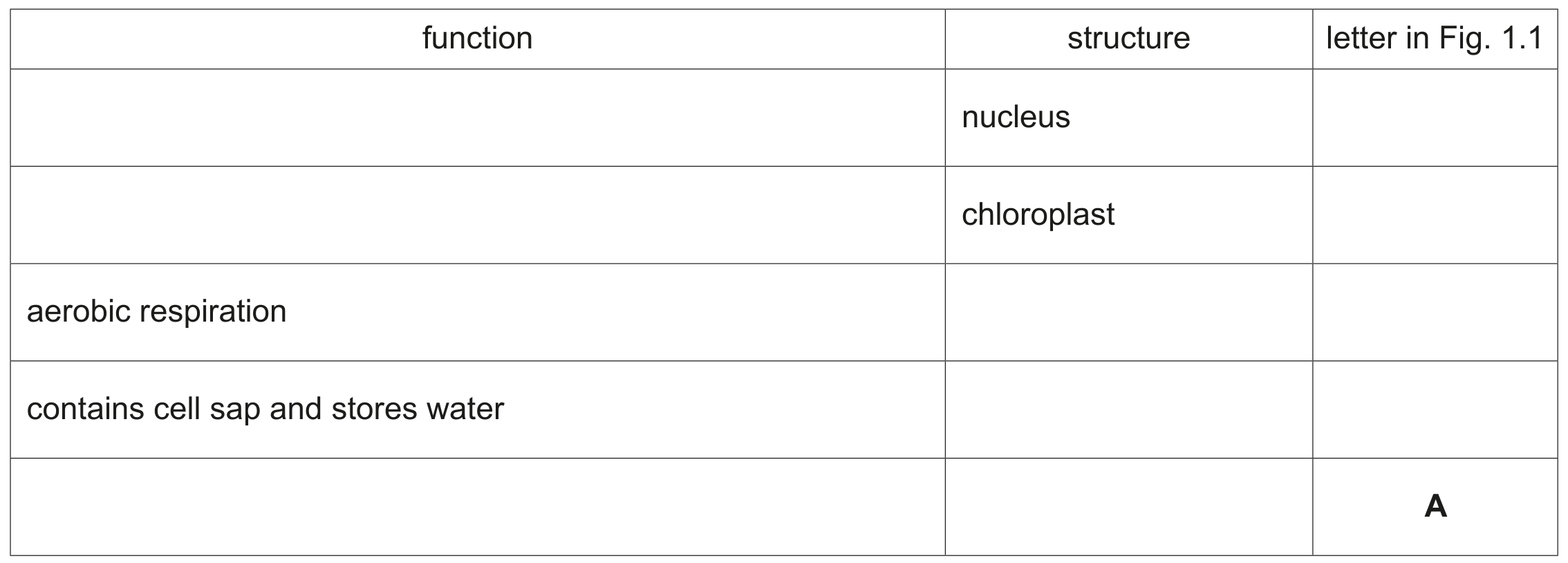
Table 1.1
Fig. 1.1 shows several villi from the ileum, which is part of the small intestine.
Complete Table 1.1 by identifying the level of organisation of each structure.
Choose your answers from the list. Each word or phrase may be used once, more than once or not at all.
cell cell structure organ organ system organism tissue

Table 1.1
The gas exchange system is one of the organ systems of the human body.
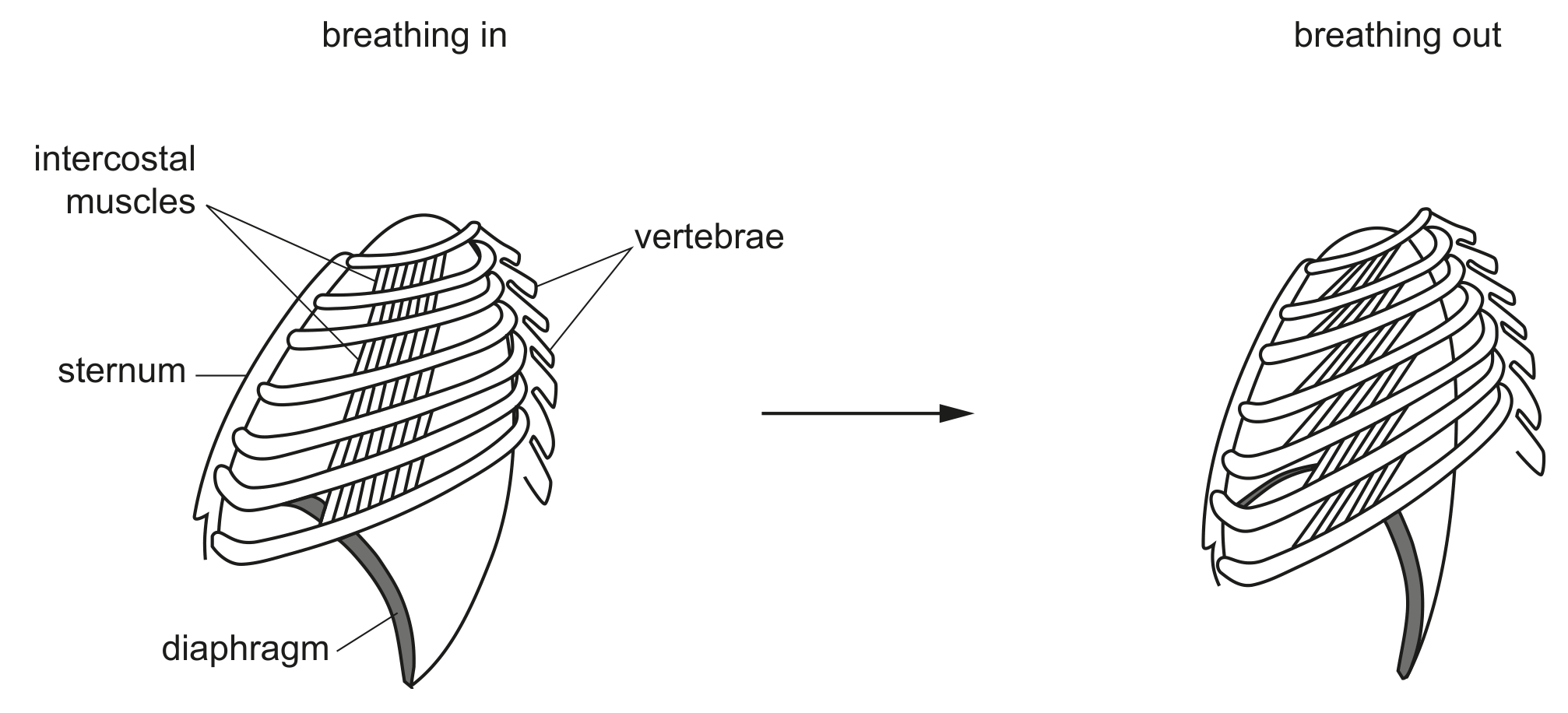
Fig. 1.1 shows parts of the gas exchange system during breathing in and breathing out.
The cells labelled X on Fig. 1.2 form a tissue.
Define the term tissue.
Bacteria are classified in the Prokaryote kingdom.
State two features of animal cells that are not found in bacteria.
1
2
Red pandas, Ailurus fulgens, and humans have a similar arrangement of teeth.
Fig. 1.1 shows a section through one tooth of a red panda. Fig. 1.2 shows the side view of the lower jaw of a red panda.

Fig. 1.1
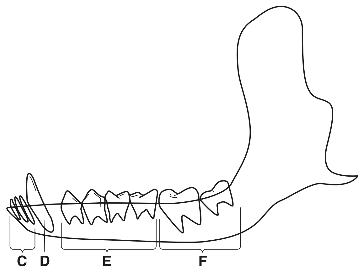
Fig. 1.2
State the names of the structures labelled A to F in Fig. 1.1 and Fig. 1.2.
A
B
C
D
E
F
The lungs and the kidneys are part of the excretory system of mammals.
The filtrate which is formed from the blood in the kidneys contains many useful substances, which are reabsorbed into the blood.
Fig. 1.1 is a photomicrograph of a cross-section of some of the cells that carry out reabsorption.
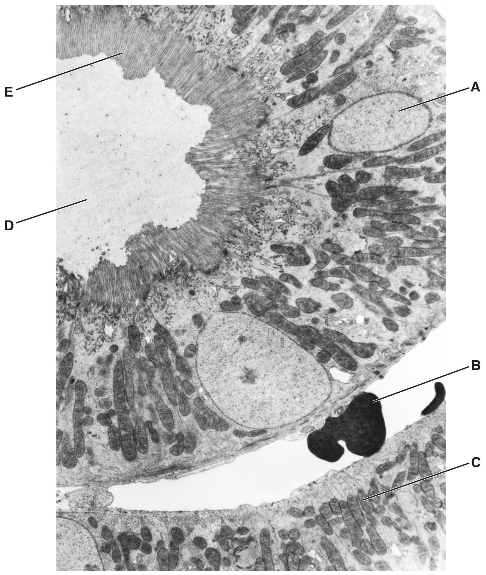
Fig. 1.1
Complete the table by stating the letter in Fig. 1.1 that identifies each structure.
State one function of the nucleus.
State the name of one part of the mammalian body other than the kidney that has cells with microvilli.
The cells that line the kidney tubules, such as those in Fig. 1.1, absorb many compounds from the filtrate.
Use Fig. 1.1 to explain how the cells are adapted for absorption.
A researcher used a light microscope to observe epithelial cells from a human cheek.
Fig. 1.1 is a photograph that the researcher made of these cells.
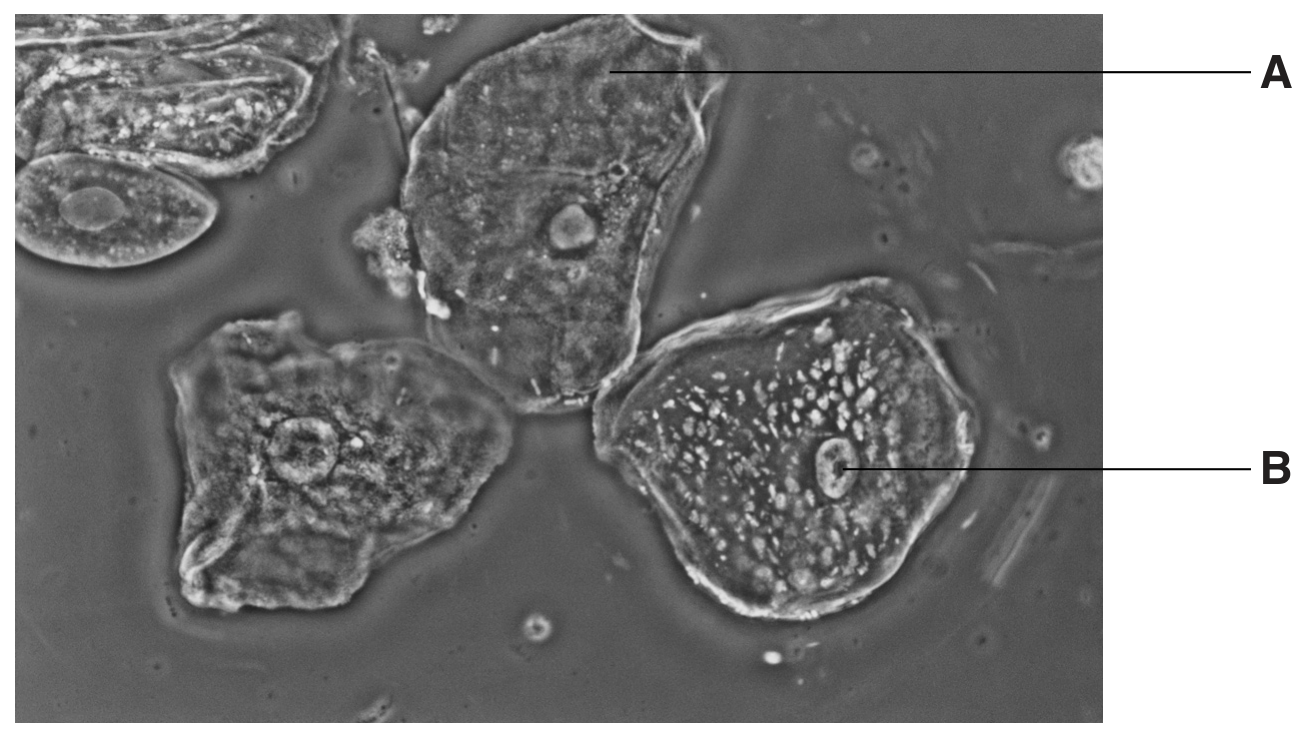
Fig. 1.1
Name the parts labelled A and B.
A
B
The cells in Fig. 1.1 each have a cell membrane.
State one of the functions of a cell membrane.
State how the shape of the cells shown in Fig. 1.1 differs from the shape of a palisade mesophyll cell in a leaf.
Fig. 1.2 shows an electron micrograph of a mitochondrion.
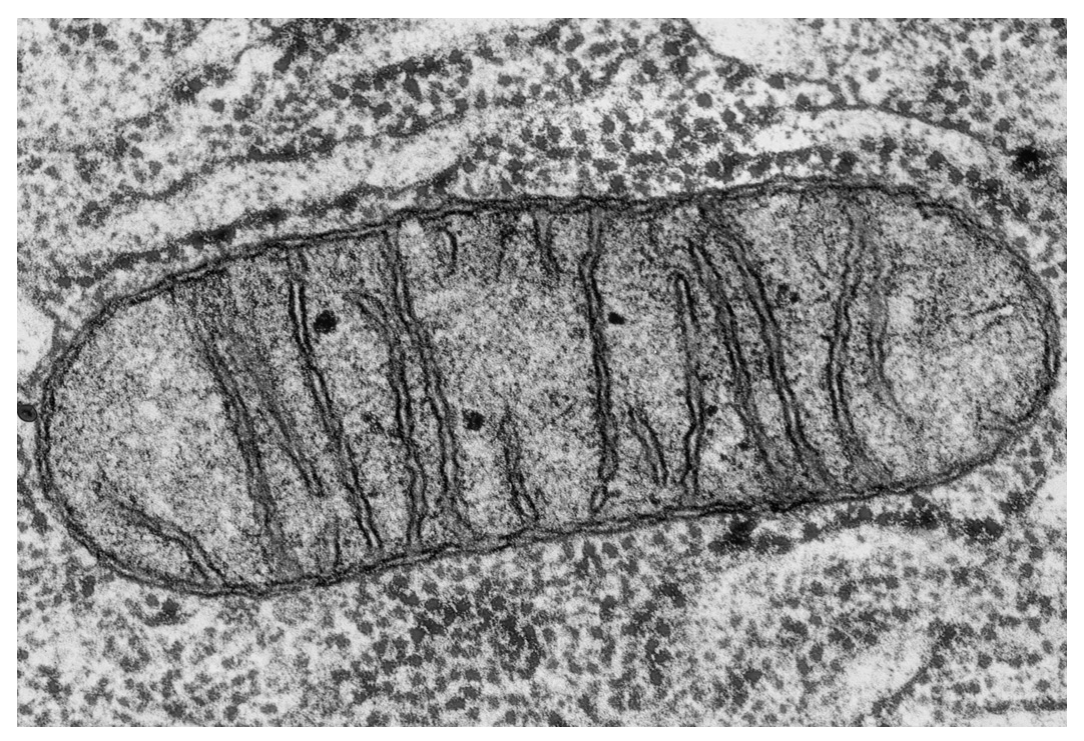
Fig. 1.2
Mitochondria have two membranes, an inner membrane and an outer membrane. The inner membrane is folded and used in respiration.
Suggest why the inner membrane of mitochondria is folded.
Table 1.1 shows different specialised cells and the average number of mitochondria each cell contains.
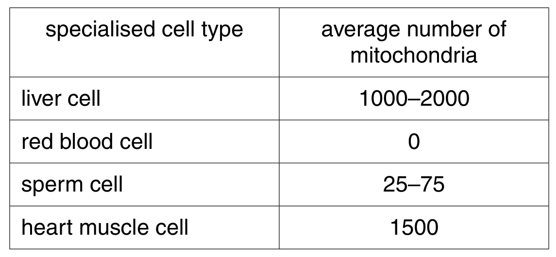
Table 1.1
Explain the differences between the average numbers of mitochondria in the cells shown in Table 1.1.
Baker's yeast, Saccharomyces cerevisiae, is a single-celled organism that is classified in the kingdom Fungi.
Fig. 1.1 is a drawing of a section through a yeast cell.
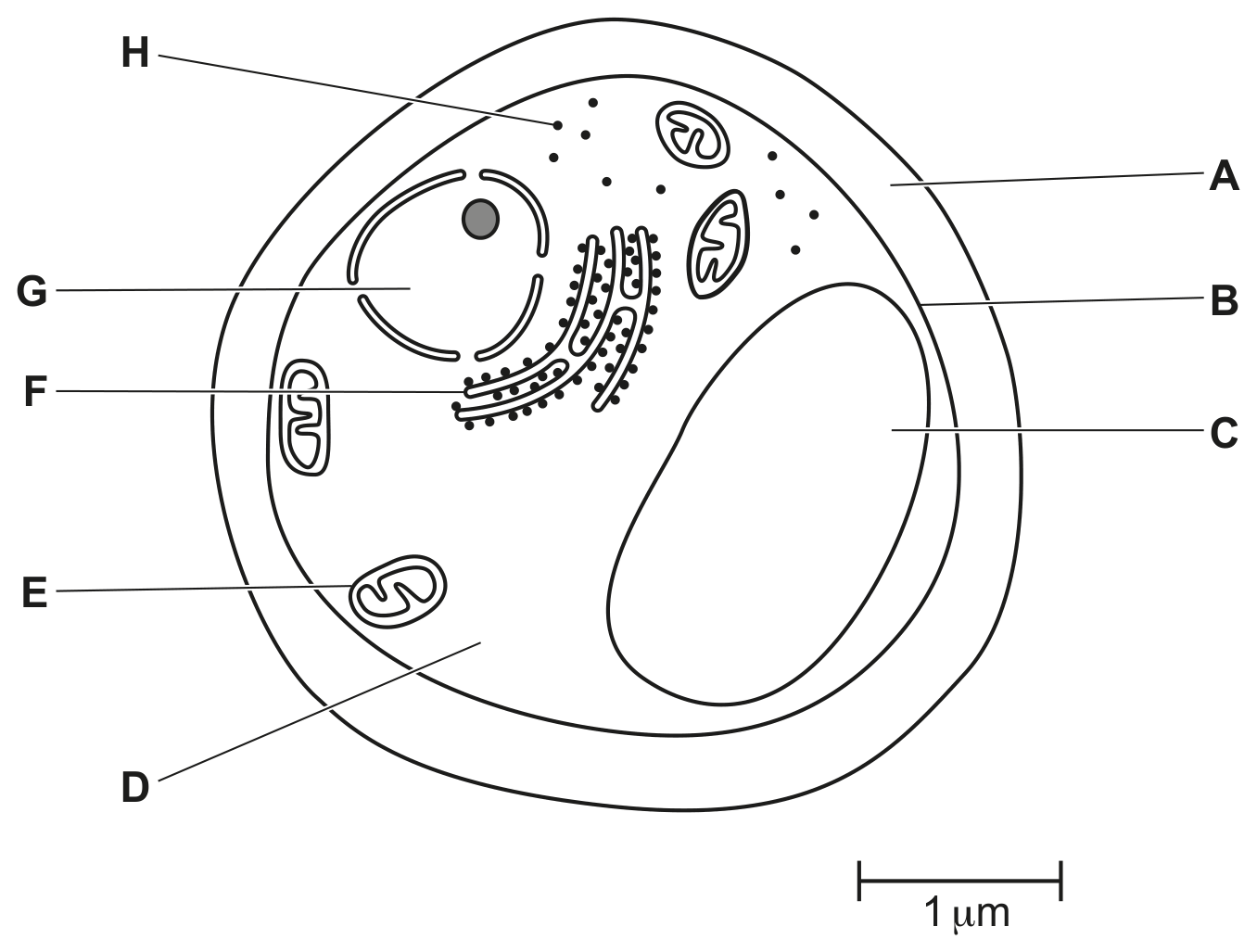
Fig. 1.1
Table 1.1 shows some cell functions.
Complete Table 1.1 by naming the cell structure responsible for each cell function and give the letter that identifies each cell structure in Fig. 1.1.
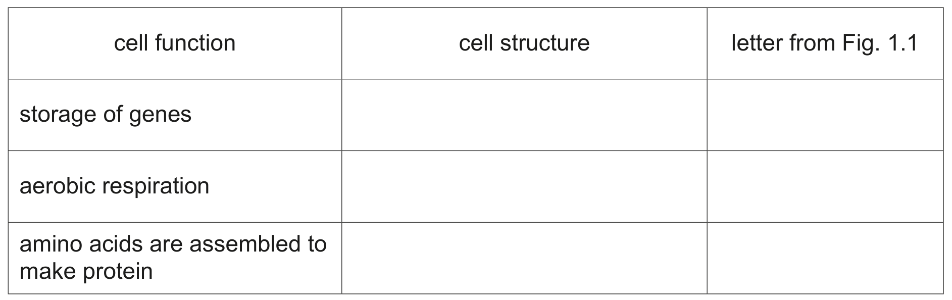
Table 1.1
Describe the similarities and differences between the structure of the yeast cell and the structure of the bacterial cell.
Use the information in Fig. 1.1 and Fig. 1.2 in your answer.