EduNinja
[Maximum number: 3]

Fig. 3.1 is a photomicrograph of a section through lung tissue.
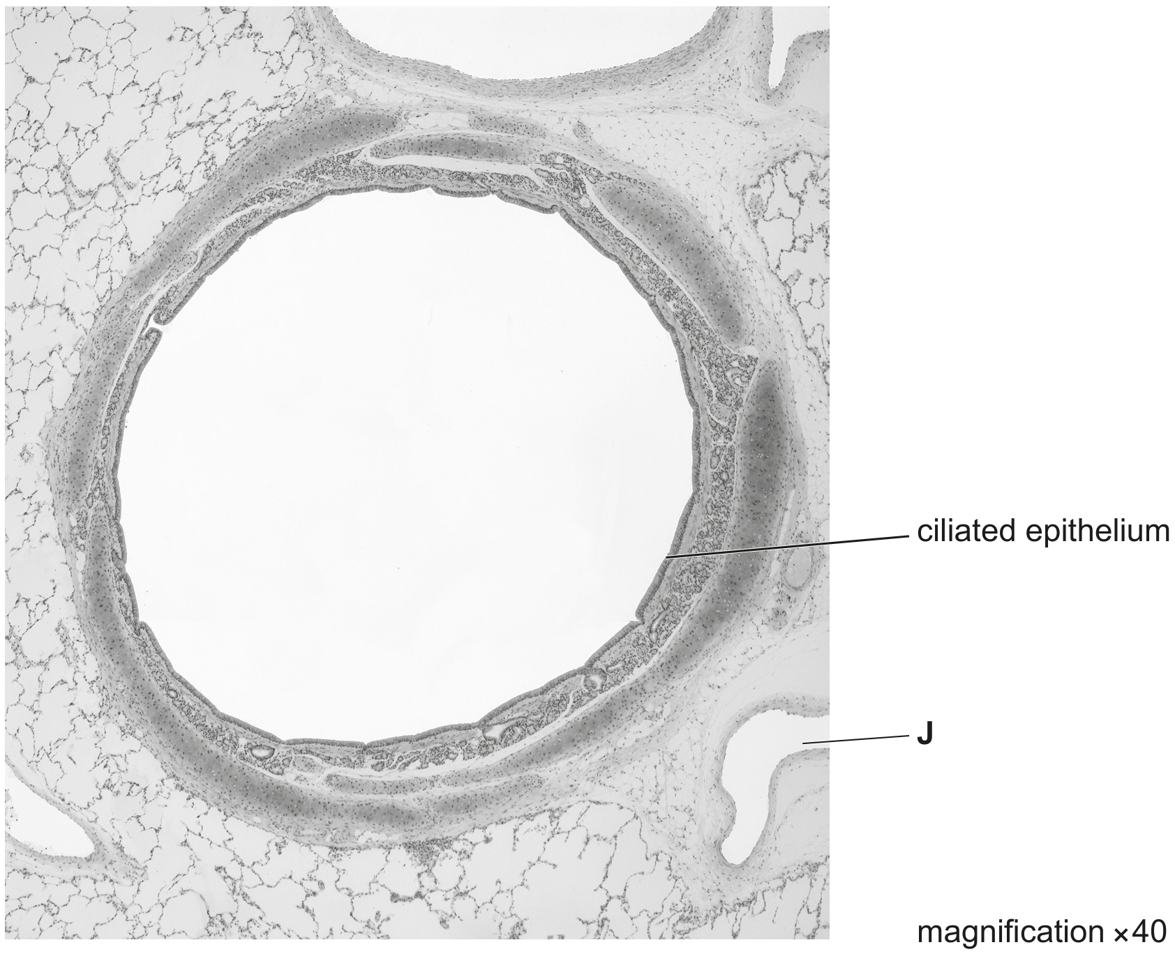
Fig. 3.1

Fig. 3.1

(a)

State the feature visible in Fig. 3.1 that identifies the structure in the centre of the image as the bronchus and list other visible features that help to confirm this identification.
feature to identify the bronchus
other features

[ 3 ]
(a)

When a section of lung tissue is viewed using a light microscope, it is possible to identify the trachea, the bronchus, the bronchioles and the alveoli.

Other than differences in their diameters, describe one structural difference visible between:
- the trachea and a bronchus
- a bronchus and a bronchiole
- a bronchiole and an alveolus.

[ 3 ]
[Maximum number: 1]

Which row shows the tissues that are present in the wall of the trachea and in the wall of the bronchus?

cartilage

squamous epithelium

goblet cells

key

×\boldsymbol{\times}

✓ = present

χ\boldsymbol{\chi}

χ=\boldsymbol{\chi}= not present

×

x

[Maximum number: 1]

A student viewed three slides at both low magnification and high magnification. Each slide was a section through a different airway of the gas exchange system.

The student observed three features in each slide.

Table

Which row is the correct set of identifications for the three slides?

slide 1

slide 2

slide 3

bronchus

bronchiole

trachea

bronchus

trachea

bronchiole

trachea

bronchiole

bronchus

trachea

bronchus

bronchiole

[Maximum number: 1]

Which row shows the tissues that are present in the wall of the trachea and the wall of the bronchus?

smooth muscle

squamous epithelium

goblet cells

key

✓ = present

x = not present

χ\boldsymbol{\chi}

χ\boldsymbol{\chi}

x

χ\boldsymbol{\chi}

[Maximum number: 1]

What identifies the structures present in a bronchus?

Question image
0