EduNinja
[Maximum number: 2]

Fig. 1.1 is a transmission electron micrograph of a cell from the stem of sago pondweed, Stuckenia pectinata.
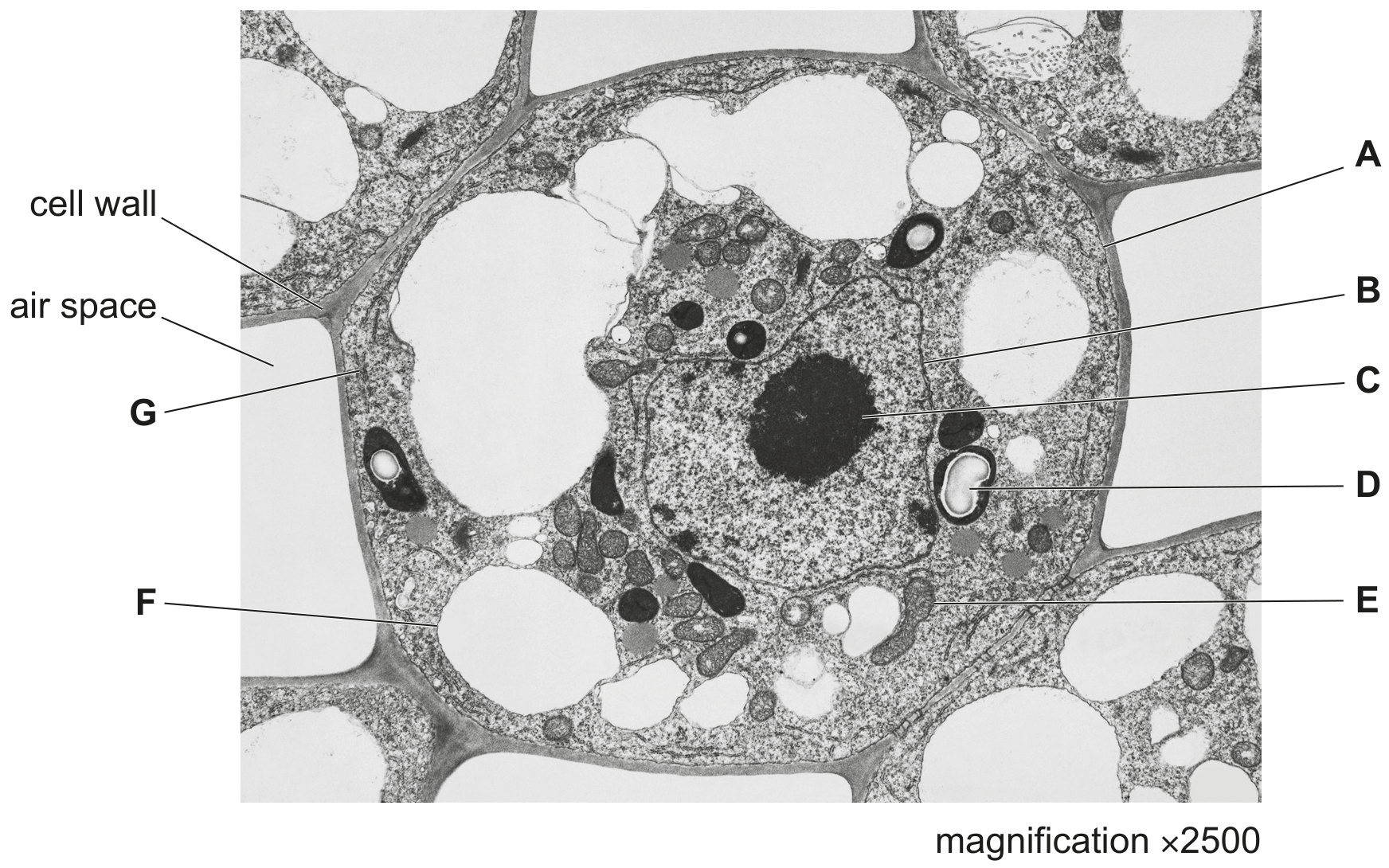
Fig. 1.1

Fig. 1.1

(a)

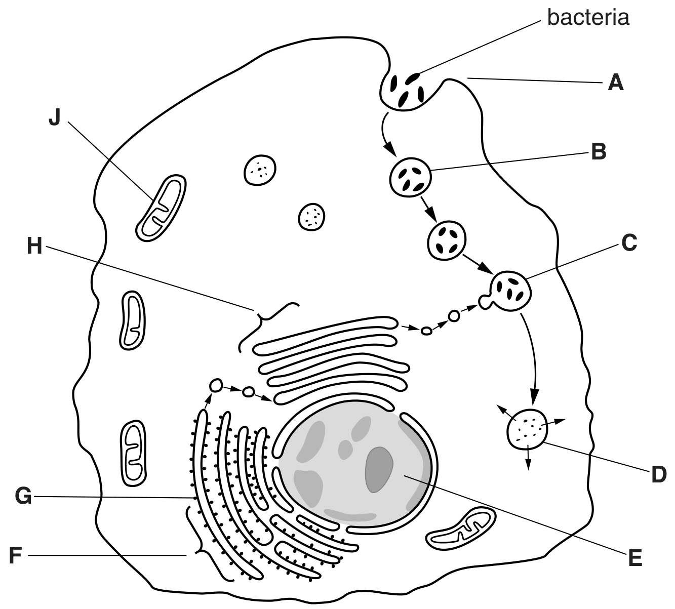
Small vacuoles in S. pectinata may have roles similar to lysosomes in animal cells. Describe the role of lysosomes in animal cells in defence against pathogens.

Question 2 is on page 6.

[ 2 ]
[Maximum number: 4]

Fig. 1.1 is a diagram of an antibody molecule.

Fig. 1.1

Fig. 1.1

(a)

When a pathogen enters the body, a primary immune response occurs. This response includes the production of antibodies.

Describe the stages in the immune response that lead to antibody being produced against a specific antigen.

[ 4 ]
[Maximum number: 1]

Name as precisely as you can the structure described in each of the following statements.

(a)

The cell that ingests and digests cell debris and bacteria in the lungs.

D

[ 1 ]
[Maximum number: 2]

Fig. 1.1 is a scanning electron micrograph of part of the wall of the bronchus of a healthy human.

Fig. 1.1

Fig. 1.1

(a)

Suggest why a person with chronic bronchitis is more likely than a healthy person to suffer from infectious diseases of the gas exchange system.

[ 2 ]
[Maximum number: 6]

Macrophages synthesise intracellular enzymes.
Fig. 2.1 is a summary diagram of events that occur in a macrophage.

Fig. 2.1

Fig. 2.1

(a)
(i)

Name the process at A.

[ 1 ]
(b)

Describe what happens to the bacteria between C and D.

[ 3 ]
(c)

Macrophages are antigen presenting cells (APCs). Antigens from pathogens such as the bacteria shown in Fig. 2.1 are presented to helper T-lymphocytes as shown in Fig. 2.2.

Fig. 2.2

Fig. 2.2

Very few helper T-lymphocytes respond to the presence of APCs by binding in the way shown in Fig. 2.2.

Suggest why this is so.

[ 2 ]
[Maximum number: 4]

Some of the steps in the production of monoclonal antibodies are shown in Fig. 2.1.
step 1
A mouse is injected with an antigen, A.
step 2
The mouse is left for a few weeks to allow an immune response to occur.
step 3
Plasma cells (effector B lymphocytes) are extracted from the mouse's spleen.
step 4
Hybridoma cells are formed.
step 5
Each hybridoma cell is isolated and allowed to grow and divide.
step 6
The hybridoma cells producing anti-A antibodies are identified and cultured on a large scale.
Fig. 2.1

(a)

With reference to Fig. 2.1, explain:

[ 4 ]
(i)

what happens during an immune response (step 2)

[ 4 ]
[Maximum number: 4]

Human cytomegalovirus (HCMV) is a common virus affecting humans. In people with a fully functioning immune system, infection by HCMV usually causes no, or only mild, symptoms.

Fig. 2.1 A is a diagram of a section through HCMV. In Fig. 2.1B, only the outer part of HCMV is sectioned.

Fig. 2.1

Fig. 2.1

The viral DNA shown in Fig. 2.1 contains genes that code for proteins important in viral replication and viral structure, including viral DNA polymerase and proteins known as tegument proteins.

Viruses can only replicate in host cells as they need to use processes and contents of the host cell. Complete viral particles that are released from the host cell are known as virions.

(a)

After a person has been infected with HCMV, the virus remains in a dormant state in the body for life.

If the virus becomes active again (reactivates), the virus will only cause serious illness if the person has a weak immune system at that time.

Explain why the response to reactivation of HCMV is more likely to cause serious illness in a person who has a weak immune system.

[ 4 ]
[Maximum number: 5]

The infectious disease cholera is caused by a bacterium.

(a)

Using genetic engineering, it is possible to produce a form of choleragen consisting of only subunit B . This can be combined with inactivated bacterial cells to produce a vaccine against cholera.

[ 5 ]
(i)

Outline how this vaccine can give protection against cholera.

[ 5 ]
[Maximum number: 5]

A vaccine, NicVAX, is being developed to help people stop smoking tobacco. Injection of NicVAX into the body causes production of antibody molecules that bind to nicotine.

(a)

Outline the immune response that leads to the production of these anti-nicotine antibodies.

[ 5 ]
[Maximum number: 1]

B-lymphocytes are activated to form plasma cells during immune responses.
Fig. 2.1 is a drawing of a plasma cell made from a transmission electron micrograph.

Fig. 2.1

Fig. 2.1

(a)
(i)

State the name of the process that is occurring at T.

[ 1 ]
0