[Maximum number: 1]
The trachea of the gas exchange system branches into two airways, each of which enters a lung.
(a)
Name the airways that branch from the trachea to enter the lungs.
[ 1 ]
 EduNinja
The trachea of the gas exchange system branches into two airways, each of which enters a lung.
Name the airways that branch from the trachea to enter the lungs.
Fig. 1.1 and Fig. 1.2 are photomicrographs showing the distribution of tissues in the lungs.
Fig. 1.1 is a photomicrograph of a section through part of the lungs.
Fig. 1.2 is a high-power view of the area indicated on Fig. 1.1.
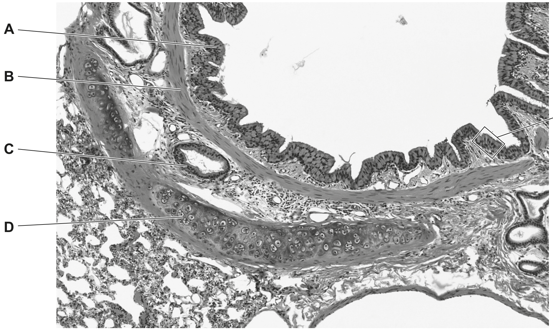
Fig. 1.1
section enlarged in Fig. 1.2
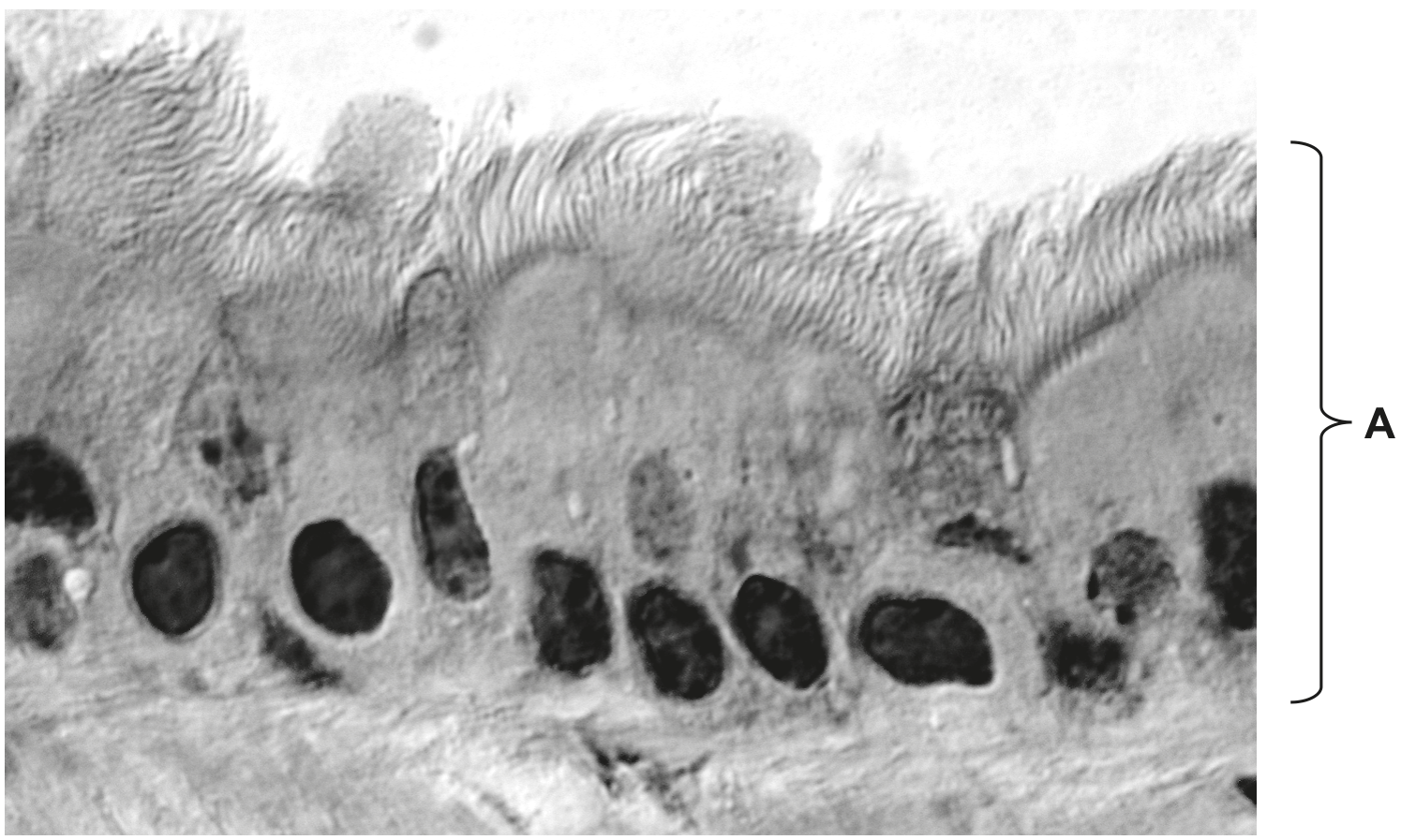
Fig. 1.2
State the names of the tissues A, B and D.
A
B
D
Fig. 1.1 is an image of a transverse section through the trachea of the gas exchange system.
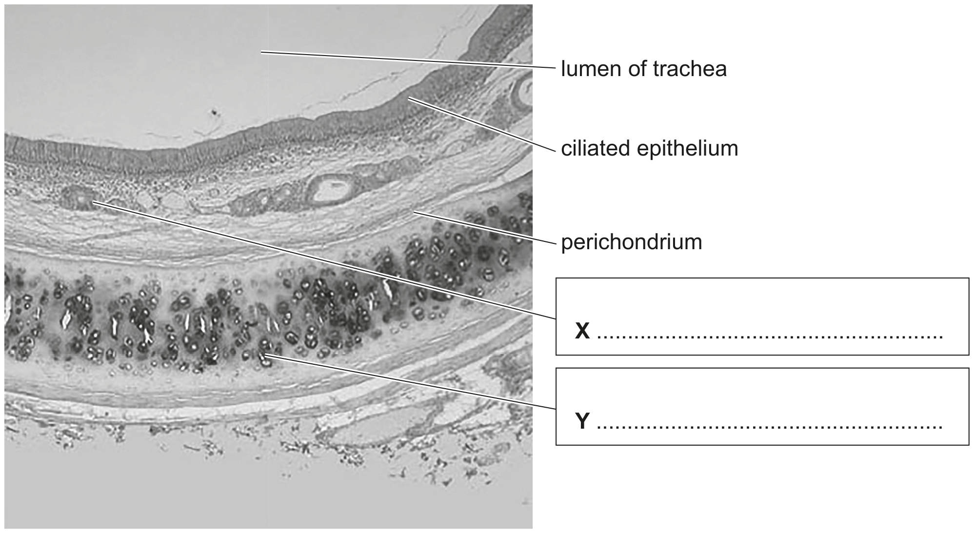
Fig. 1.1
Write the names of structures X and Y on Fig. 1.1 in the boxes provided.
Fig. 1.1 A is a photomicrograph of healthy lung tissue.
Fig. 1.1B is a photomicrograph of lung tissue from a person with emphysema, a chronic obstructive pulmonary disease (COPD). The images are both at magnification .
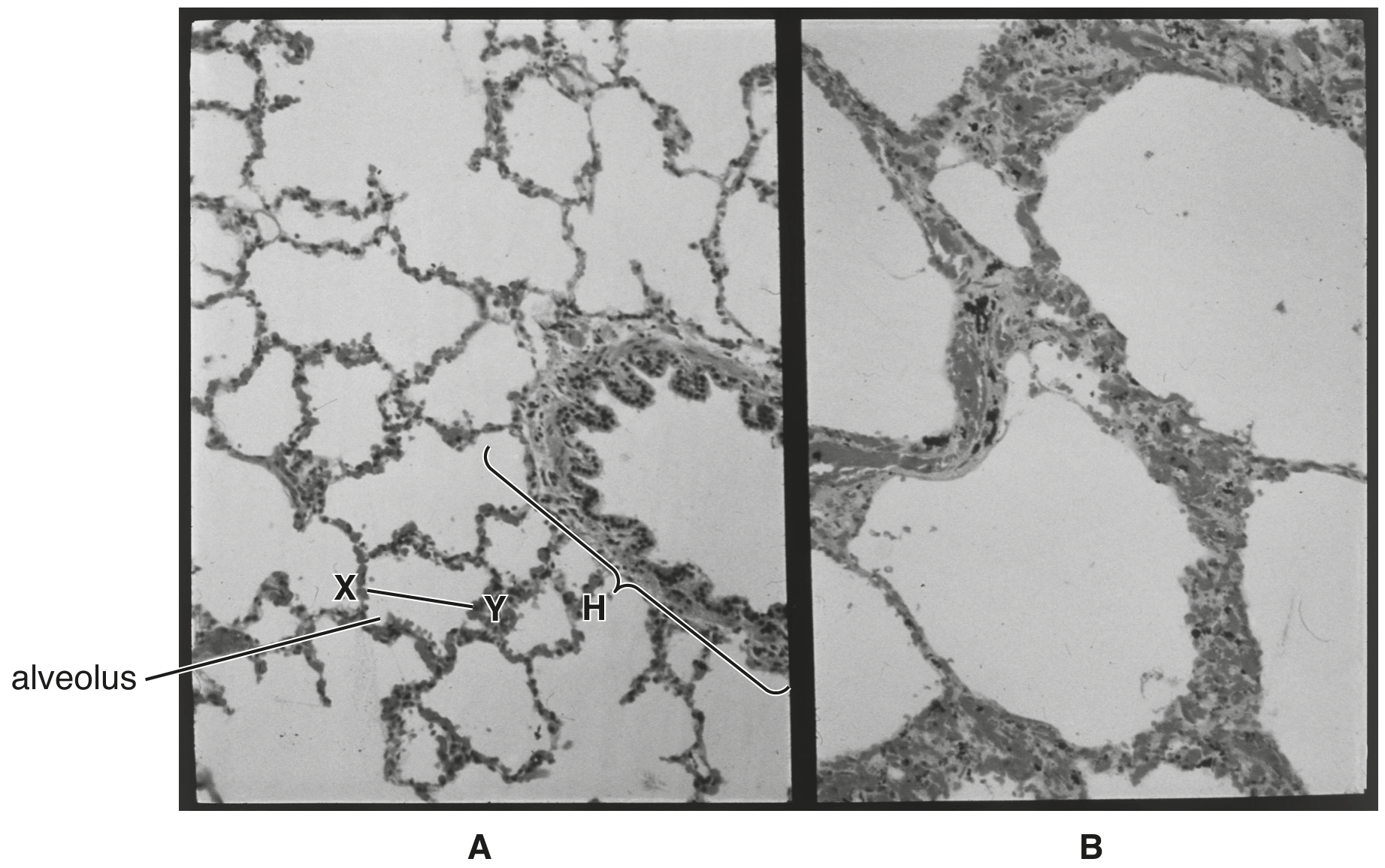
Fig. 1.1
Name the structure labelled H in Fig. 1.1A.
Fig. 1.1 shows the human gas exchange system.
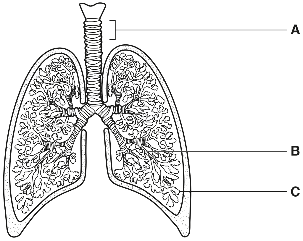
Fig. 1.1
Name the structures labelled A, B, and C in Fig. 1.1.
A
B
C
Name a non-infectious disease that affects the human gas exchange system.
Fig. 1.1 is a photomicrograph of epithelial cells in the bronchus.
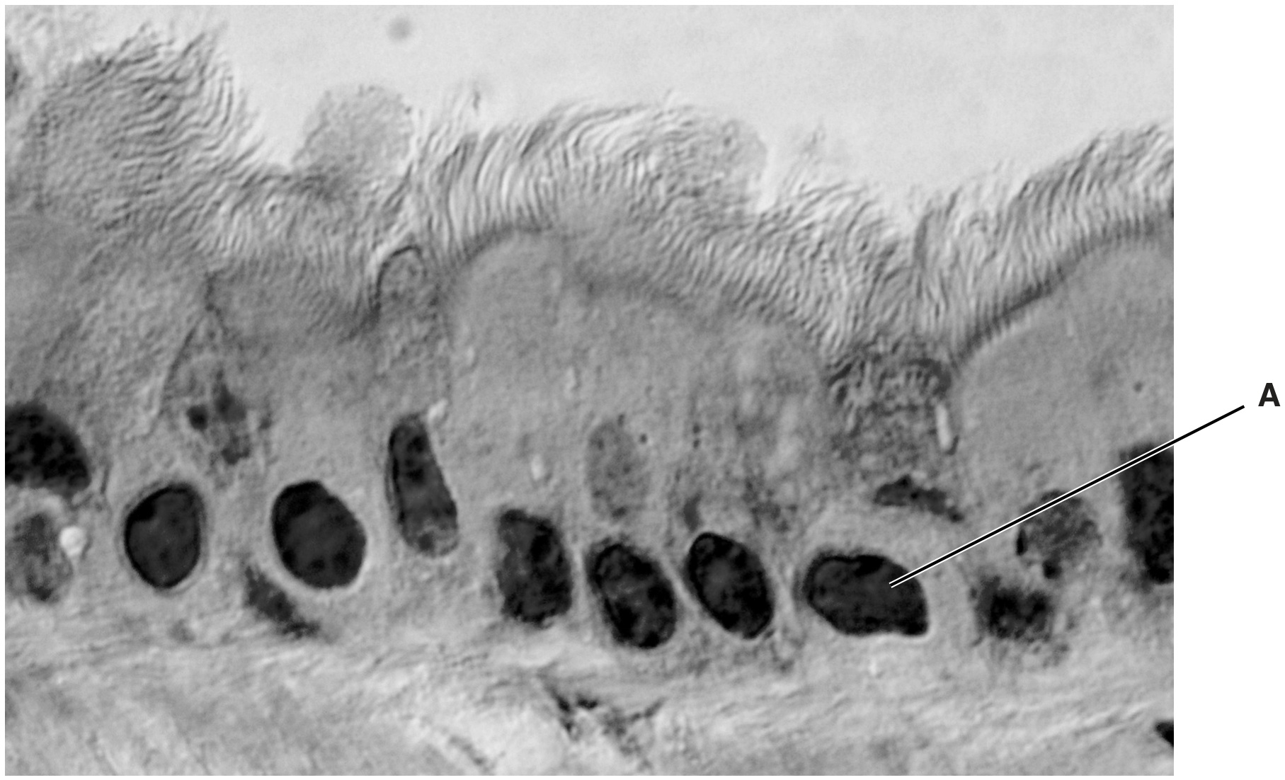
Fig. 1.1
Write a letter X on Fig. 1.1 to show the lumen of the bronchus.
Fig. 1.1 is a light micrograph of a section through part of the gas exchange system.
A, B and C are three different types of tissue.
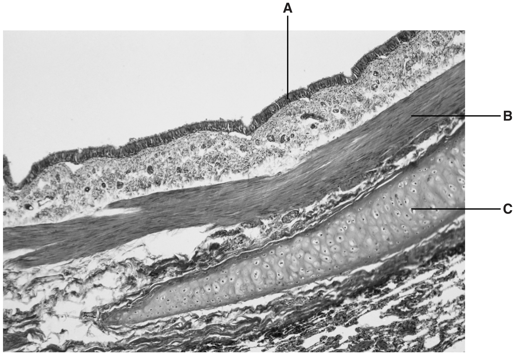
Fig. 1.1
Name the parts of the gas exchange system where tissue C is distributed.
Fig. 1.1 is an electron micrograph of cells from the lining of the small intestine.
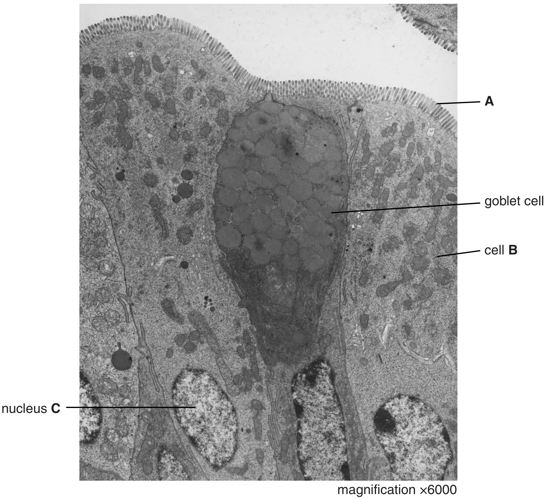
Fig. 1.1
State two ways in which the cells lining the alveoli in the lungs differ from cell B shown in Fig. 1.1.
1.
Fig. 1.1 is a scanning electron micrograph of part of the wall of the bronchus of a healthy human.
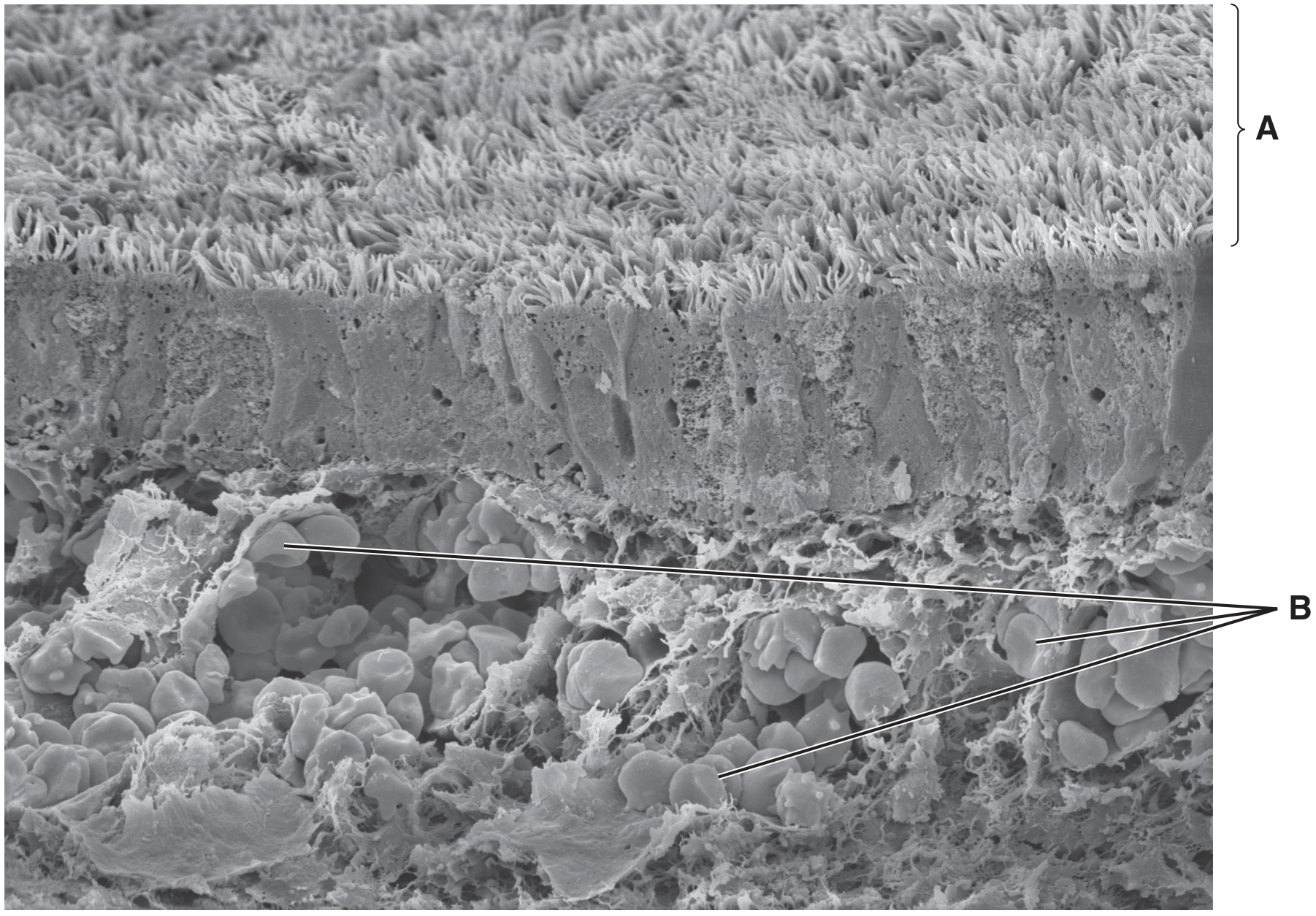
Fig. 1.1
Name the structures labelled A.