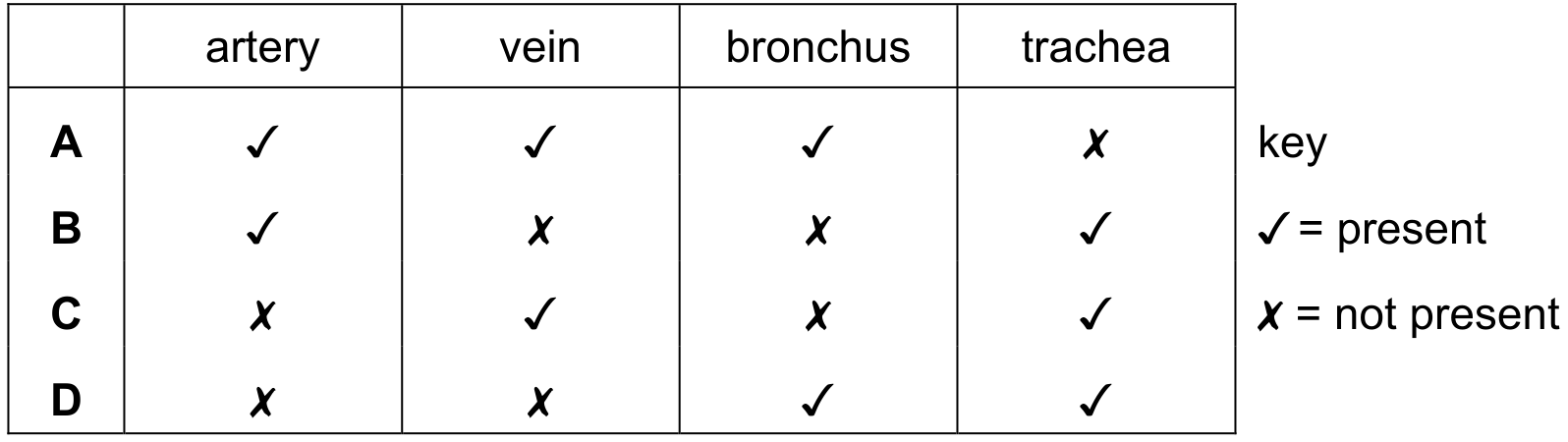
[Maximum number: 3]
The trachea of the gas exchange system branches into two airways, each of which enters a lung.
(a)
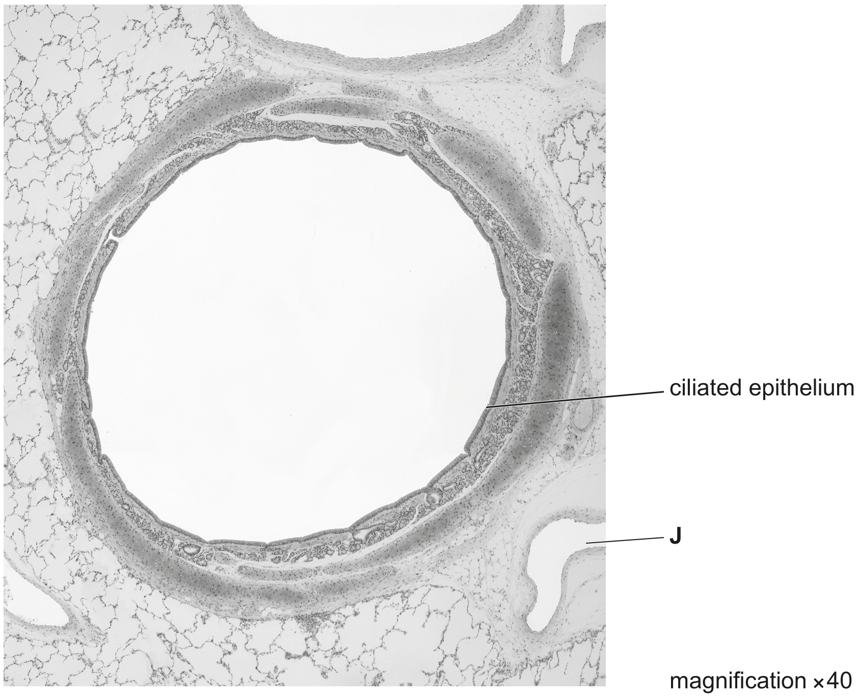
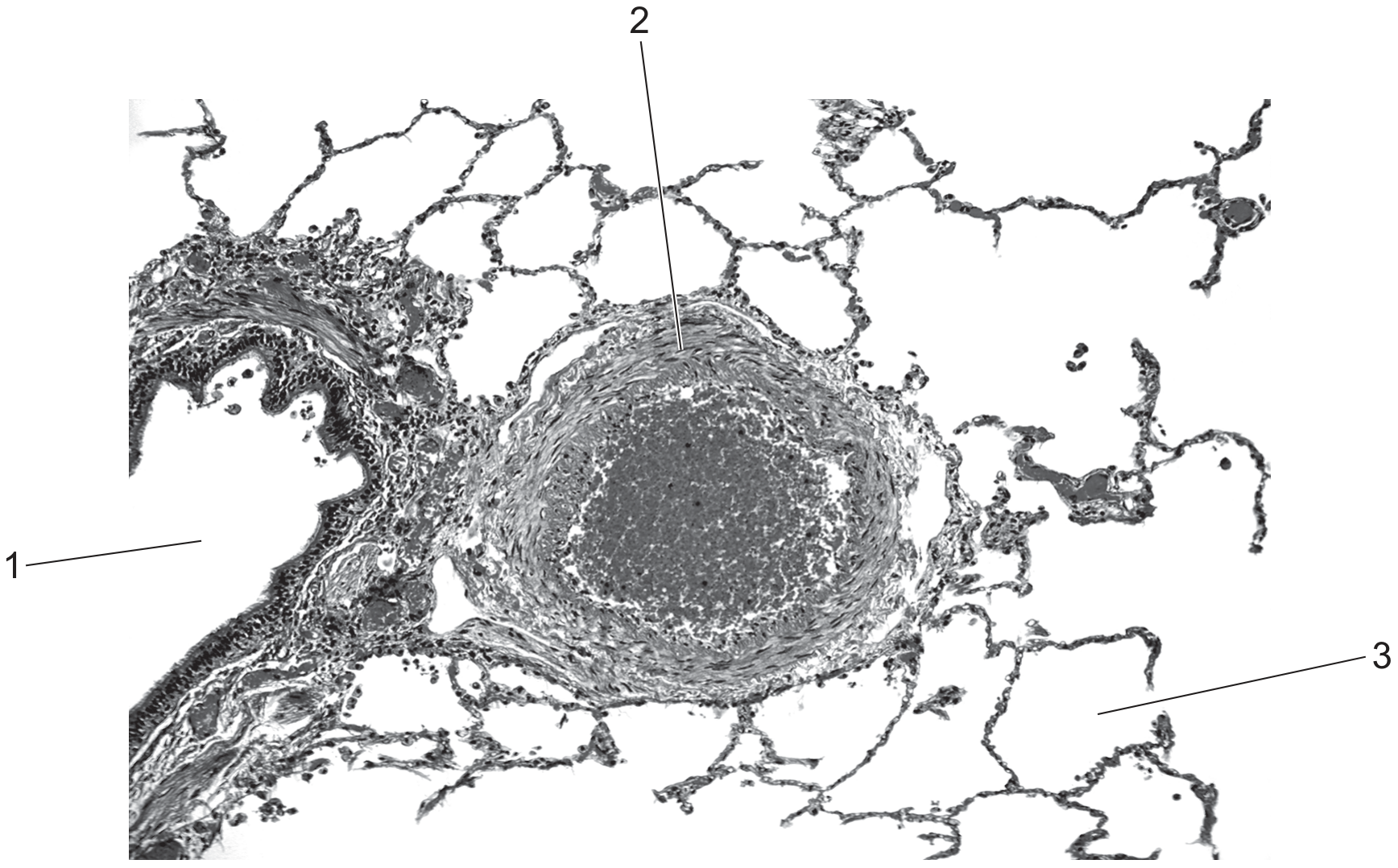
Fig. 1.1 is a photomicrograph of a section through part of the trachea.
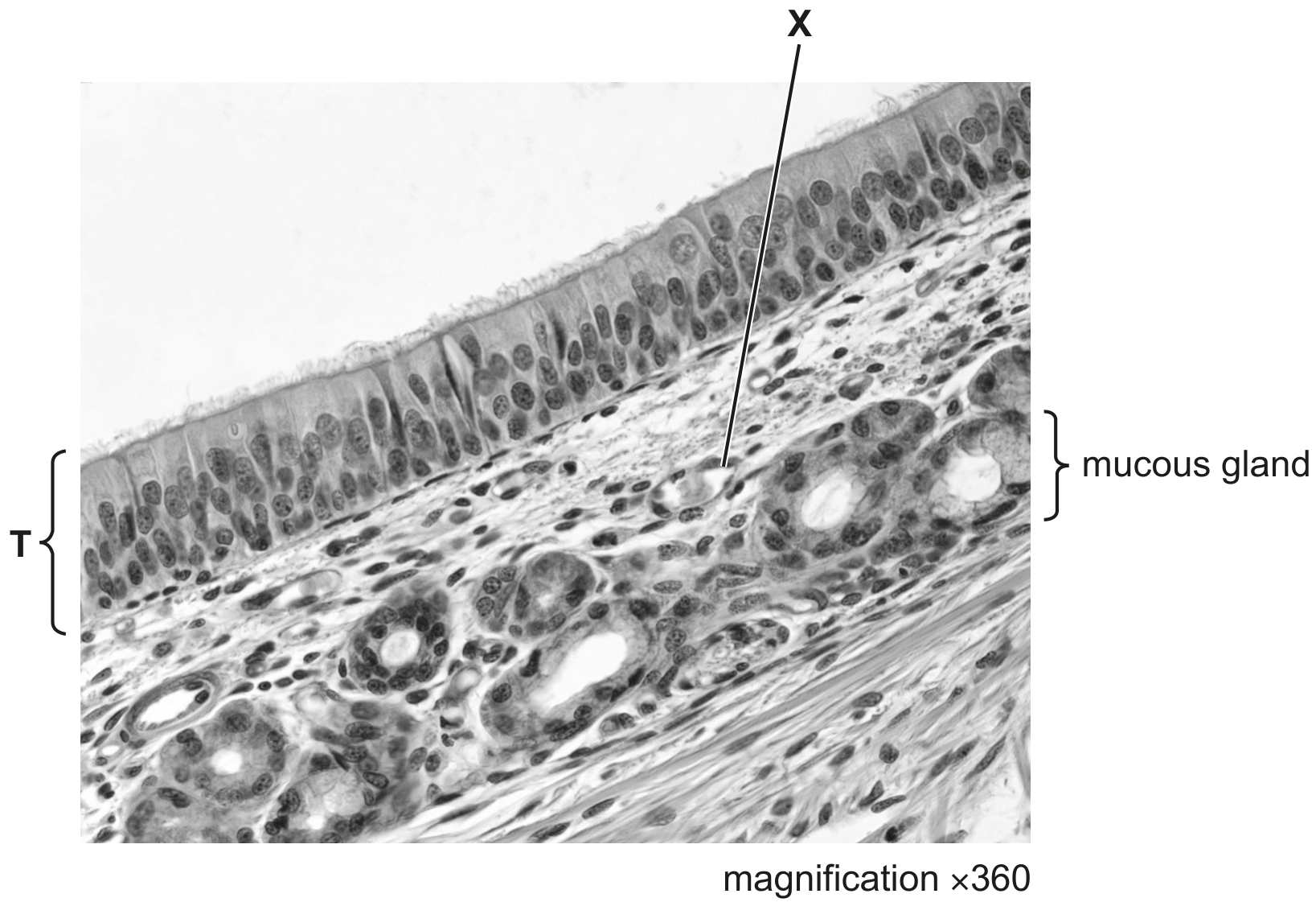
Fig. 1.1
[ 3 ]
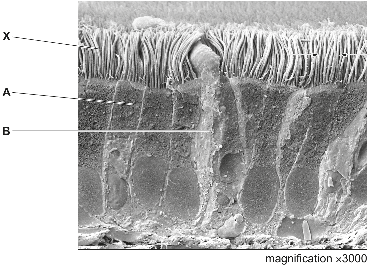
(i)
Identify structure X in Fig. 1.1 and outline the features that helped your identification. structure X=
[ 3 ]