[Maximum number: 5]
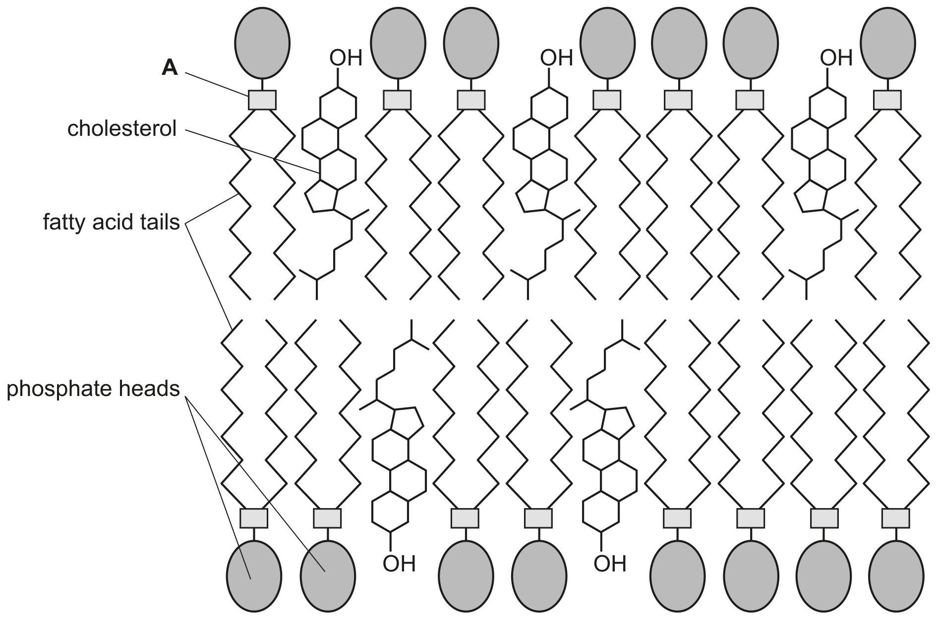
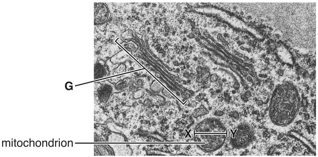
Fig. 1.1 is a diagram showing part of a cell surface membrane of an animal cell.
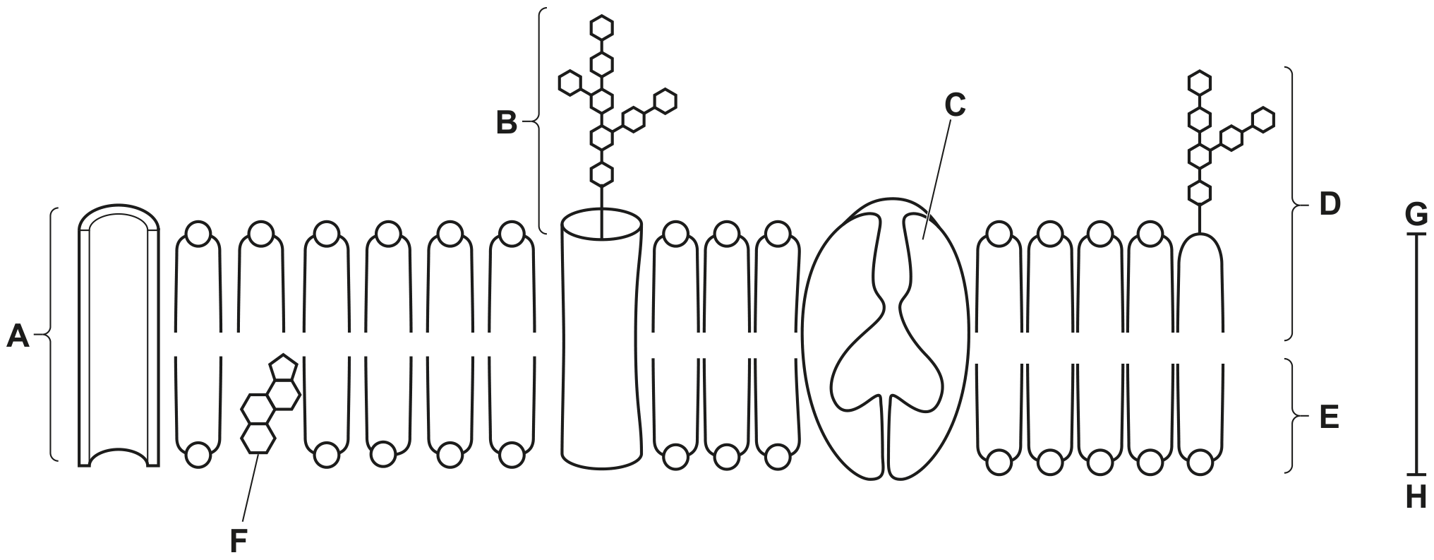
Fig. 1.1
(a)
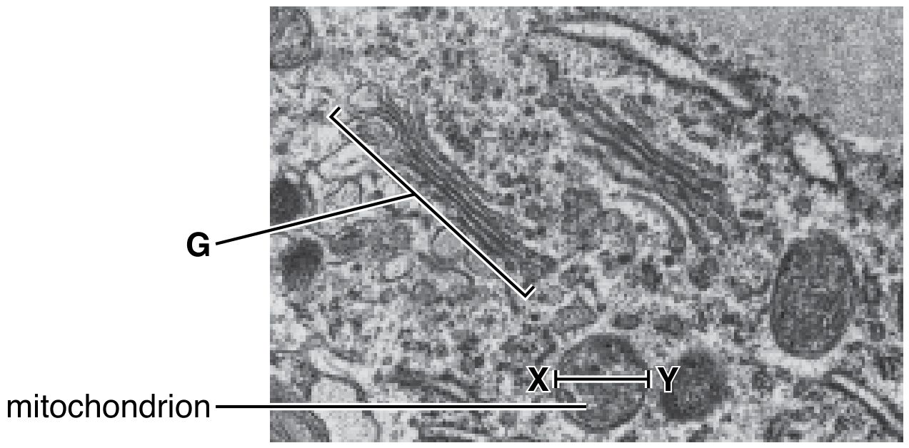
(i)
State the approximate thickness of the membrane as shown by the line G-H.
[ 1 ]
(ii)
Complete Table 1.1 to show:
- the names and functions of the components of the cell surface membrane
- the letters of the labels in Fig. 1.1 that identify each component.
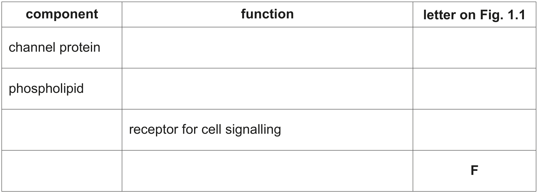
Table 1.1
[ 4 ]