EduNinja
(a)

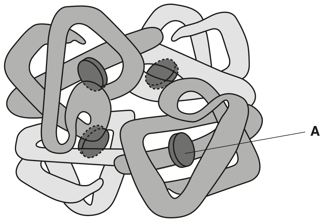
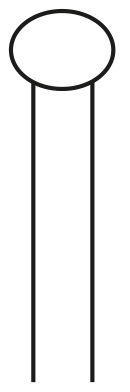
Fig. 1.1 is a diagram representing part of the phospholipid bilayer of a cell surface membrane.
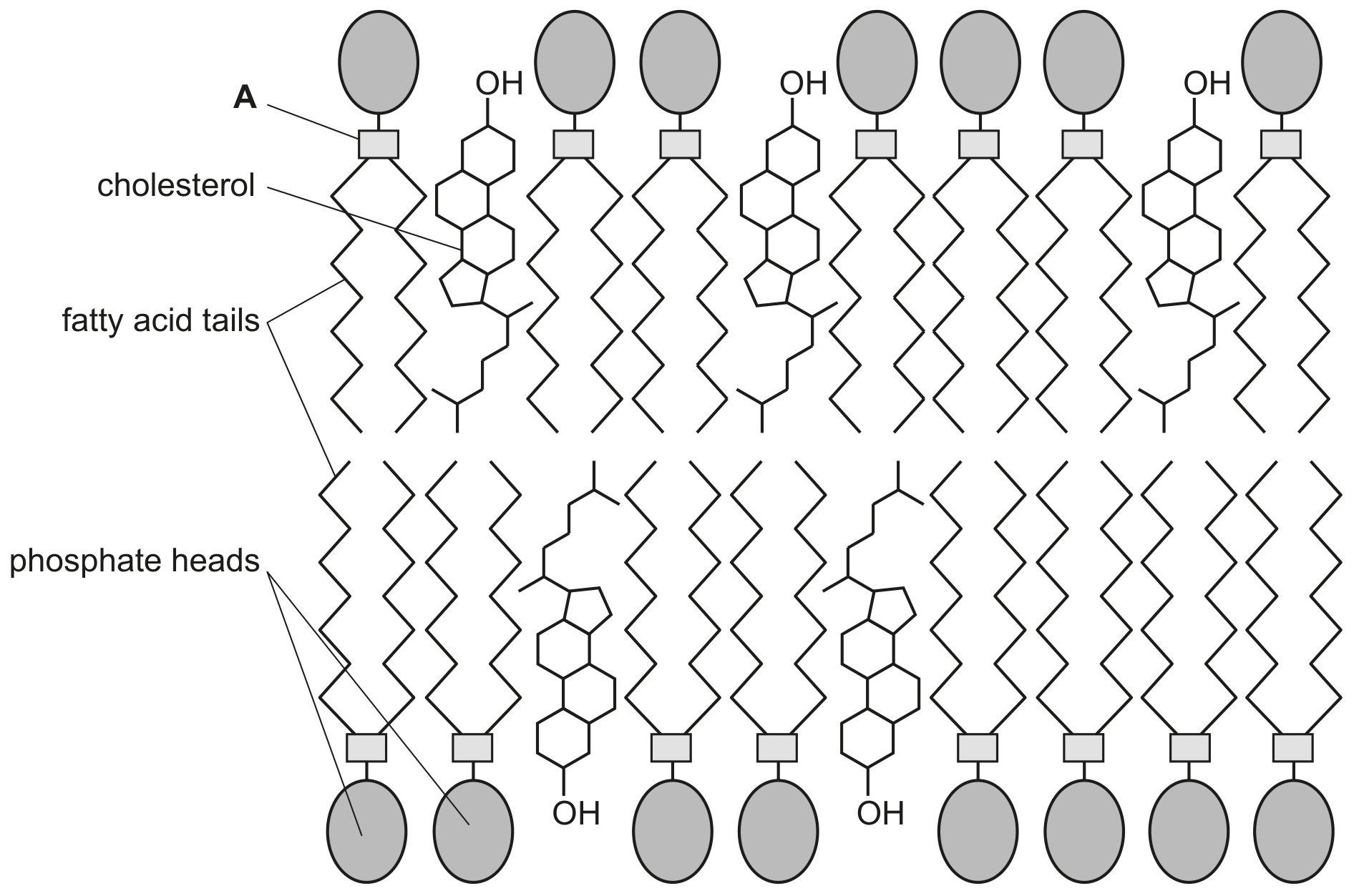
Fig. 1.1

Fig. 1.1

[ 1 ]
(i)

Identify the part of a phospholipid molecule, labelled A in Fig. 1.1, that forms bonds with the phosphate heads and with the fatty acid tails.

[ 1 ]
(a)
(i)

Starch granules contain amylose and amylopectin.

Describe the similarities and differences between the structure of amylose and the structure of amylopectin.

[ 4 ]
[Maximum number: 2]

The Golgi body, rough endoplasmic reticulum (RER) and smooth endoplasmic reticulum (SER) form part of the internal membrane system of a cell. The membranes have a fluid mosaic structure.

Fig. 1.1 is a transmission electron micrograph of one area of a liver cell showing a region with RER and a region with SER. Mitochondria are also visible in the image.

Fig. 1.1

Fig. 1.1

(a)

Phospholipids are one of the main components of membranes.

Describe the structure of a phospholipid molecule.

[ 2 ]
[Maximum number: 4]

Fig. 1.1 shows five biological molecules.

Fig. 1.1

Fig. 1.1

(a)

State the name of the bond in molecule A indicated by the arrow.

[ 1 ]
(b)

Molecule B is described as a saturated fatty acid.

State why molecule B is described as a saturated fatty acid.

[ 1 ]
(c)

Molecule D is a polymer.

State the name of the monomer that is used to synthesise this polymer.

[ 1 ]
(d)

Molecule A and molecule C dissolve in water. Molecule B does not dissolve in water.

Explain why molecule A and molecule C dissolve in water, but molecule B does not dissolve in water.

[ 1 ]
[Maximum number: 2]

Fig. 1.1 is a diagram of a molecule of haemoglobin.

Fig. 1.1

Fig. 1.1

(a)

The gene H B B codes for the β\beta-globin polypeptide.

State why a polypeptide, such as β\beta-globin, is described as a polymer.

[ 2 ]
(a)

Table 1.1 shows some features of four biological molecules that are all polymers.
Complete Table 1.1 by using a tick ()(\checkmark) to indicate the features that apply to each polymer.

Table 1.1

Table 1.1

[ 4 ]
(b)

Fig. 1.1 is a simple diagram of a phospholipid molecule.

Explain how the structure of a phospholipid molecule makes it suitable for its function in cell membranes. You may label and annotate Fig. 1.1 as part of your answer.

Fig. 1.1

Fig. 1.1

[ 3 ]
[Maximum number: 4]

Fig. 1.1 shows the structures of four biological molecules A, B, C and D.

Fig. 1.1

Fig. 1.1

(a)

Give the letter, A to D, of the molecule in Fig. 1.1 which:

[ 3 ]
(i)

contains ester bonds.

[ 3 ]
(b)

Some of the molecules in Fig. 1.1 can form polymers.

[ 1 ]
(i)

Name a polymer which can be formed only from many molecules of C.

[ 1 ]
[Maximum number: 2]

Fig. 1.1 is a labelled diagram of a leaf palisade mesophyll cell, as seen with a high quality light microscope.

Fig. 1.1

Fig. 1.1

An electron micrograph of the same leaf mesophyll cell at the same magnification would show more detail than is shown in Fig. 1.1.

(a)

In Fig. 1.1, starch granules are visible within the chloroplasts. Starch is the most common storage compound of plants. It is composed of amylopectin and amylose.

[ 2 ]
(i)

Describe the structural differences between amylopectin and amylose.

[ 2 ]
[Maximum number: 4]

Fig. 1.1 is a drawing made from an electron micrograph of a mammalian liver cell.

Fig. 1.1

Fig. 1.1

(a)

As shown in Fig. 1.1, liver cells contain many storage granules of glycogen.

Describe the molecular structure of glycogen and explain how this structure makes it suitable for storage.

[ 4 ]
[Maximum number: 1]

Fig. 1.1 is an electron micrograph of three cells of the same species of bacterium, Erwinia carotovora.

Fig. 1.1

Fig. 1.1

(a)

E. carotovora causes a disease in carrot and potato plants.

The bacteria release an enzyme called pectinase which hydrolyses the polysaccharide pectin. Pectin helps plant cells to attach to each other.

[ 1 ]
(i)

Name the type of chemical bond which will be hydrolysed by pectinase.

[ 1 ]
0