EduNinja
[Maximum number: 2]

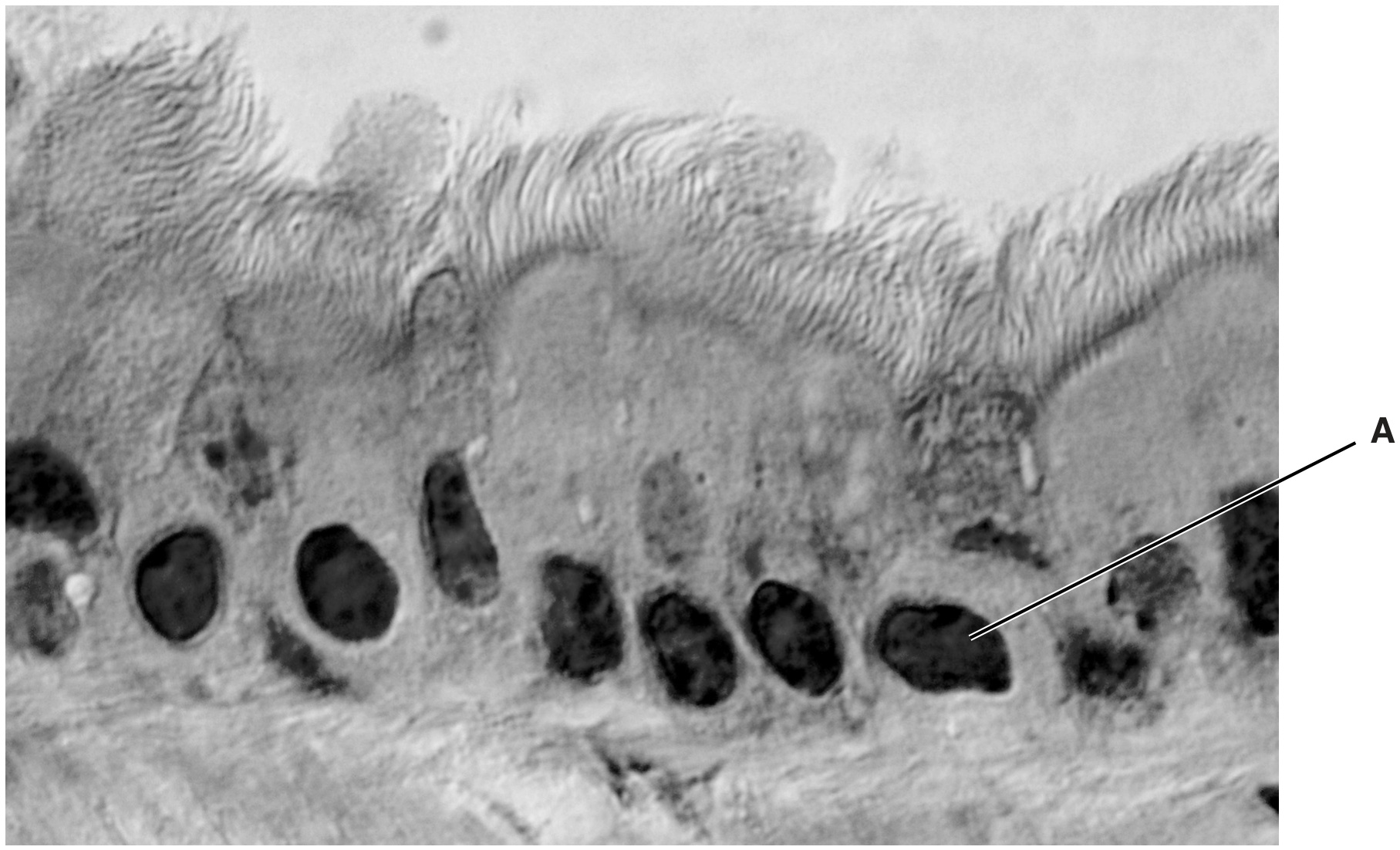
Smooth muscle is a tissue composed of smooth muscle cells. The cells contain cytoplasm packed with proteins that are involved in contraction and relaxation.

(a)

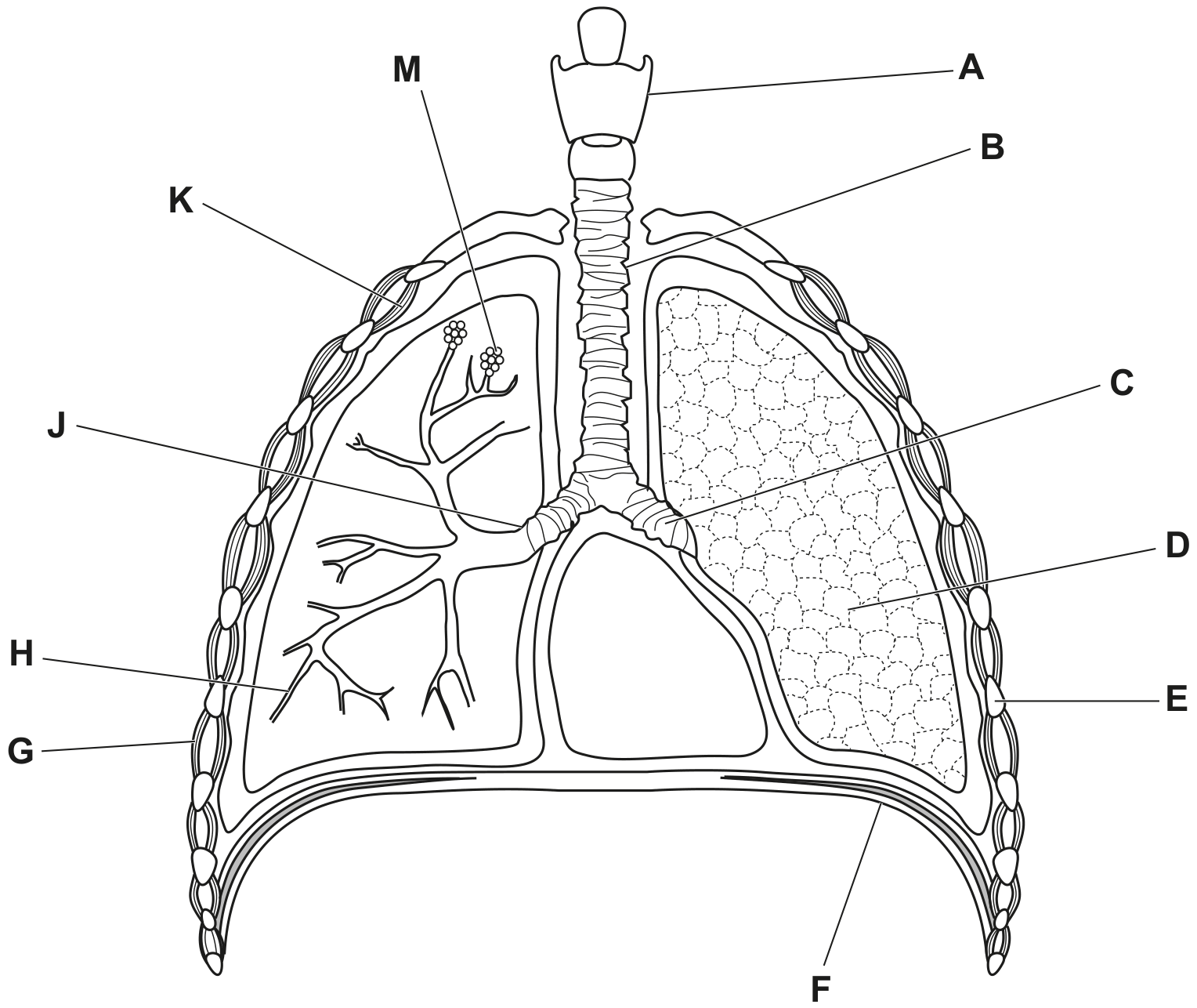
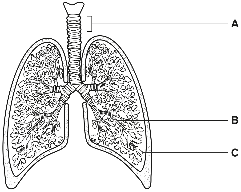
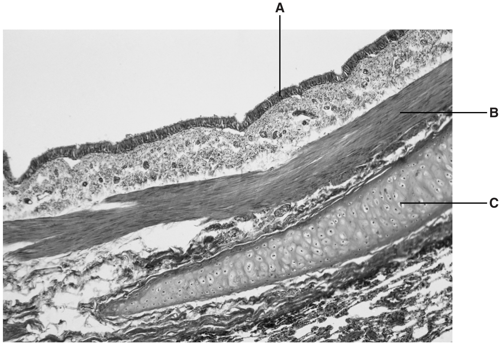
Smooth muscle is present in the airways of the gas exchange system.

Explain how smooth muscle cells in the walls of the bronchioles contribute to the function of these airways.

[ 2 ]
[Maximum number: 7]

The trachea of the gas exchange system branches into two airways, each of which enters a lung.

(a)

Name the airways that branch from the trachea to enter the lungs.

[ 1 ]
(b)

Fig. 1.1 is a photomicrograph of a section through part of the trachea.

Fig. 1.1

Fig. 1.1

[ 6 ]
(i)

In Fig. 1.1, one of the tissues in the trachea is labelled T.

Describe the structural features of tissue T visible in Fig. 1.1.

[ 3 ]
(ii)

Identify structure X in Fig. 1.1 and outline the features that helped your identification. structure X=

[ 3 ]
[Maximum number: 6]

Fig. 1.1 and Fig. 1.2 are photomicrographs showing the distribution of tissues in the lungs.
Fig. 1.1 is a photomicrograph of a section through part of the lungs.
Fig. 1.2 is a high-power view of the area indicated on Fig. 1.1.

Fig. 1.1

Fig. 1.1

section enlarged in Fig. 1.2

Fig. 1.2

Fig. 1.2

(a)

State the names of the tissues A, B and D.

A

B
D

[ 3 ]
(b)

Describe the role of the glands labelled C in Fig. 1.1 in maintaining the health of the gas exchange system.

[ 3 ]
(a)

Fig. 1.1 is an image of a transverse section through the trachea of the gas exchange system.

Fig. 1.1

Fig. 1.1

Write the names of structures X and Y on Fig. 1.1 in the boxes provided.

[ 2 ]
(b)

Some structures in the gas exchange system are listed in alphabetical order in Table 1.1.
- Write YES in the box provided if the structure contains smooth muscle.
- Write NO in the box provided if the structure does not contain smooth muscle.

Table 1.1

Table 1.1

[ 1 ]
[Maximum number: 4]

Fig. 1.1 shows a section through the human chest. The gross structure of the human gas exchange system is shown.

Fig. 1.1

Fig. 1.1

(a)

Descriptions of some parts of the gas exchange system are shown in Table 1.1.

Complete Table 1.1 to:
- state the name of the part described
- write a letter from Fig. 1.1 to identify each part.

Table 1.1

Table 1.1

[ 4 ]
[Maximum number: 5]


Fig. 1.1 A is a photomicrograph of healthy lung tissue.
Fig. 1.1B is a photomicrograph of lung tissue from a person with emphysema, a chronic obstructive pulmonary disease (COPD). The images are both at magnification ×40\times 40.

Fig. 1.1

Fig. 1.1

(a)

Name the structure labelled H in Fig. 1.1A.

[ 1 ]
(b)

Gas exchange in the lungs occurs in the alveoli.

Describe the process of gas exchange in healthy lungs and suggest how this compares with gas exchange in the diseased lung shown in Fig. 1.1B.

[ 4 ]
[Maximum number: 4]

Fig. 1.1 shows the human gas exchange system.

Fig. 1.1

Fig. 1.1

(a)

Name the structures labelled A, B, and C in Fig. 1.1.

A
B
C

[ 3 ]
(b)

Name a non-infectious disease that affects the human gas exchange system.

[ 1 ]
[Maximum number: 5]

Fig. 1.1 is a photomicrograph of epithelial cells in the bronchus.

Fig. 1.1

Fig. 1.1

(a)
(i)

Write a letter X on Fig. 1.1 to show the lumen of the bronchus.

[ 1 ]
(ii)

Name the structure in Fig. 1.1 labelled A.

[ 1 ]
(iii)

State one feature of the cells, visible in Fig. 1.1, which indicates that these are not epithelial cells from the alveolus.

(b)

Epithelial cells are replaced when they are damaged.

[ 3 ]
(i)

The cells shown in Fig. 1.1 are from a non-smoker.

Smoking causes damage to the epithelial cells of the lungs.
Describe the appearance of the lining of the bronchus in a long-term smoker.

[ 3 ]
[Maximum number: 5]

Fig. 1.1 is a light micrograph of a section through part of the gas exchange system.
A, B and C are three different types of tissue.

Fig. 1.1

Fig. 1.1

(a)

The cell types in tissue A have different functions.

Describe how the cell types work together to maintain the health of the gas exchange system.

[ 3 ]
(b)

Suggest why the cells in tissue B have many mitochondria.

[ 1 ]
(c)

Name the parts of the gas exchange system where tissue C is distributed.

[ 1 ]
0