[Maximum number: 2]
Fig. 1.1 is a scanning electron micrograph of part of the wall of the bronchus of a healthy human.
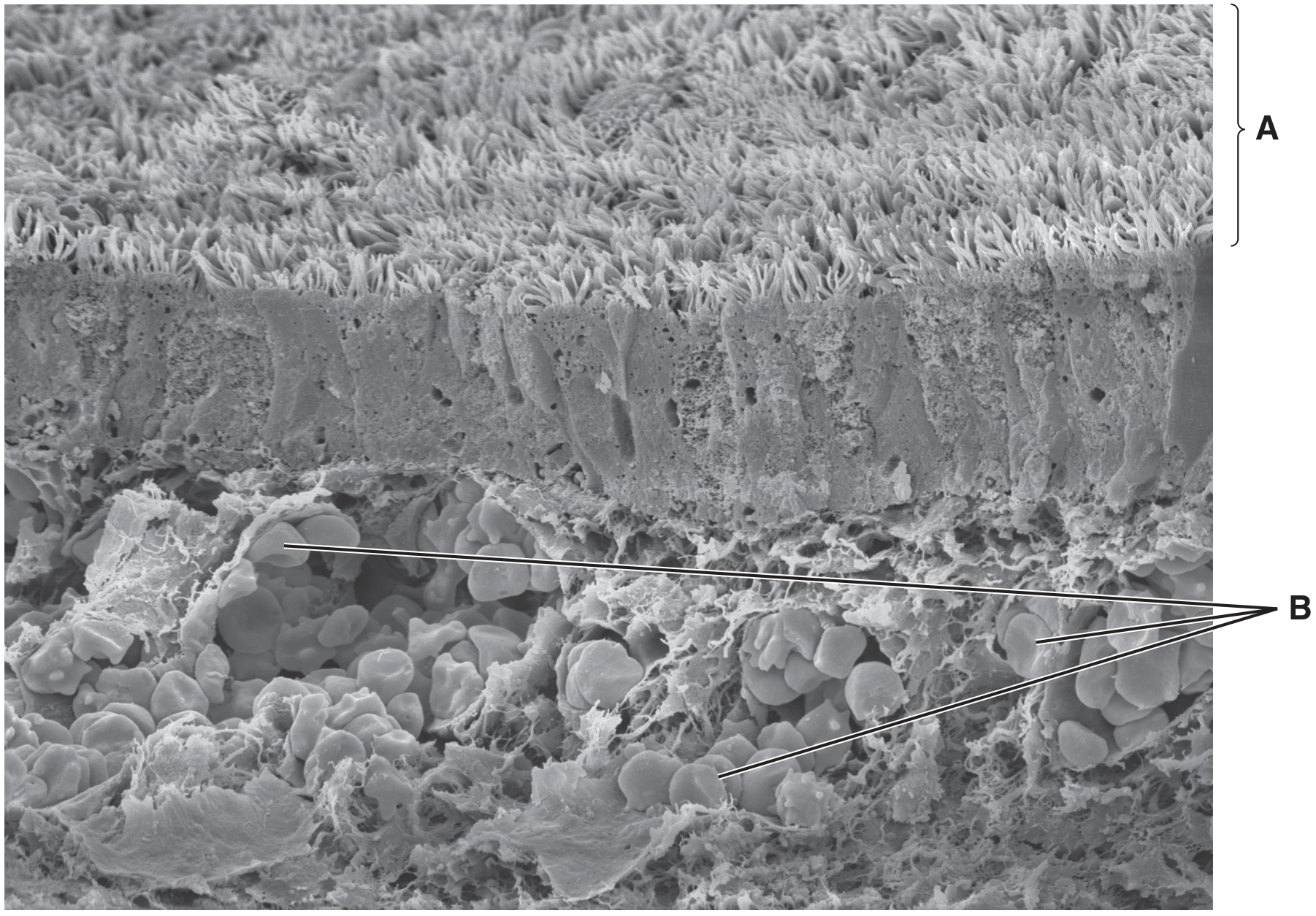
Fig. 1.1
(a)
Name two tissues found in the wall of the bronchus that are not visible in Fig. 1.1.
1.
[ 2 ]
 EduNinja
Fig. 1.1 is a scanning electron micrograph of part of the wall of the bronchus of a healthy human.
Fig. 1.1
Name two tissues found in the wall of the bronchus that are not visible in Fig. 1.1.
1.
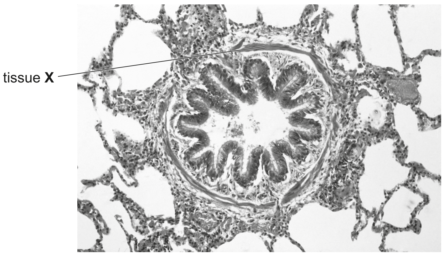
Fig. 1.1 is a light micrograph of a section through part of the gas exchange system.
A, B and C are three different types of tissue.
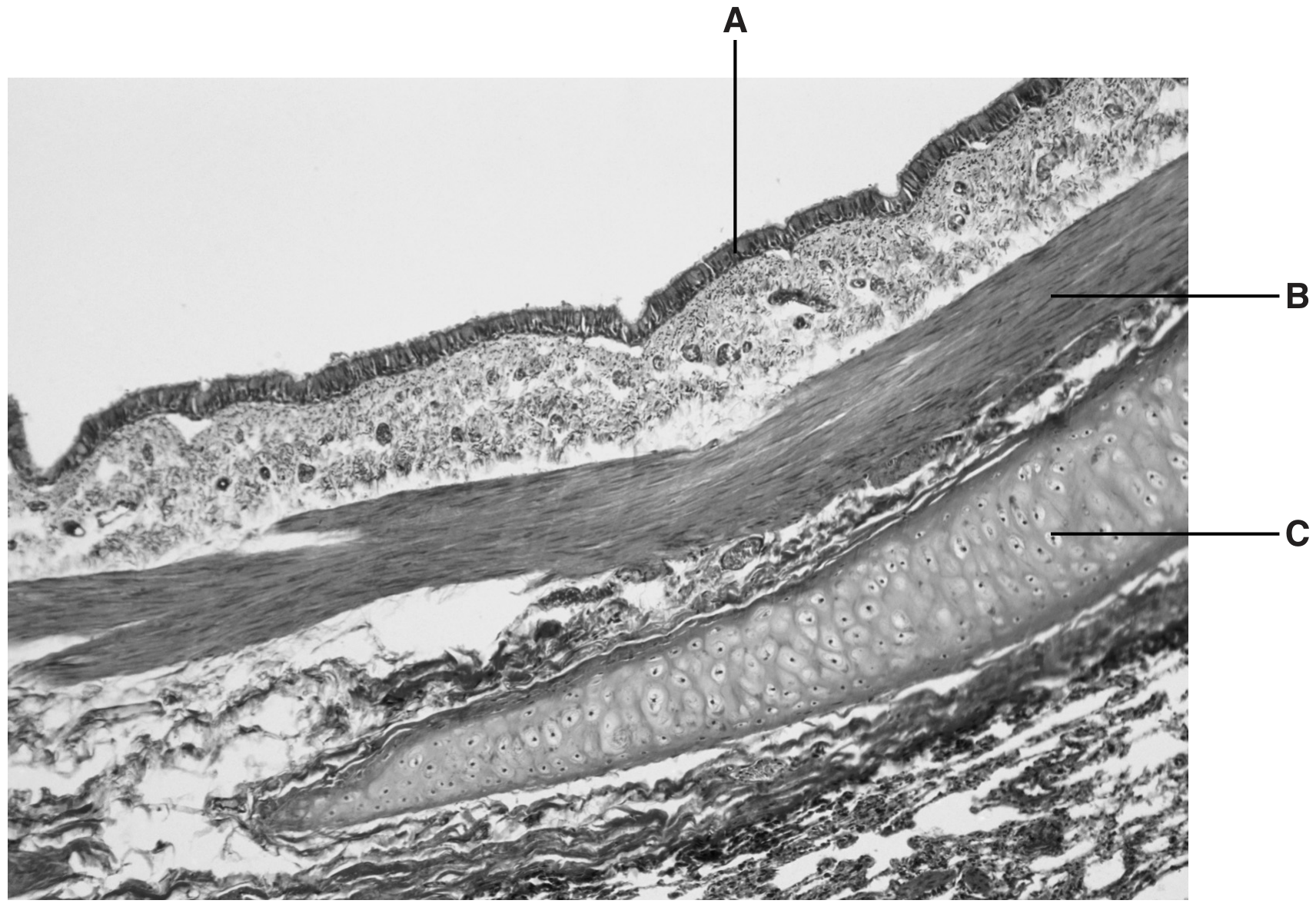
Fig. 1.1
Suggest why the cells in tissue B have many mitochondria.
Fig. 1.1 is a photomicrograph of epithelial cells in the bronchus.
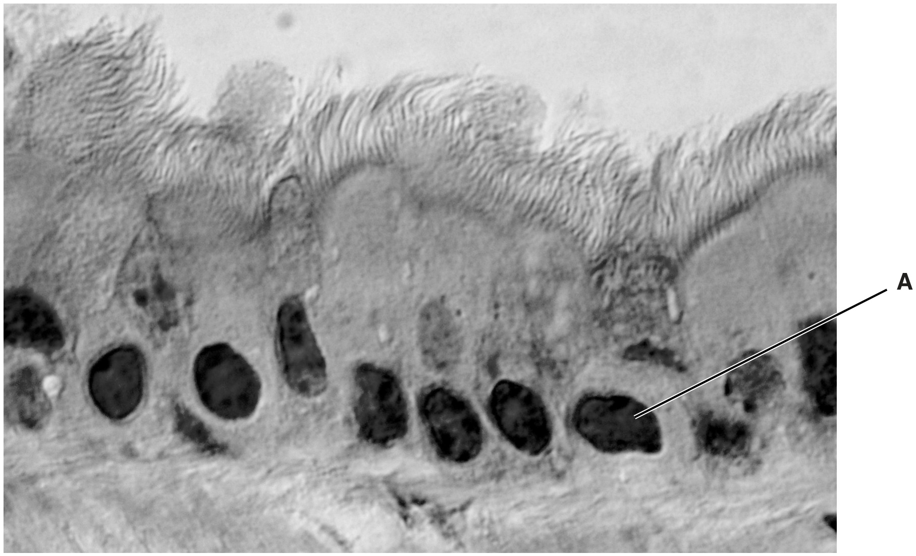
Fig. 1.1
Name the structure in Fig. 1.1 labelled A.
Some structures in the gas exchange system are listed in alphabetical order in Table 1.1.
- Write YES in the box provided if the structure contains smooth muscle.
- Write NO in the box provided if the structure does not contain smooth muscle.
Table 1.1
The trachea of the gas exchange system branches into two airways, each of which enters a lung.
Fig. 1.1 is a photomicrograph of a section through part of the trachea.
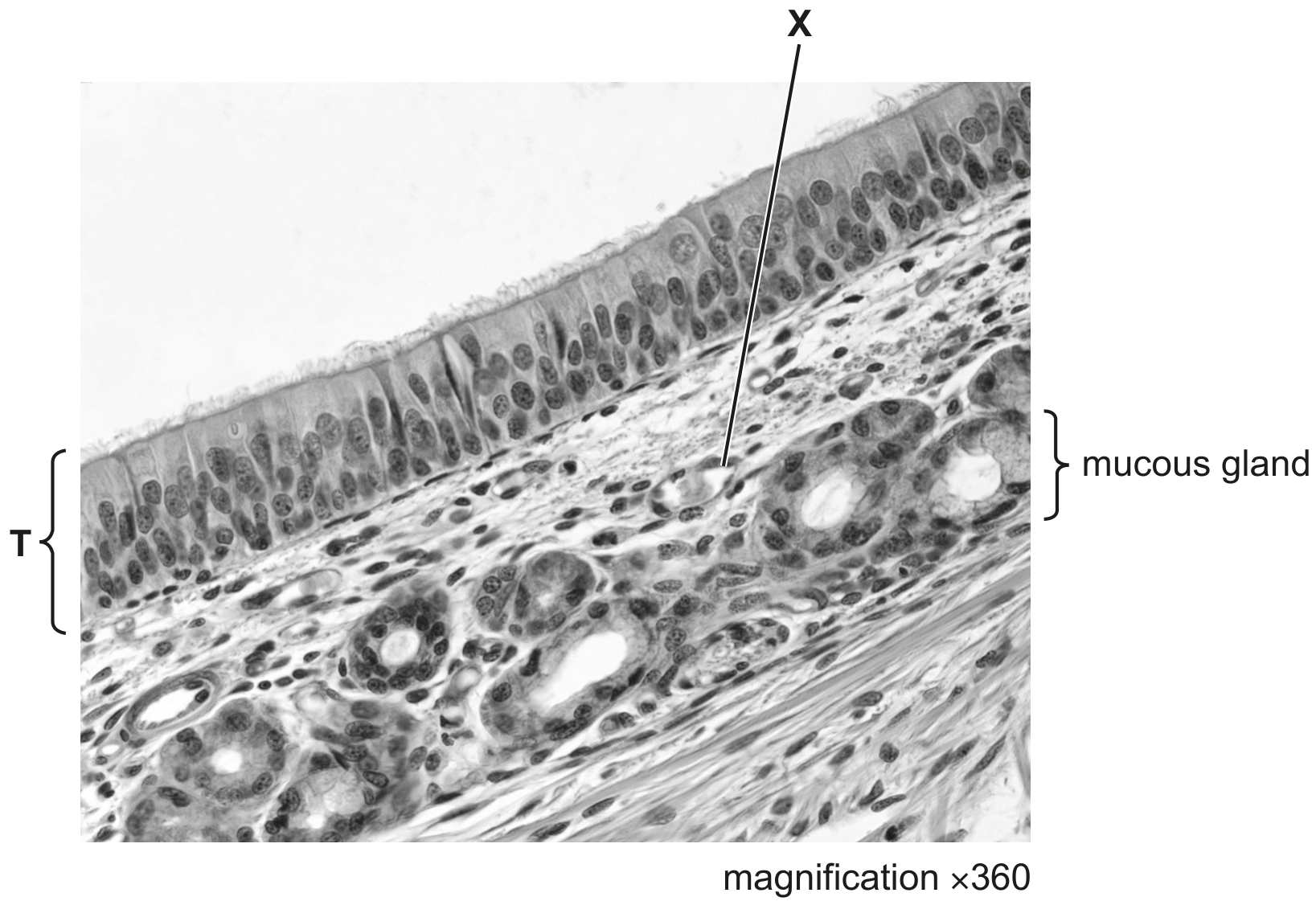
Fig. 1.1
In Fig. 1.1, one of the tissues in the trachea is labelled T.
Describe the structural features of tissue T visible in Fig. 1.1.
The airways of the gas exchange system are lined with epithelium. Gradual changes in the structural features of this epithelium occur as the airways branch and become increasingly narrow.
Fig. 4.1 is a photomicrograph of a section through a bronchiole, which is surrounded by alveoli.
magnification \(\times 40\)
There are structural differences between the epithelium of the bronchiole and the epithelium of an alveolus.
Describe the differences between the epithelium of bronchioles and the epithelium of alveoli, other than differences in the number of goblet cells.
Tissue X, shown in Fig. 4.1, is located in the wall of the bronchiole. Name tissue X and outline the function of tissue X in the bronchiole. tissue X =
Fig. 4.1 is a scanning electron micrograph showing the tissue that lines the bronchi in the gas exchange system.
Fig. 4.2 is a transmission electron micrograph of a horizontal section made at the position indicated by the two arrows in Fig. 4.1.
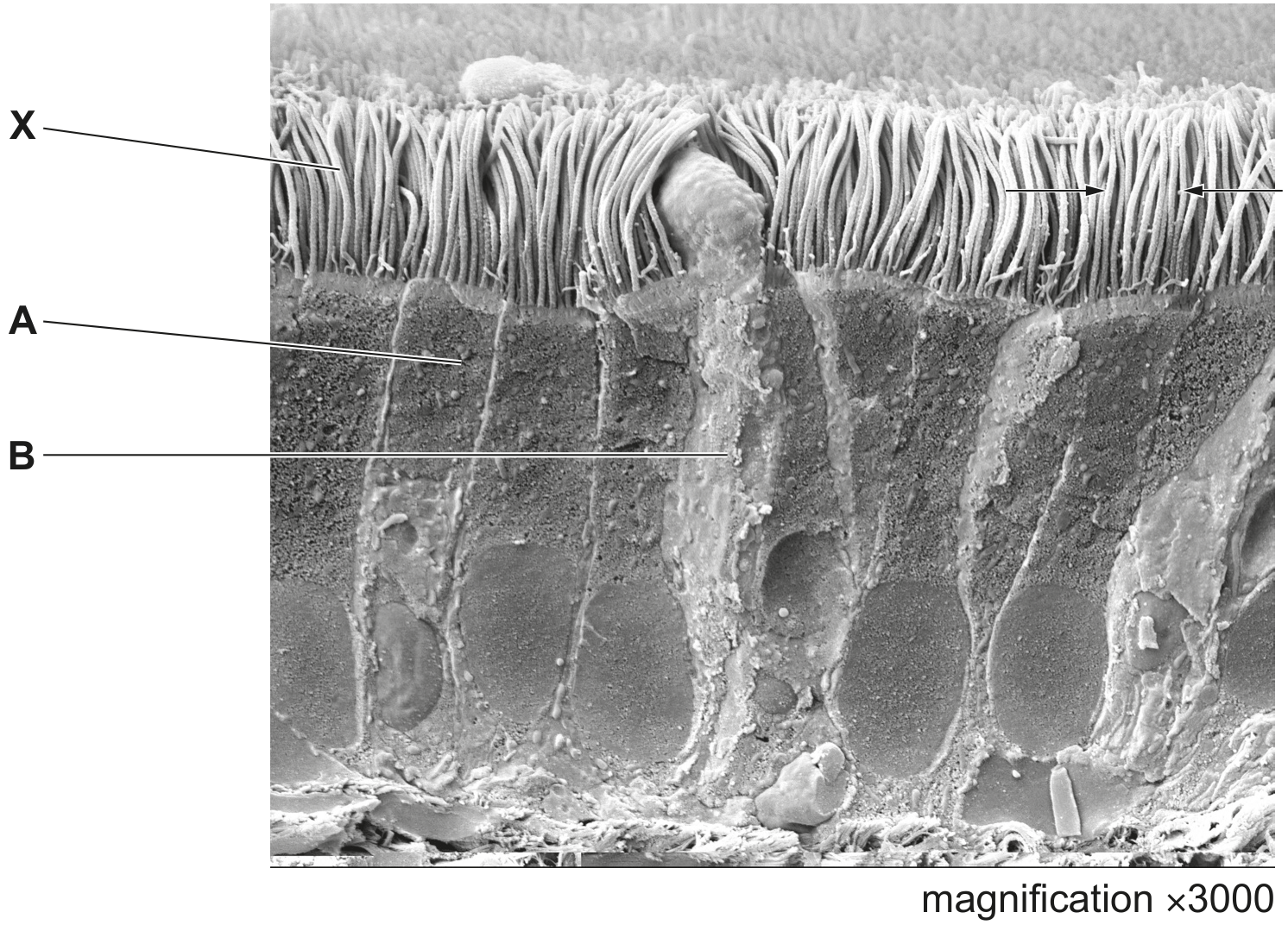
Fig. 4.1
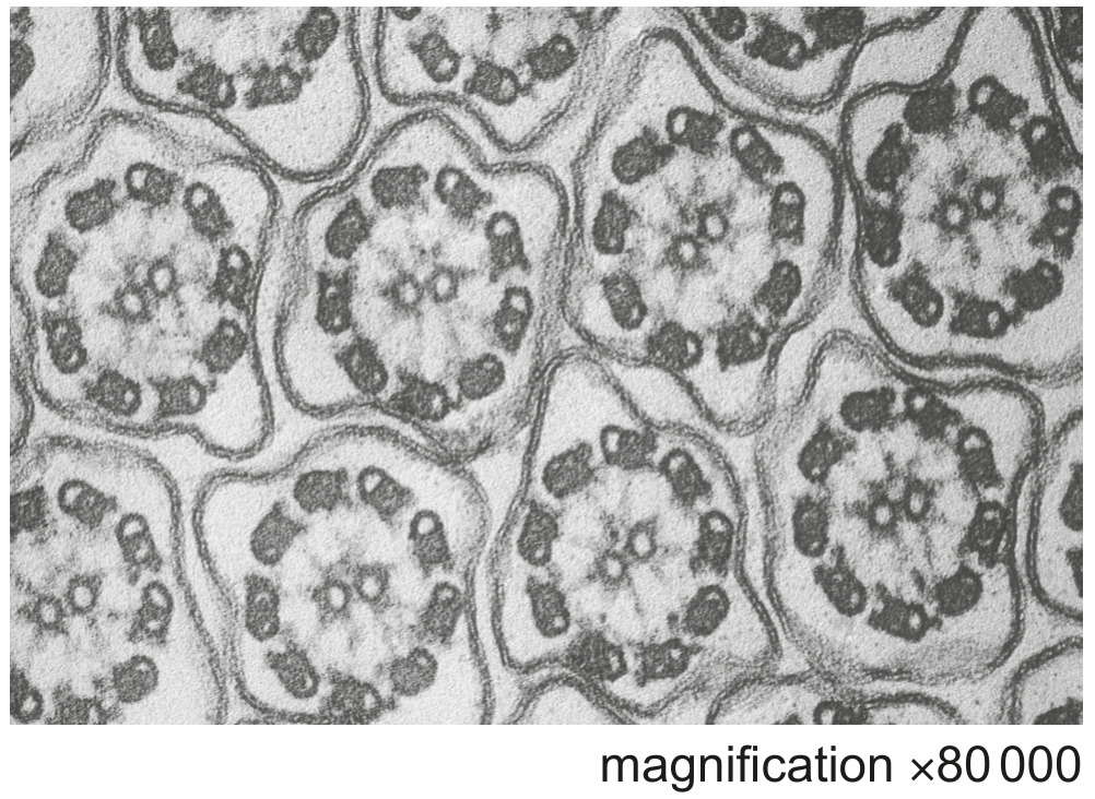
Fig. 4.2
The structures labelled X in Fig. 4.1 have a characteristic internal appearance, as seen in Fig. 4.2.
Describe the internal appearance of the structures labelled X.
Explain how Fig. 4.2 shows that each of the structures labelled X are intracellular.
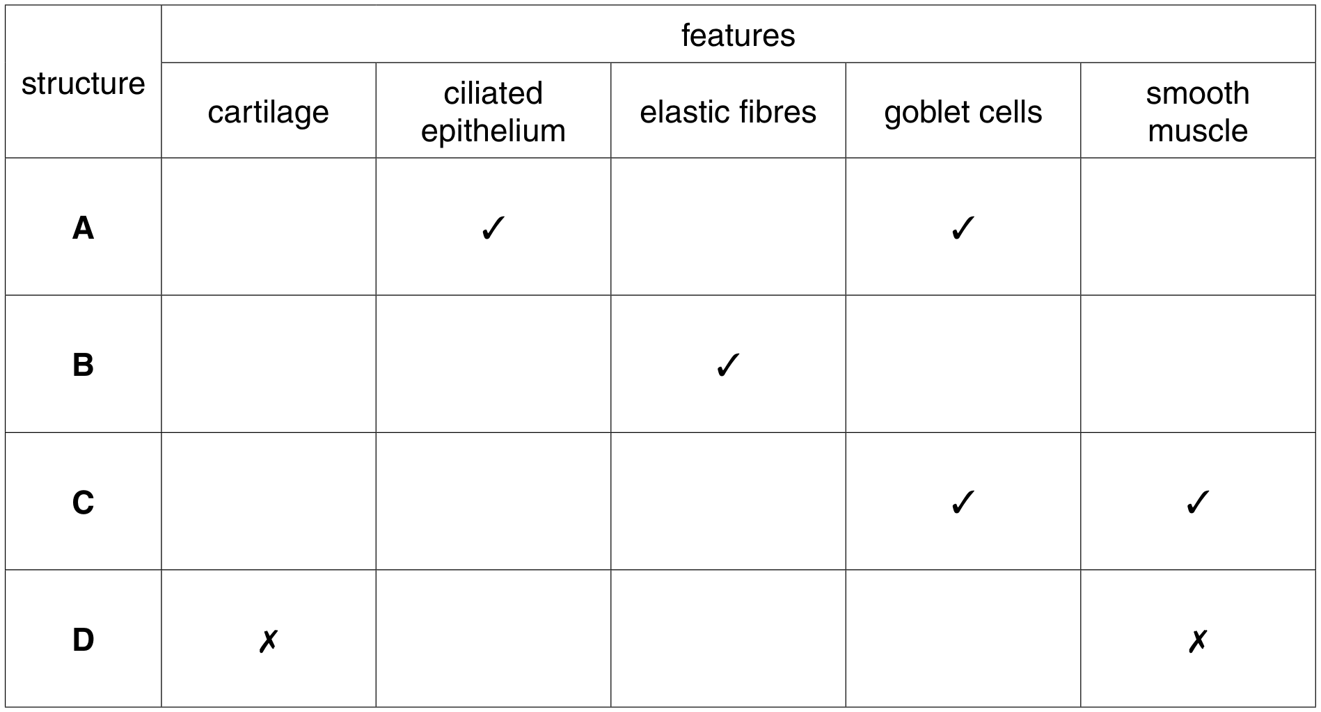
Fig. 5.1 is a diagram of part of the human gas exchange system.
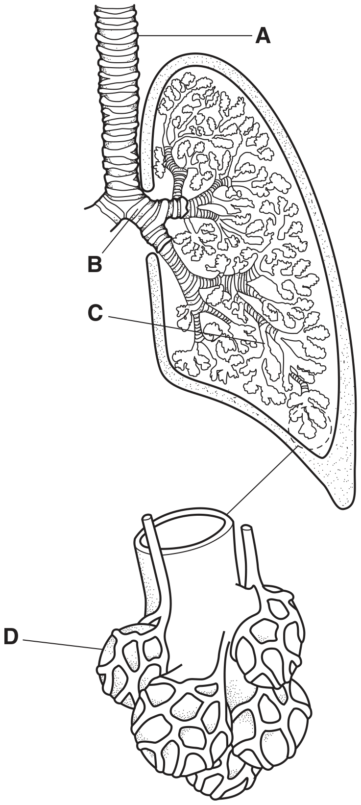
Fig. 5.1
Complete the table to show the distribution of the structural features within the parts of the gas exchange system, A to D, shown in Fig. 5.1.
Use a tick if the feature is present and a cross (X) if the feature is absent. Some of the boxes have been completed for you.
Smooth muscle and cartilage are two of the tissues found in the walls of structures of the gas exchange system of mammals.
Complete Fig. 5.1 to show the distribution of these tissues in the gas exchange system of mammals.
Choose from the four structures listed below.
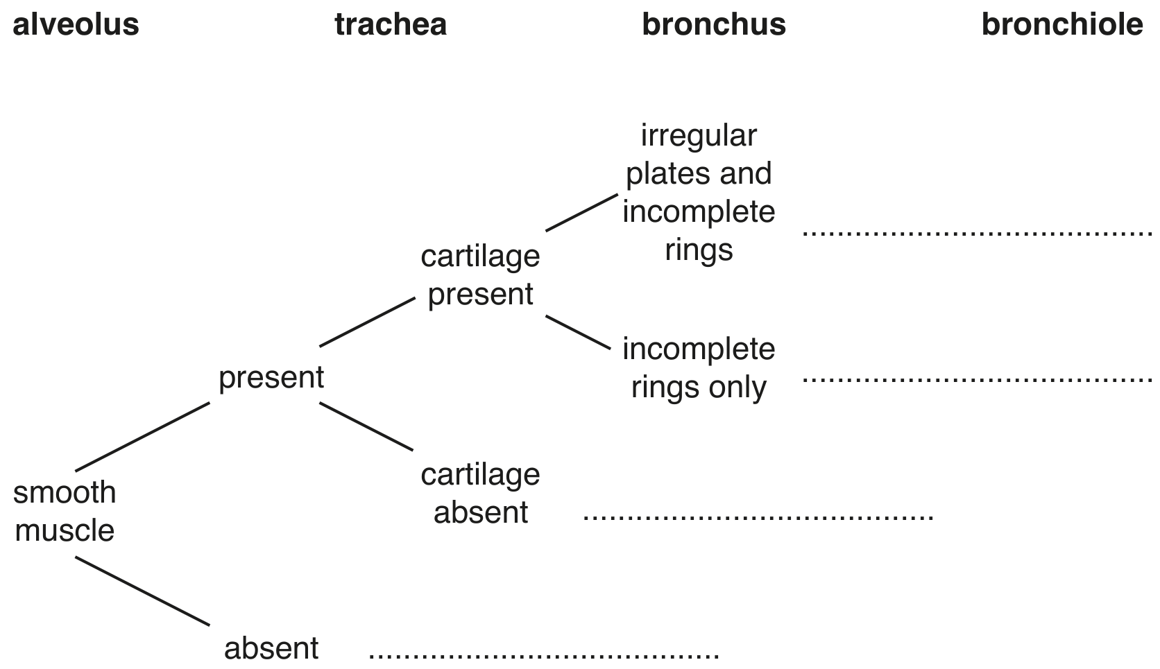
Fig. 5.1
Tobacco smoke is known to be one of the causes of lung cancer and chronic obstructive pulmonary disease (COPD).
Fig. 5.2 shows a section through the wall of one part of the gas exchange system in a person with COPD.
The tissue in the section of the wall labelled X is the result of changes to the original healthy tissue lining the lumen of the gas exchange system. The tissue shown is not scar tissue and is not a tumour.
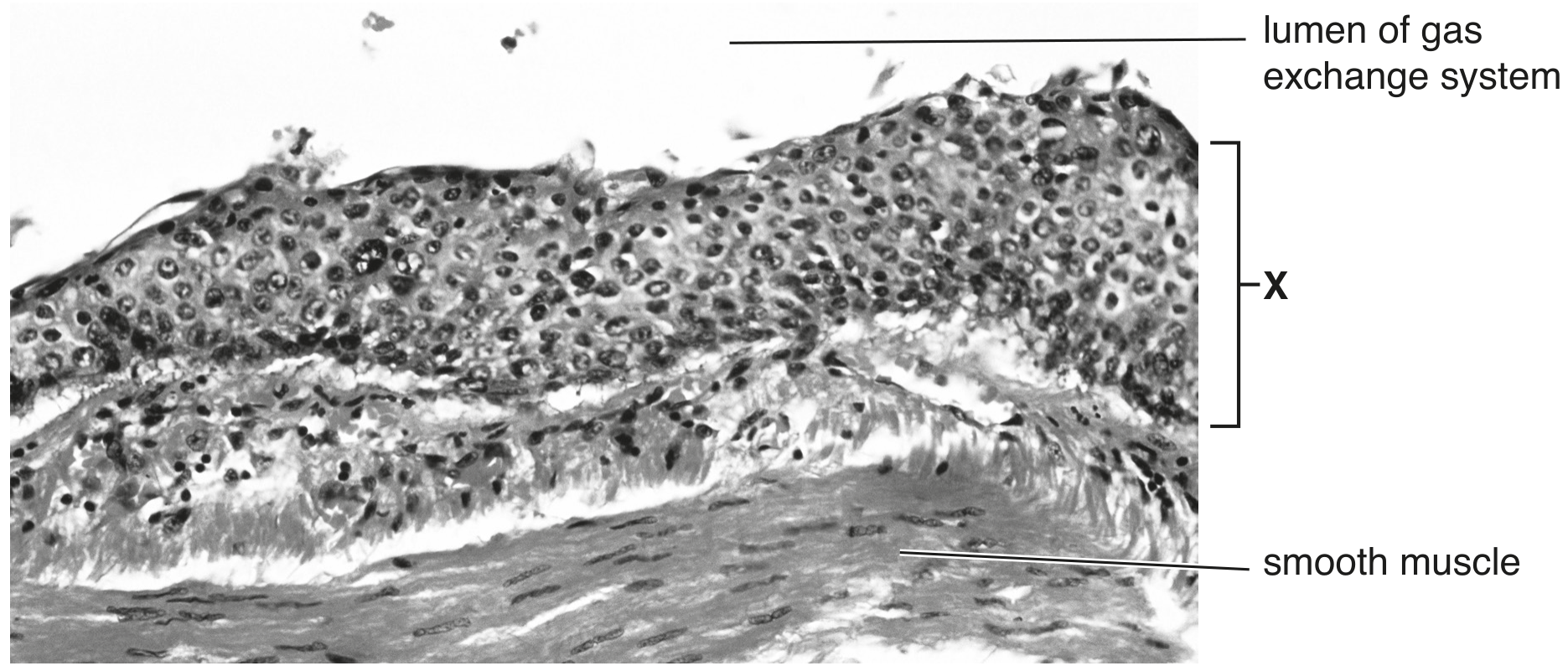
Fig. 5.2
The area labelled X on Fig. 5.2 is different in appearance to the original healthy tissue in the same part of the gas exchange system.
Describe these differences.
Fig. 5.1 is a photomicrograph of a transverse section of a bronchus in the lungs.
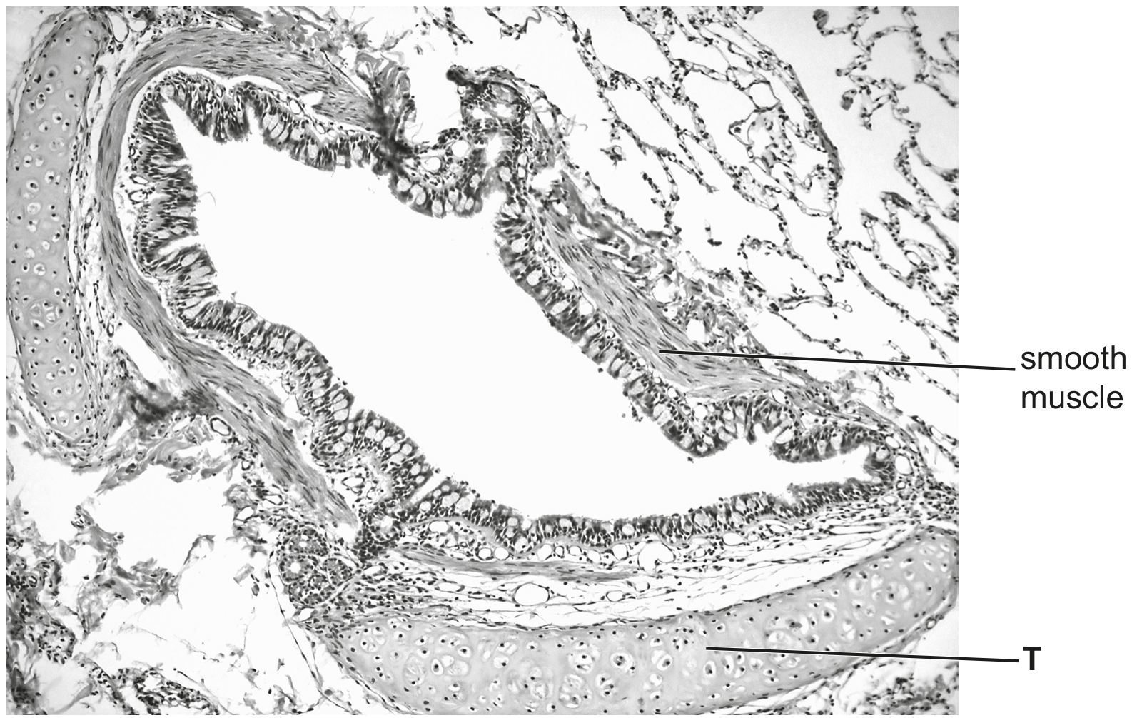
Fig. 5.1
Identify the tissue labelled T in Fig. 5.1.