EduNinja
[Maximum number: 1]

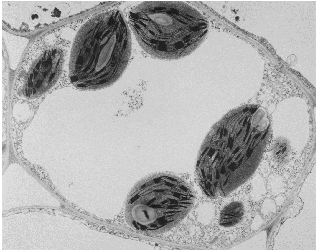
The electron micrograph shows onion root cells prepared using a freeze-fracture technique. The cells were quickly frozen and then physically broken apart. Freeze fracture breaks apart cells along weak areas, such as membranes and the surfaces of organelles.
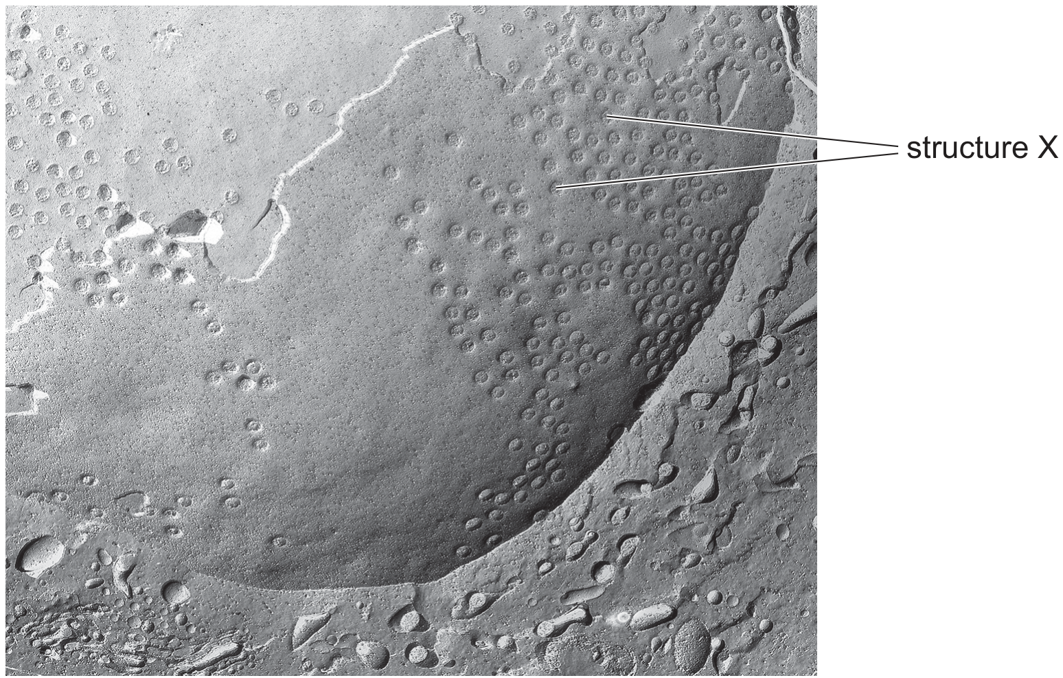
\(\times 20000\)

\(\times 20000\)

Which statement best explains the appearance of the electron micrograph?

A

The cells were broken apart at the endoplasmic reticulum; structure X is a ribosome.

B

The cells were broken apart at the nuclear envelope; structure X is a nuclear pore.

C

The cells were broken apart at the nuclear envelope; structure X is a ribosome.

D

The cells were broken apart at the tonoplast; structure X is a plasmodesma.

[Maximum number: 1]

Which row shows the correct order of size of these cell structures?

1 width of a mitochondrion
2 width of a ribosome
3 width of a cell surface membrane
4 width of a chloroplast

largest

smallest

1

4

2

3

1

4

3

2

4

1

2

3

4

2

1

3

[Maximum number: 1]

The photomicrograph shows the ultrastructure of part of a cell.

Question image

Which statement about the type of cell shown in the photomicrograph is correct?

A

It is a plant cell because it has both chloroplasts and a nucleus.

B

It is a plant cell because it has chloroplasts.

C

It is an animal cell because it has a cell membrane.

D

It is an animal cell because it has mitochondria.

[Maximum number: 1]

Which cell structure contains cytoplasm?

A

chloroplasts

B

mitochondria

C

plasmodesmata

D

smooth endoplasmic reticulum

[Maximum number: 1]

A student made notes describing photomicrographs of four cells.
cell 1 Grey cytoplasm at edge of cell contains many black lines and spots. Large white area in centre of cell.
cell 2 Grey cytoplasm contains many black lines and spots which fill the entire cell.
cell 3 Pale blue cytoplasm surrounds a single dark blue spot.
cell 4 Many green structures are enclosed within a rectangular shape with visible boundaries.

Which table identifies the type of cell and the type of microscope used to take each photograph?

A
Question image
B
Question image
C
Question image
D
Question image
0