EduNinja
[Maximum number: 6]

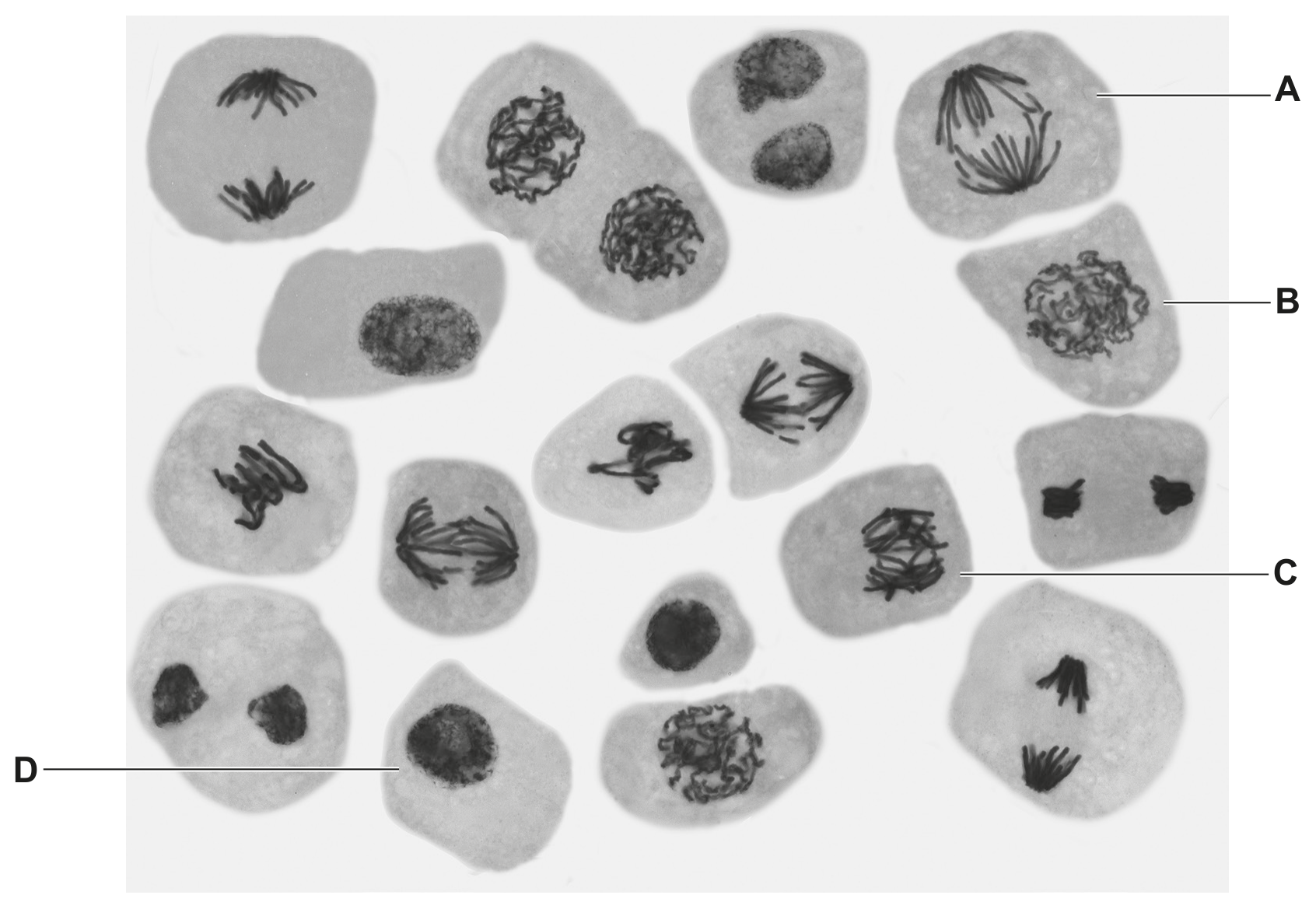
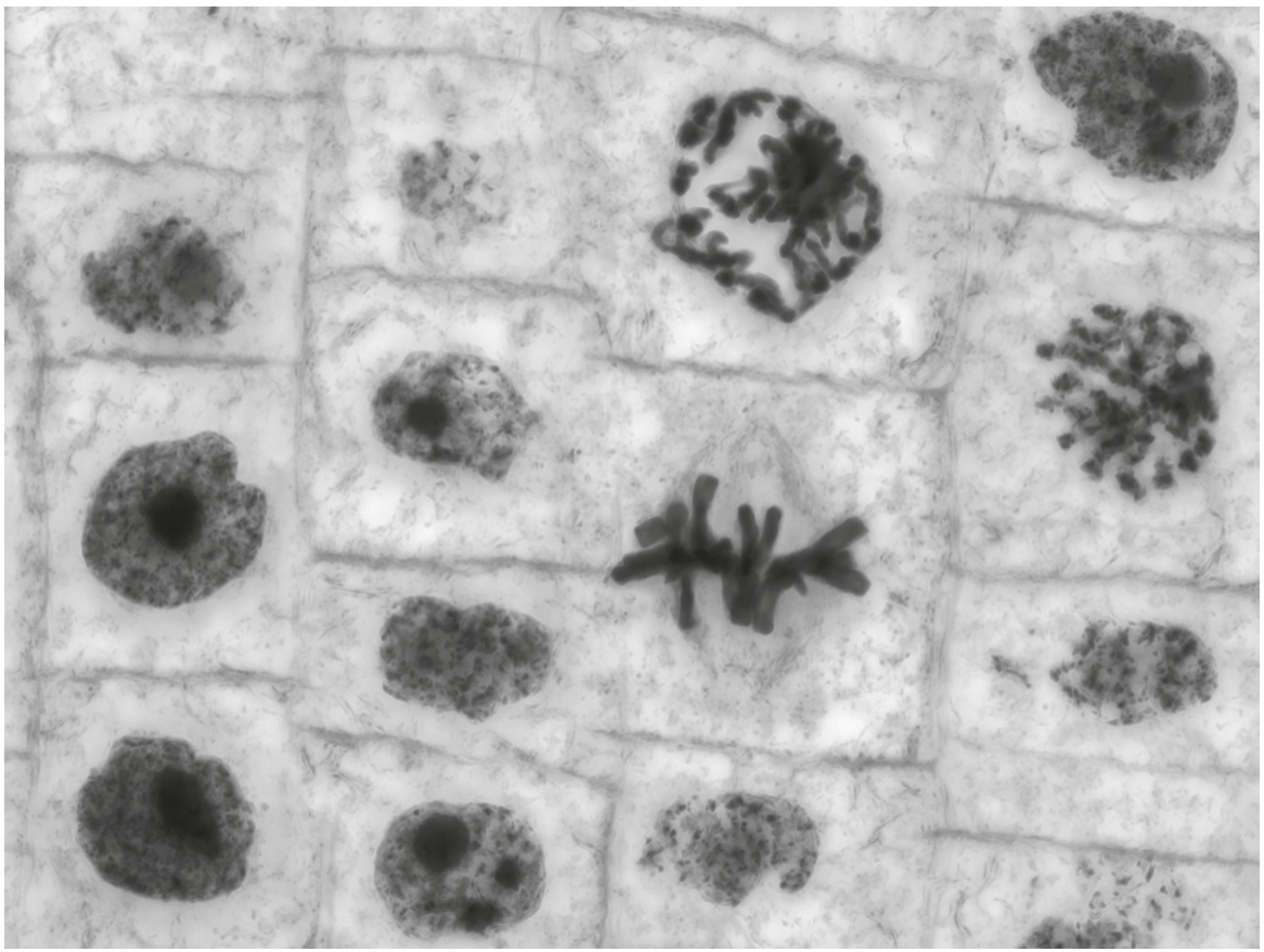
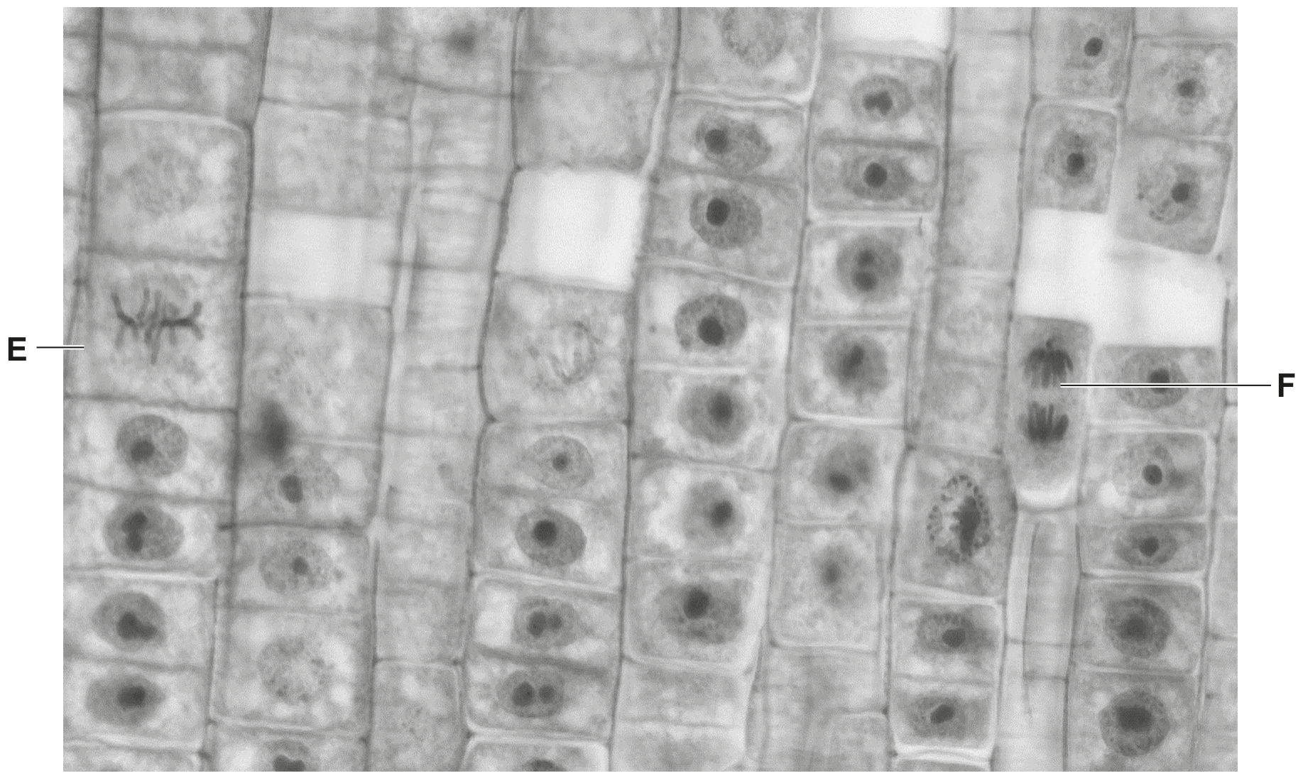
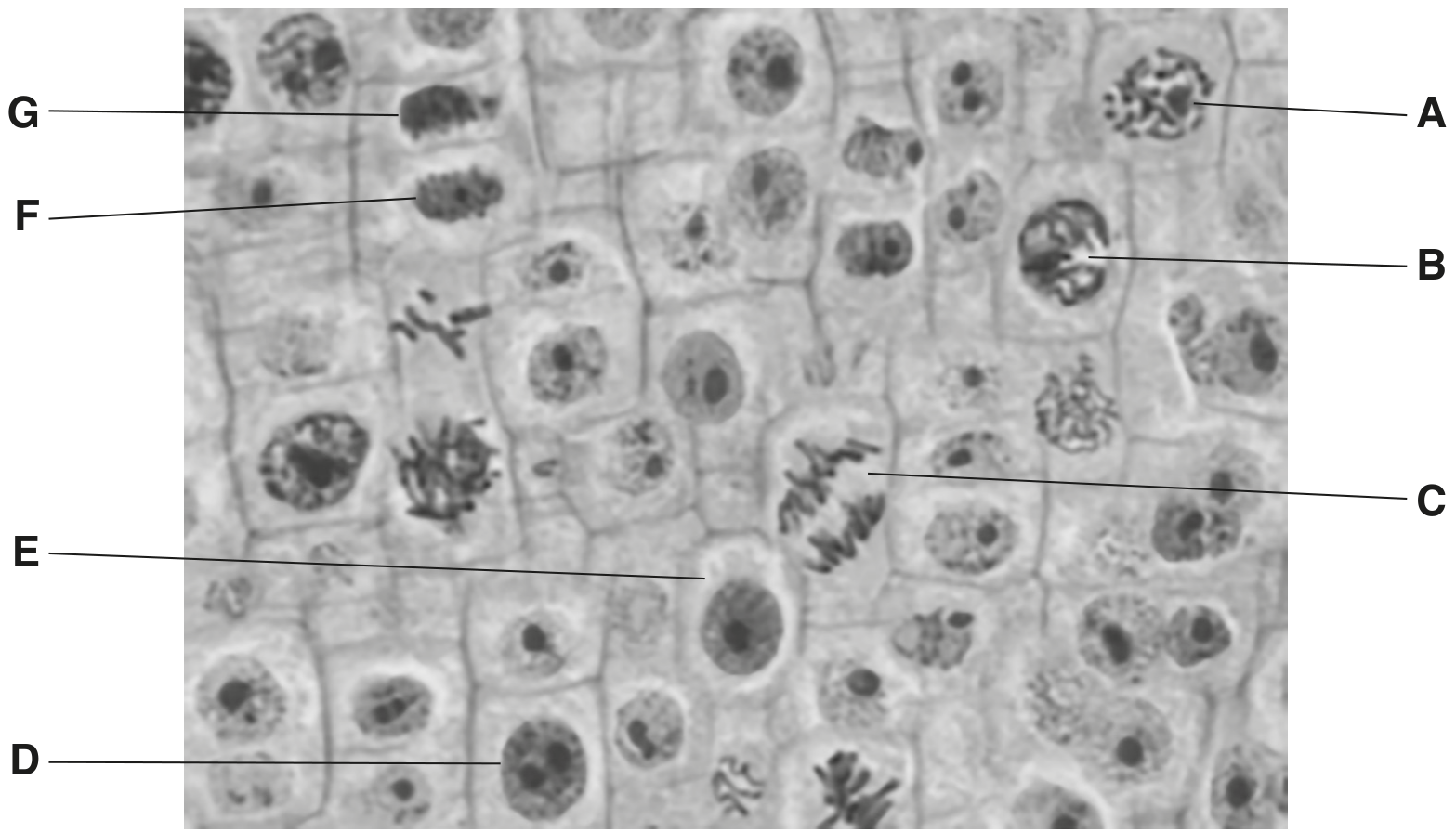
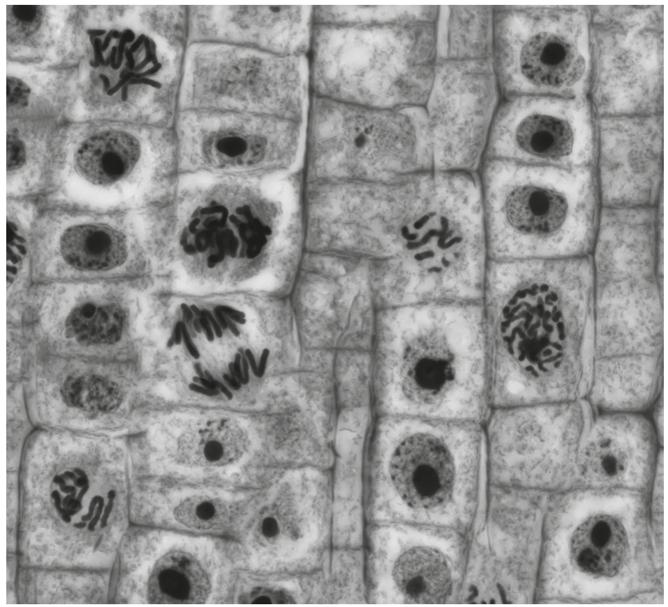
During interphase and mitosis of the cell cycle, the chromosomes within a cell go through a number of changes. Each chromosome is composed of DNA complexed with proteins.
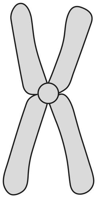
(a)

When viewed through a microscope, a chromosome is most clearly visible during the metaphase stage of mitosis.

Complete Fig. 1.1 to produce a labelled diagram of the metaphase stage of mitosis in an animal cell with two chromosomes.

Fig. 1.1

Fig. 1.1

[ 3 ]
(b)

Outline the changes that occur to the structure and behaviour of a chromosome:
- from the start of the S phase to the end of interphase
- during prophase of mitosis.

[ 3 ]
[Maximum number: 5]

The cells in a tissue are often at different stages of the cell cycle.

(a)

Fig. 1.1 shows cells at different stages of the cell cycle.

Fig. 1.1

Fig. 1.1

[ 5 ]
(i)

Identify the stages of mitosis occurring in the cells labelled B and C in Fig. 1.1.

B
C

[ 2 ]
(ii)

Describe the behaviour of the chromosomes in the stage of mitosis shown in cell A in Fig. 1.1.

[ 3 ]
[Maximum number: 2]

There are two types of cell, prokaryotic and eukaryotic. Bacterial cells are prokaryotic and plant cells are eukaryotic.

(a)

Fig. 1.2 shows plant cells in a root tip where cell division by mitosis is taking place.

Fig. 1.2

Fig. 1.2

Identify two cells in Fig. 1.2 that are in different stages of mitosis.
Draw a label line to each cell and add the name of the stage of mitosis that is shown by the cell.

[ 2 ]
(a)

Fig. 1.1 is a photomicrograph of a region of eukaryotic tissue. Some of the cells are in stages of mitosis.

Fig. 1.1

Fig. 1.1

[ 2 ]
(i)

Identify which stage of mitosis is shown in cell E and in cell F in Fig. 1.1.

E
F

[ 2 ]
(a)

Fig. 1.1 is a diagram of a human chromosome at a stage in mitosis.

Fig. 1.1

Fig. 1.1

[ 1 ]
(i)

State one stage during mitosis when the chromosome would appear as shown in Fig. 1.1.

[ 1 ]
[Maximum number: 5]

Fig. 1.1 is a photomicrograph of root tip meristem. Different stages of the cell cycle are visible. Some cells are in the same stage of the cell cycle and some are in the same stage of mitosis.

Fig. 1.1

Fig. 1.1

(a)

Name the stage of mitosis occurring in each of cells A, B and C.
cell A
cell B
cell C

[ 3 ]
(b)

Cells F and G are newly formed cells. Cytokinesis has occurred with the formation of a cell plate.

Describe the events that have occurred in the stage of mitosis immediately before cytokinesis.

[ 2 ]
[Maximum number: 3]

The root apical meristem is a region of undifferentiated cells in the root tips of plants. Mitosis occurs in this region.

(a)

Fig. 1.1 is an image of the root tip of Allium as observed using a microscope with a low-power objective lens.

Fig. 1.1

Fig. 1.1

State which region, B, C, or D, should be chosen in order to observe the highest proportion of cells in stages of mitosis.

[ 1 ]
(b)
(i)

Describe the behaviour of the nuclear envelope during mitosis.

[ 2 ]
[Maximum number: 1]

Fig. 1.1 is a photomicrograph of plant cells showing stages in cell division.

Fig. 1.1

Fig. 1.1

(a)

On Fig. 1.1, draw a circle around a cell in anaphase.

[ 1 ]
[Maximum number: 1]

A diagram of a chromosome from a dividing cell is shown in Fig. 1.1.

Fig. 1.1

Fig. 1.1

(a)

The chromosome shown in Fig. 1.1 consists of one long DNA molecule associated with histone proteins.

Name one stage of mitosis in which a chromosome would have the same general structure as the chromosome shown in Fig. 1.1.

[ 1 ]
[Maximum number: 2]

Fig. 1.1 is a transmission electron micrograph of part of an animal cell.

Fig. 1.1

Fig. 1.1

(a)

Cells such as that in Fig. 1.1 can divide by mitosis.

Describe the role of centrioles in mitosis.

[ 2 ]
0