EduNinja
[Maximum number: 3]

The malarial pathogen, Plasmodium falciparum, enters red blood cells after a person becomes infected. After some time, each cell of P. falciparum divides to form daughter cells.

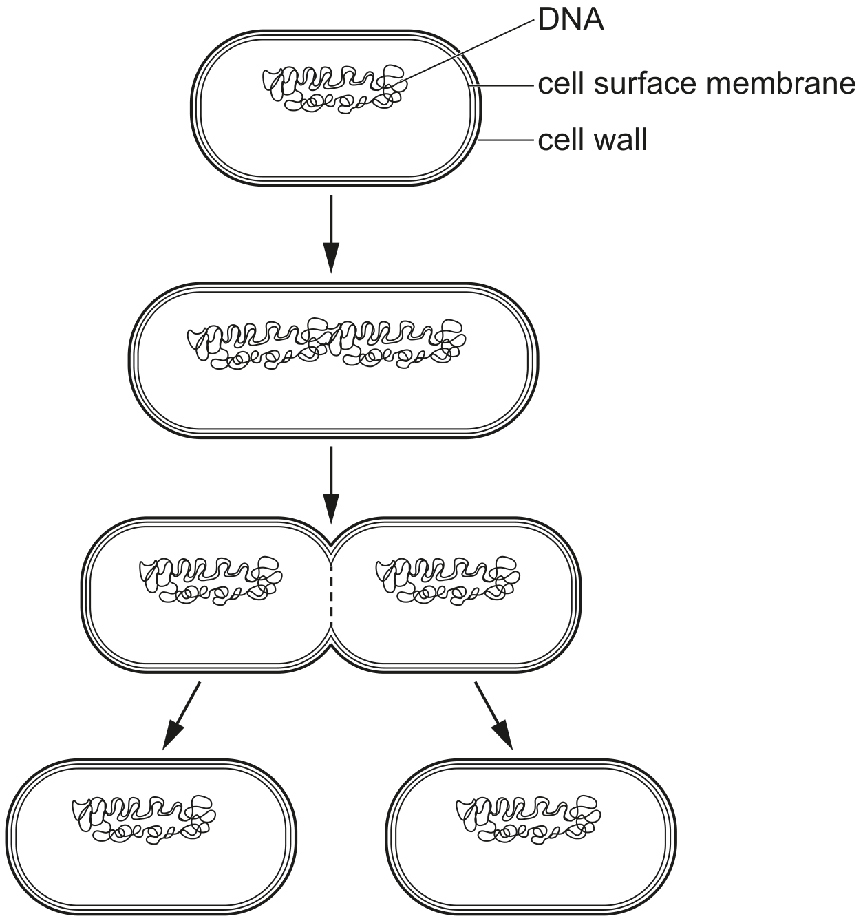
Fig. 1.1 shows a cell of P. falciparum that is forming many daughter cells.
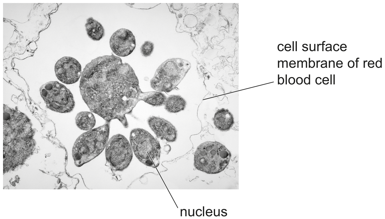
Fig. 1.1

Fig. 1.1

(a)

When P. falciparum divides there is unequal division of the cytoplasm to form small, genetically identical daughter cells.

Outline the events that occur in the cell of P. falciparum to form the daughter cells shown in Fig. 1.1.

In 2013, the World Health Organization (WHO) set a target for researchers to create a vaccine for malaria. WHO required the vaccine to show 75\% efficacy and be ready for use by 2030 .

Efficacy is a measure of the effectiveness of a vaccine in reducing the number of new cases of malaria.

A trial of the R21/Matrix-M TM{ }^{\mathrm{TM}} vaccine in Burkina Faso in 2020 achieved a 77 % efficacy over a 12-month period. A control group received a vaccine for rabies.

Vaccines stimulate an immune response with the production of antibodies.

[ 3 ]
[Maximum number: 3]

The cells in a tissue are often at different stages of the cell cycle.

(a)

Telomerase is an enzyme that is active during interphase in some cells.

Telomerase helps to maintain telomeres present on chromosomes.

[ 3 ]
(i)

State the location of telomeres on a chromosome.

[ 1 ]
(ii)

Some people with a long lifespan have cells showing a higher than normal activity of telomerase.

Suggest why a long lifespan could result from a higher telomerase activity.

[ 2 ]
(a)

Mitogens are short chains of amino acids that function as cell-signalling molecules. Mitogens are released from secretory cells and travel in the blood to target cells, where the mitogens bind to cell surface receptors. The target cells respond by progressing from the G1\mathrm{G}_{1} phase to the S phase of the mitotic cell cycle.

[ 3 ]
(i)

Outline what happens in the G1\mathrm{G}_{1} phase and S phase of the mitotic cell cycle.
G1\mathrm{G}_{1} phase

S phase

[ 2 ]
(ii)

As a result of mutation, the production and release of mitogens into the blood can be greatly increased.

Suggest a possible consequence for target cells of increased concentrations of mitogens in the blood.

[ 1 ]
[Maximum number: 4]

Epithelial cells in the small intestine have cell structures known as microvilli. The microvilli of these cells are found only on the surface that borders the gut lumen.

Fig. 1.1 shows images of microvilli of intestinal epithelial cells. These images have been obtained using a scanning electron microscope and a transmission electron microscope.

Fig. 1.1 A is at a different magnification to Fig. 1.1B.

Fig. 1.1

Fig. 1.1

(a)

Stem cells are also located in the wall of the small intestine. These cells divide by mitosis continuously.

Suggest and explain the importance of mitosis by stem cells in the small intestine.

[ 4 ]
(a)

Fig. 1.1 is a diagram of a human chromosome at a stage in mitosis.

Fig. 1.1

Fig. 1.1

[ 5 ]
(i)

The paragraph describes the structure of the chromosome shown in Fig. 1.1.

Complete the paragraph using the most appropriate term in each space provided.
The chromosome shown in Fig. 1.1 has two genetically identical joined at a The chromosome is composed of two DNA molecules, each wrapped around proteins known as proteins.

[ 3 ]
(ii)

Suggest the role of ATP in the process of mitosis.

[ 2 ]
(b)

Prokaryotes divide by a process known as binary fission.

Fig. 1.2 shows some of the stages in binary fission.

Fig. 1.2

Fig. 1.2

With reference to Fig. 1.2, identify two events that occur during binary fission that do not occur during mitosis in human cells.

[ 2 ]
[Maximum number: 2]

Fig. 1.1 is a diagram showing the structure of a section of a DNA molecule.

Fig. 1.1

Fig. 1.1

(a)

The telomere is a region found at the end of a chromosome.

Outline the function of telomeres.

[ 2 ]
[Maximum number: 1]

Fig. 1.1 is a photomicrograph of root tip meristem. Different stages of the cell cycle are visible. Some cells are in the same stage of the cell cycle and some are in the same stage of mitosis.

Fig. 1.1

Fig. 1.1

(a)

Cell D and cell E are in the same stage of the cell cycle.

State the difference between the nucleus of cell D and cell E.

[ 1 ]
[Maximum number: 3]

The root apical meristem is a region of undifferentiated cells in the root tips of plants. Mitosis occurs in this region.

(a)
(i)

Draw a labelled diagram to show the structure of a chromosome at late prophase of mitosis.

[ 3 ]
[Maximum number: 3]

Fig. 1.1 is a photomicrograph of plant cells showing stages in cell division.

Fig. 1.1

Fig. 1.1

(a)

Mitosis is important in producing more cells for plant growth.

Describe three other ways in which mitosis is important in plants.

[ 3 ]
[Maximum number: 2]

A diagram of a chromosome from a dividing cell is shown in Fig. 1.1.

Fig. 1.1

Fig. 1.1

(a)

A dividing cell is at risk of losing genetic material each time DNA replication occurs.

On Fig. 1.1, add a label line and the letter G to show the location on the chromosome of an area that helps to prevent the loss of genes.

[ 1 ]
(b)

Name the stage in the mitotic cell cycle during which the cytoplasm and the cell divide to produce two genetically identical daughter cells.

[ 1 ]
0