EduNinja
[Maximum number: 4]

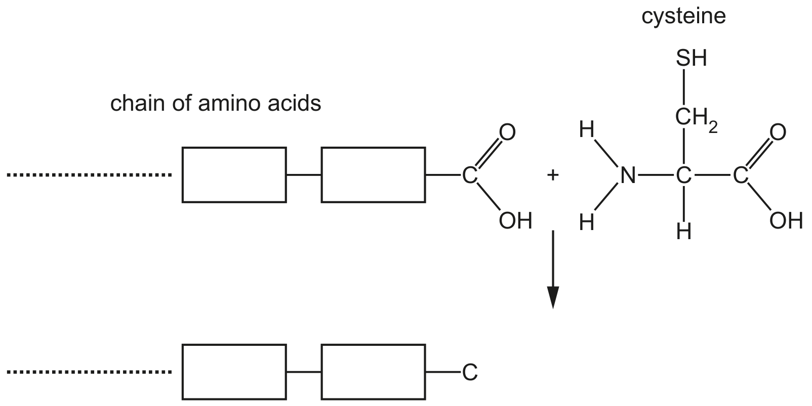
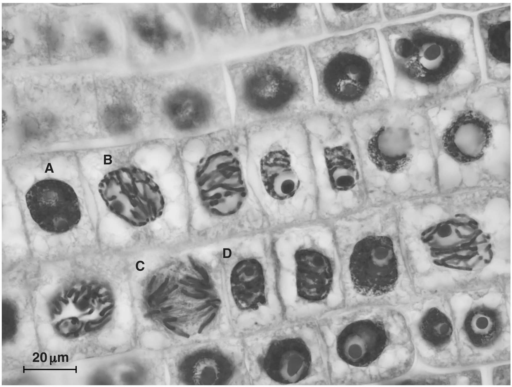
Smooth muscle is a tissue composed of smooth muscle cells. The cells contain cytoplasm packed with proteins that are involved in contraction and relaxation.

(a)

Caldesmon is a large protein with a number of binding sites to attach to other proteins.

Caldesmon exists in two different forms, H-caldesmon and L-caldesmon.
H-caldesmon helps to regulate contraction and relaxation in smooth muscle cells.
L -caldesmon is found in some non-muscle cells, where it also acts as a regulatory protein.
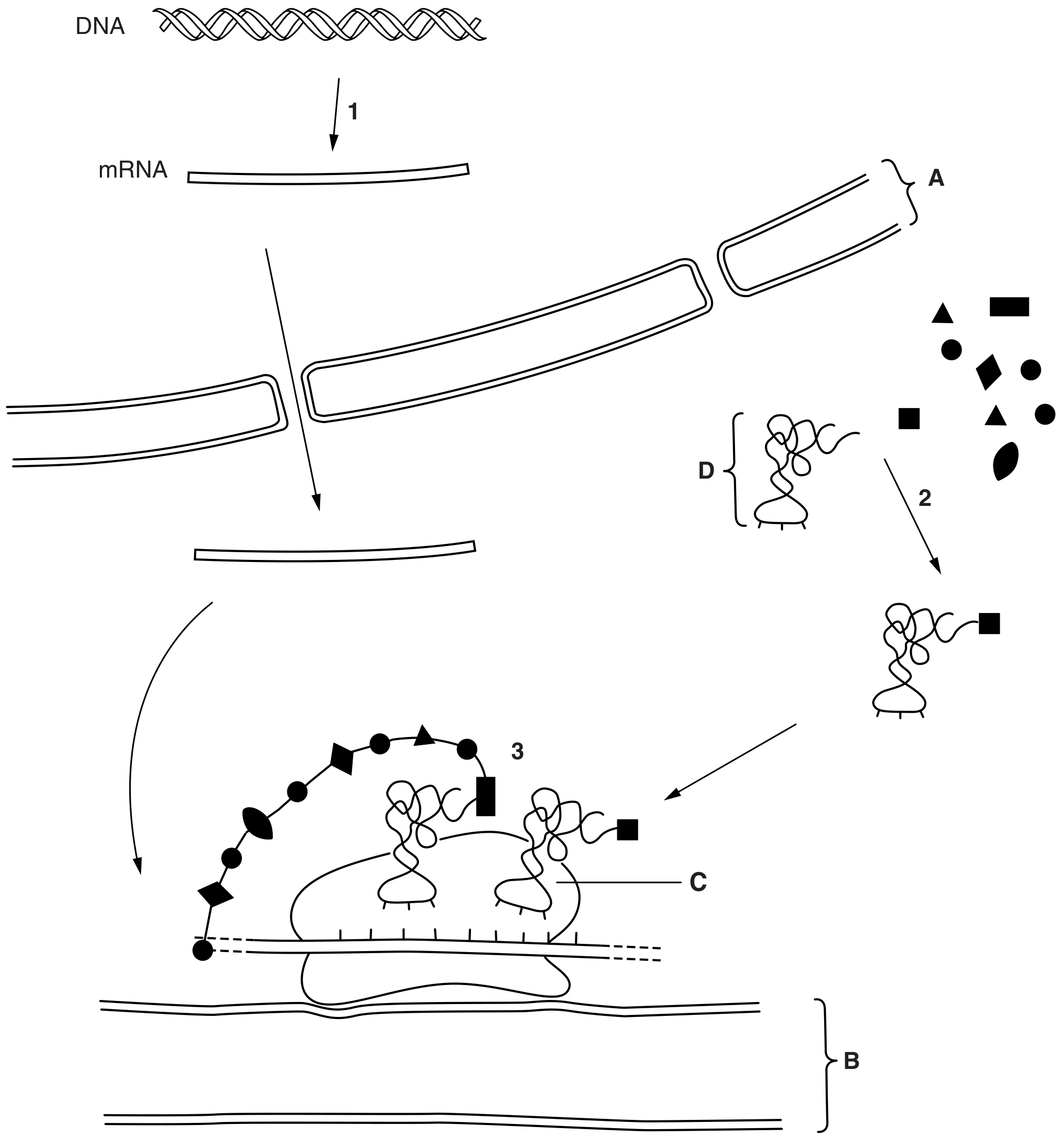
- Caldesmon is coded for by a gene known as CALD1.
- CALD1 has 17 exons.
- The primary structure of H-caldesmon has a repeating sequence in the middle of the amino acid chain that is not present in L-caldesmon.

[ 4 ]
(i)

Researchers have discovered that a gene mutation is not the cause of the two different forms of caldesmon.

Explain what is meant by a gene mutation.

[ 2 ]
(ii)

Researchers now know that the two different forms of caldesmon are the result of events occurring directly after transcription of DNA. Changes occur to the primary transcript that is formed by DNA transcription.

Suggest how the smooth muscle cells and non-muscle cells can produce different forms of caldesmon from the same primary transcript.

[ 2 ]
(a)

During translation, a polypeptide is synthesised when amino acids are added to a growing chain of amino acids.

Fig. 1.1 shows part of a growing chain of amino acids and the amino acid cysteine.

Fig. 1.1

Fig. 1.1

[ 3 ]
(i)

Complete Fig. 1.1 by showing the formation of the bond between cysteine and the growing chain of amino acids in the process of translation.

[ 3 ]
[Maximum number: 5]

Fig. 1.1 is a diagram showing a stage in protein synthesis.

Fig. 1.1

Fig. 1.1

(a)
(i)

Name the stage of protein synthesis that is shown in Fig. 1.1.

[ 1 ]
(ii)

Identify A, B and C in Fig. 1.1.

A
B
C

[ 3 ]
(iii)

State the base sequences at D and E.

D

E

[ 1 ]
[Maximum number: 1]

Fig. 1.1 is a transmission electron micrograph of cells from duckweed, Spirodela oligorrhiza.

magnification \(\times 4275\)

magnification \(\times 4275\)

(a)
(i)

Name a cell structure that produces mRNA.

[ 1 ]
[Maximum number: 6]

Antibodies are secreted by activated B-lymphocytes known as plasma cells.
Fig. 1.1 is a diagram showing the cellular processes involved in the production of a polypeptide of an antibody molecule (not drawn to scale).

Fig. 1.1

Fig. 1.1

(a)
(i)

Name molecule D.

D

[ 1 ]
(ii)

State what is occurring at 1,2 and 3.
at 1
at 2
at 3

[ 3 ]
(b)

The genes responsible for antibody production are found on different chromosomes, such as chromosomes 2 and 14 in humans.

Explain how one antibody molecule is the product of more than one gene.

[ 2 ]
[Maximum number: 2]

Fig. 1.1 is a photomicrograph of plant root cells near the growing tip. Some of the cells are undergoing mitosis.

Fig. 1.1

Fig. 1.1

(a)
(i)

Mutations can sometimes occur in cells which are rapidly dividing.

Outline how a mutation can cause an altered polypeptide to be produced.

[ 2 ]
[Maximum number: 2]

Fig. 1.1 shows a diagram of part of a cell surface membrane.

Fig. 1.1

Fig. 1.1

(a)

Structure E is a protein composed of 588 amino acids.

Calculate the minimum number of nucleotide base pairs required in the gene coding for this protein. Show your working.

Answer =

[ 2 ]
[Maximum number: 2]

Protein production involves a complex sequence of events and a number of cell structures.

(a)

One example of protein modification is the removal of the first amino acid, methionine, from a newly formed polypeptide chain to make a functioning protein.

[ 2 ]
(i)

Suggest two other ways in which the polypeptide chain is modified to produce the functioning protein.

[ 2 ]
(a)

Fig. 1.1 shows part of a guard cell.

Fig. 1.1

Fig. 1.1

[ 1 ]
(i)

State the type of protein represented by Q.

[ 1 ]
(a)

Fig. 1.1 shows part of a cell in a growing region of a plant.

Fig. 1.1

Fig. 1.1

[ 1 ]
(i)

State the type of protein represented by A.

[ 1 ]
0