EduNinja
[Maximum number: 7]

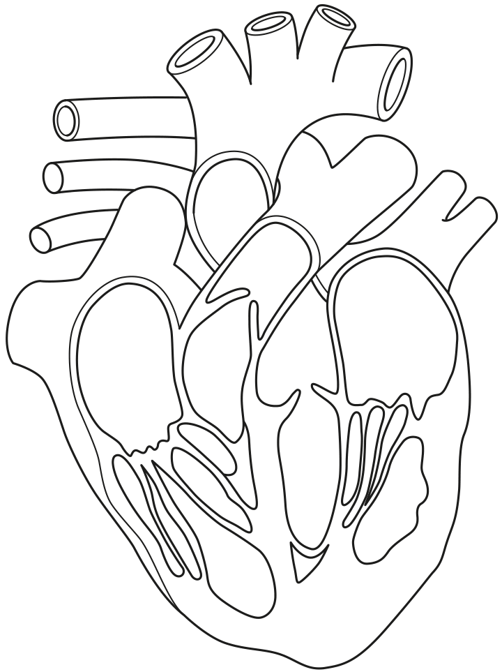
The sinoatrial node (SAN) and the atrioventricular node (AVN) have an important role in the control of the cardiac cycle. The timing of atrial and ventricular systole and diastole must be controlled so that blood passes through the heart efficiently.

(a)

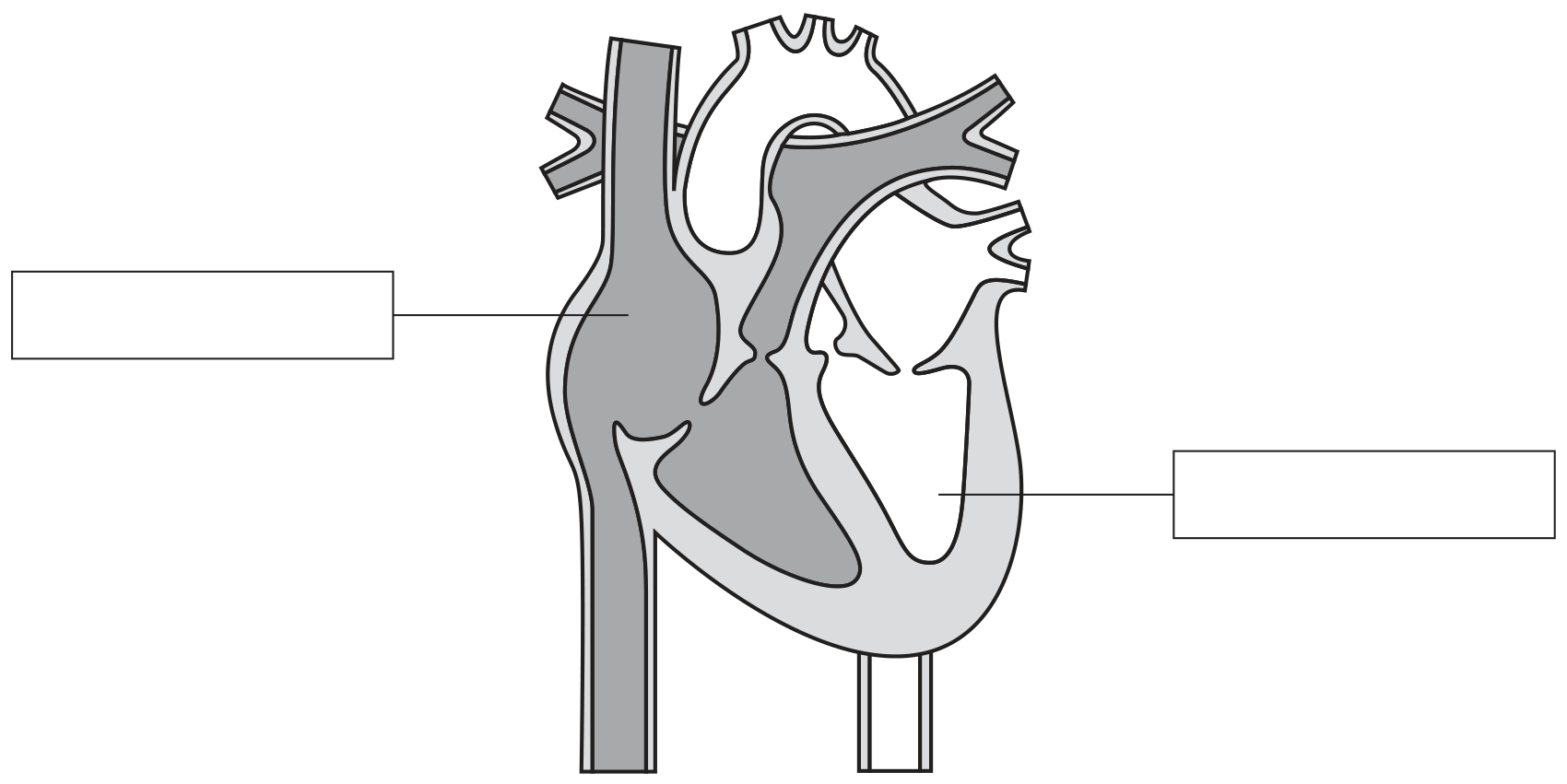
Fig. 1.1 is a summary of blood flow through the right side of the heart during one cardiac cycle. Three boxes in Fig. 1.1 are not complete.

Complete boxes 3, 5 and 7 in Fig. 1.1 using only the terms systole and diastole.

1 blood arrives at the heart in the superior vena cava and inferior vena cava
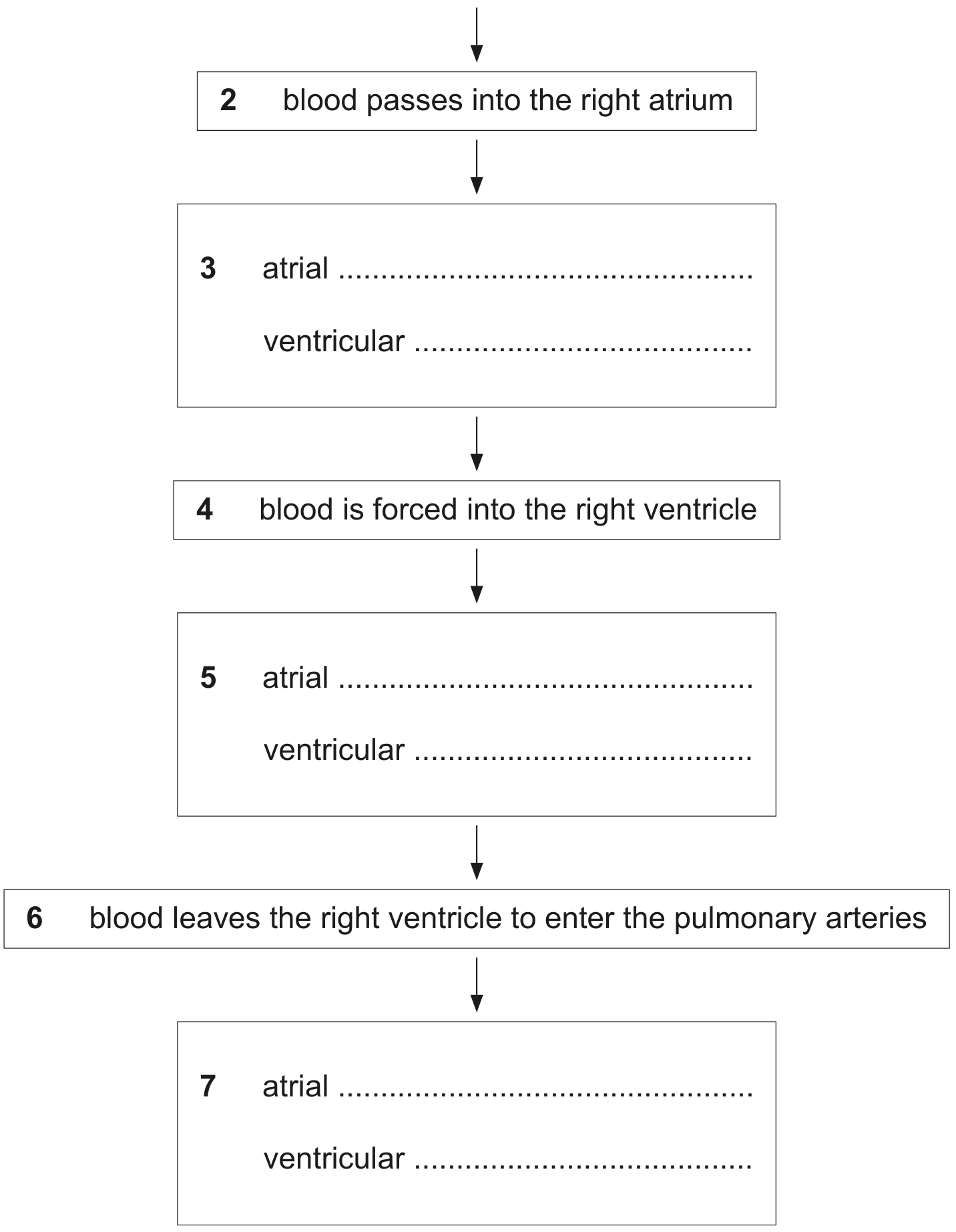
Fig. 1.1

Fig. 1.1

[ 2 ]
(b)

Impulses sent out by the SAN pass to the AVN, where there is a short delay.

With reference to Fig. 1.1, explain why it is important for the control of the cardiac cycle that there is a short delay at the AVN after impulses have been sent out by the SAN.

[ 2 ]
(c)

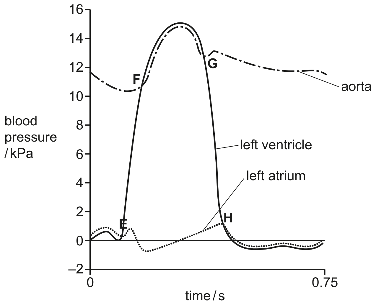
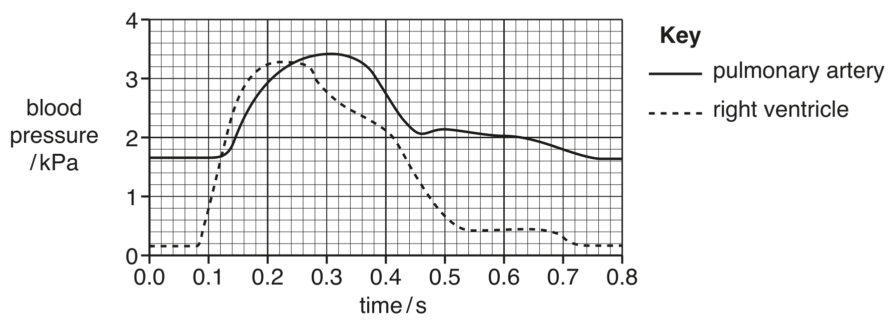
Changes in blood pressure occur in the heart during the cardiac cycle. These changes cause the opening and closing of the bicuspid and tricuspid (atrioventricular) valves and the aortic and pulmonary (semilunar) valves.

Explain how blood pressure changes:
- cause the opening of the tricuspid valve
- cause the opening of the pulmonary valve
- help the flow of blood through the heart.

[ 3 ]
[Maximum number: 2]

The mammalian circulatory system is adapted for the long-distance transport of the respiratory gases, oxygen and carbon dioxide.

The system is described as a closed double circulation.

(a)

Fig. 1.1 is a diagram of a section through the heart.

Fig. 1.1

Fig. 1.1

On Fig. 1.1:
- add a label line and the letter L to show the artery that takes blood from the heart to the lungs
- add a label line and the letter R to show the valve that closes when the right ventricle is in systole.

[ 2 ]
[Maximum number: 5]

Each of statements A to E describe a structure associated with the mammalian heart.
For each statement, identify the structure that is being described.

A

The chamber that pumps blood into the pulmonary artery.

B

A blood vessel that transports deoxygenated blood into the right atrium.

C

The specialised tissue responsible for delaying the conduction of impulses from the atria to the ventricles.

D

The blood vessels that supply cardiac muscle with oxygenated blood. E The valve that prevents the backflow of blood from the ventricle that contains oxygenated blood.

[Maximum number: 7]

Fig. 2.1 is a diagram of a vertical section through a healthy mammalian heart.

Fig. 2.1

Fig. 2.1

(a)
(i)

Label the two chambers of the heart by writing in the boxes provided on Fig. 2.1.

[ 1 ]
(b)

Match the one correct area from A to G on Fig. 2.2 with each of the congenital heart diseases.

The first one has been completed for you.
patent ductus arteriosus A
pulmonary stenosis
coarctation of the aorta
ventricular septal defect

[ 3 ]
(c)

Suggest and explain how the flow of blood in a person with patent ductus arteriosus differs from that of a person with a healthy heart.

[ 3 ]
(a)

Examination of the transplanted hearts showed that the hearts in group A had significant damage to their coronary arteries, whereas in group D these blood vessels appeared normal.

Explain the importance of the coronary arteries of the heart.

[ 3 ]
[Maximum number: 9]

In a healthy mammalian heart, contraction of the four chambers is coordinated by the action of the sinoatrial node (SAN) and atrioventricular node (AVN).

(a)

After the atria fill with blood, atrial systole (contraction) occurs.

State the events that occur to initiate and cause atrial systole.

[ 2 ]
(b)

State and explain how the structure of the heart allows the atria to contract before the ventricles.

[ 2 ]
(c)

Fig. 2.1 shows blood pressure changes that occur in the left ventricle, left atrium and aorta during one cardiac cycle.
E, F, G and H are the points at which a valve opens or closes as a result of blood pressure changes.

Fig. 2.1

Fig. 2.1

[ 5 ]
(i)

For each of the points E, F, G and H on Fig. 2.1, name the valve concerned and state whether the valve opens or closes.

E
F
G
H

[ 3 ]
(ii)

Explain how Fig. 2.1 provides evidence that the wall of the left atrium has a different thickness to the wall of the left ventricle.

[ 2 ]
[Maximum number: 4]

Woolly foxglove, Digitalis lanata, shown in Fig. 2.1A, and common oleander, Nerium oleander, shown in Fig. 2.1B, are plants grown for the attractive flowers that they produce.

Both plants are poisonous, as their leaves produce toxic organic compounds known as cardiac glycosides. Cardiac glycosides have a powerful effect on the action of cardiac muscle.

Fig. 2.1

Fig. 2.1

(a)

Digoxin, a cardiac glycoside extracted from D. lanata leaves, can be purified and used as a drug to treat some heart disorders.

Examples of these heart disorders are:
- atrial fibrillation, where the normal rhythmic cardiac cycle is disrupted
- heart failure, where cardiac muscle is contracting weakly.

[ 4 ]
(i)

Describe the sequence of events occurring in the left side of the heart during one normal cardiac cycle. Include reference to blood pressure changes.

The first event in the sequence is described for you.
The left atrium fills with blood during relaxation of the left atrium and left ventricle.

[ 4 ]
[Maximum number: 6]

Fig. 2.1 is a transmission electron micrograph of a section through a blood vessel.

Fig. 2.1

Fig. 2.1

(a)

Fig. 2.2 is a graph showing how the blood pressure in the pulmonary artery and in the right ventricle changes during one cardiac cycle.

Fig. 2.2

Fig. 2.2

Use Fig. 2.2 to state the time at which:

[ 6 ]
(i)

the valve between the right ventricle and the pulmonary artery closes

[ 1 ]
(ii)

the ventricle begins to contract.

[ 1 ]
(iii)

State and explain the similarities and differences between Fig. 2.2 and a graph showing how the blood pressure for the left ventricle changes during the same cardiac cycle.

[ 4 ]
[Maximum number: 1]

Which row correctly describes the events during the cardiac cycle?

nerve impulses from atrio-ventricular node (AVN) to

nerve impulses from Purkyne tissue (PT) to

nerve impulses from sino-atrial node (SAN) to

SAN

the ventricles

AVN

PT

the atria

PT

PT

the ventricles

AVN

SAN

the atria

PT

0