[Maximum number: 2]
Fig. 1.1 is an electron micrograph of a cross section through a blood vessel.
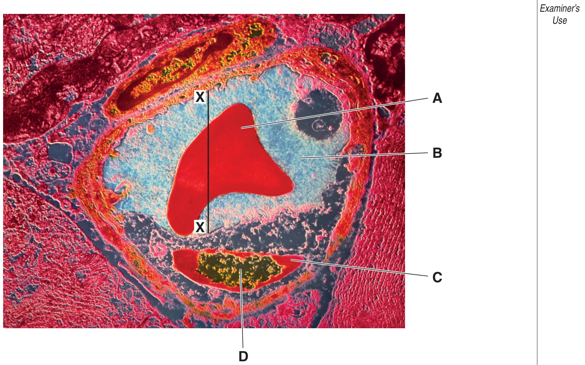
Fig. 1.1
(a)
Name:
[ 2 ]
(i)
the main component of substance B.
[ 2 ]
 EduNinja
Fig. 1.1 is an electron micrograph of a cross section through a blood vessel.
Fig. 1.1
Name:
the main component of substance B.
Fig. 1.1 is a diagram of a transverse section through a vein.
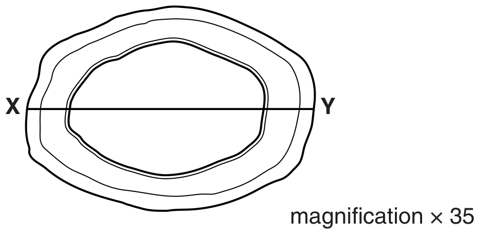
Fig. 1.1
The presence of a valve would help to confirm that the blood vessel in Fig. 1.1 is a vein and not an artery.
Describe three structural features of the blood vessel shown in Fig. 1.1 that would help to identify it as a vein and not as an artery.
1.
2.
3.
Mammals have a closed double circulation system.
Explain why arteries have thicker walls than veins.
Smooth muscle is a tissue composed of smooth muscle cells. The cells contain cytoplasm packed with proteins that are involved in contraction and relaxation.
Fig. 1.2 is a photomicrograph of smooth muscle tissue in the wall of the intestines. A capillary is visible in addition to smooth muscle cells.
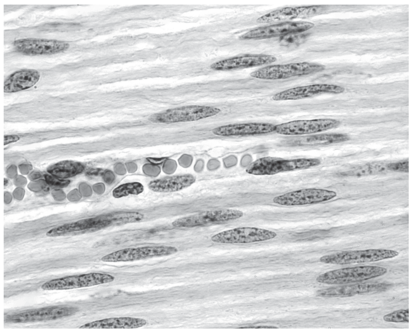
Fig. 1.2
Explain how the structure of a capillary is related to its function in smooth muscle.
Fig. 2.1 is a simplified diagram of the human circulatory system.
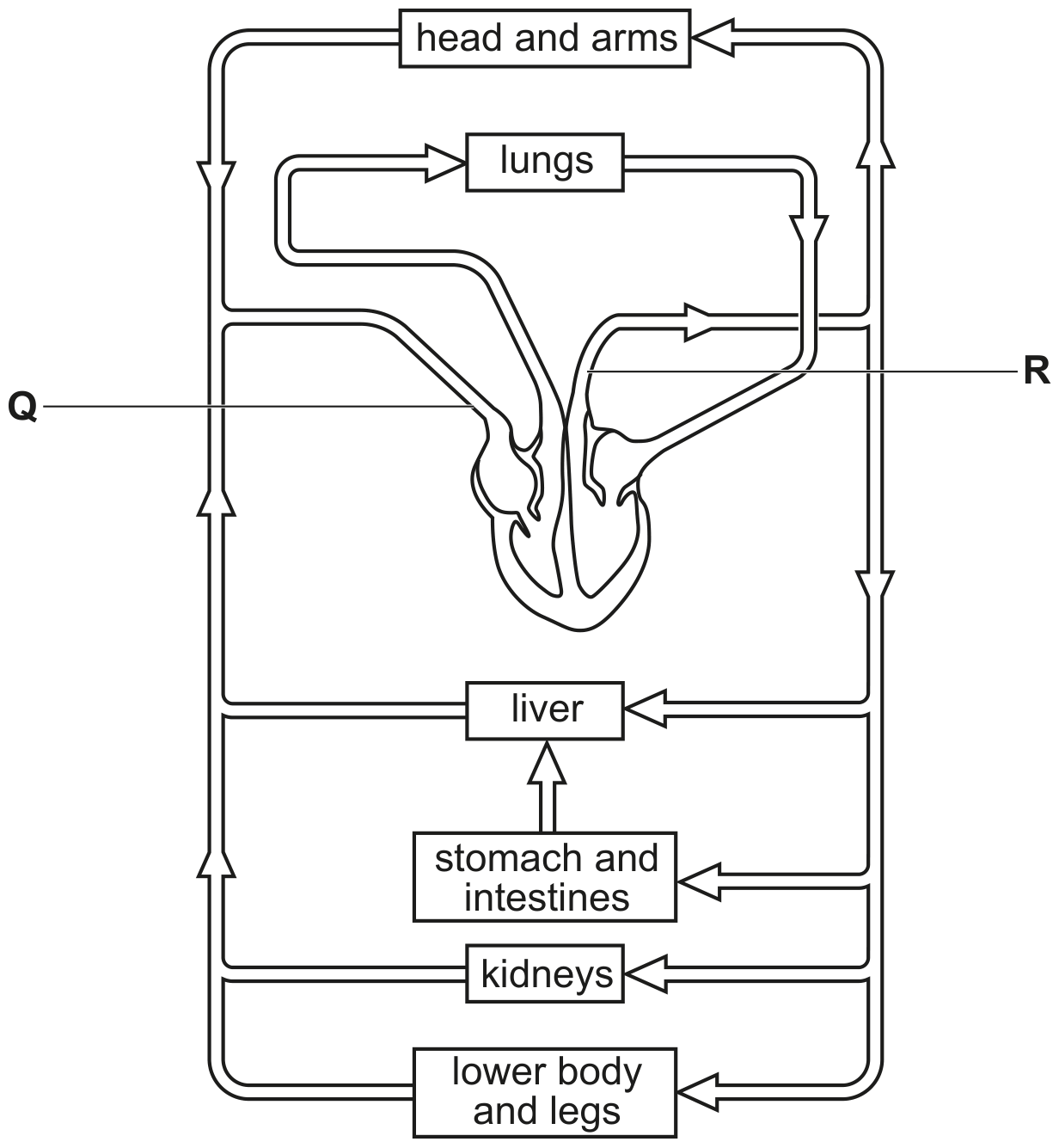
Fig. 2.1
Blood leaving the heart passes through valves before it enters blood vessel R.
Describe the differences between the structure of blood vessel Q and the structure of blood vessel R. Do not refer to valves in your answer.
Fig. 2.2 is a transmission electron micrograph of a cross-section of an arteriole. Blood flows from muscular arteries through arterioles into capillary networks.
The lining of the arteriole is folded because the arteriole has constricted. This constriction causes the blood pressure to decrease from 12.7 kPa in the muscular artery to 2.7 kPa at the end of the arteriole.
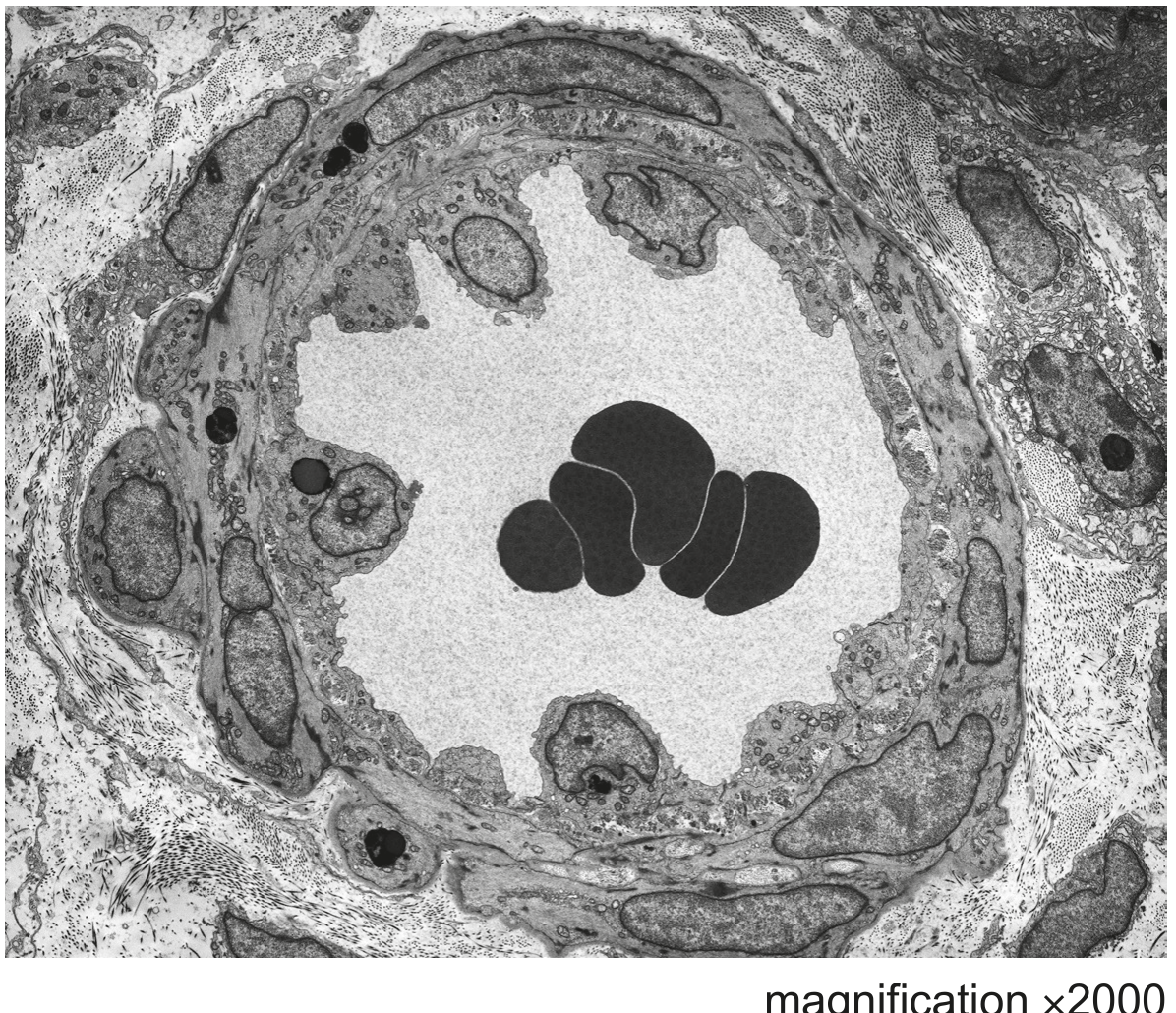
Fig. 2.2
Explain why it is important that the pressure of blood decreases as it passes through arterioles.
Compare the structure of a muscular artery with the structure of the arteriole shown in Fig. 2.2.
Outside the body, red blood cells can be maintained in an intact state by keeping the cells in a 0.9 % solution of sodium chloride. This is known as a normal saline solution.
Fig. 3.1 shows intact red blood cells.
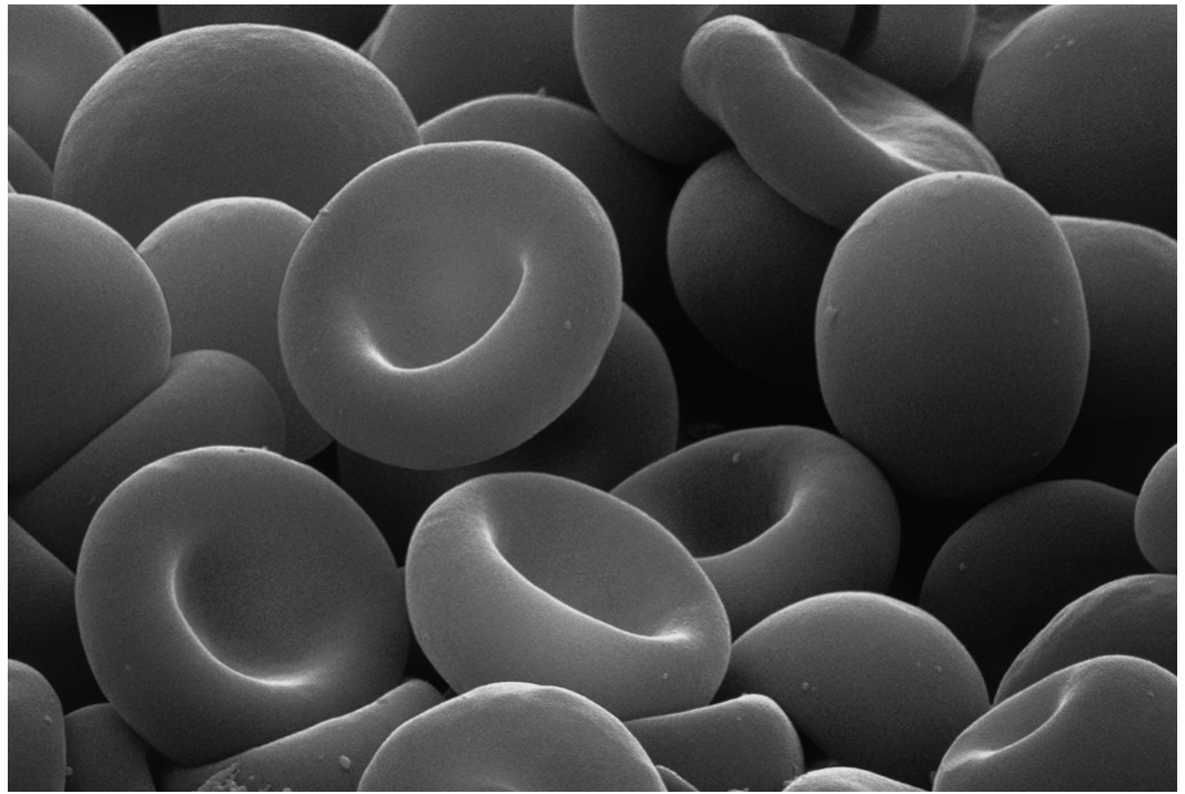
Fig. 3.1
With reference to Fig. 3.2, explain one feature that enables the surrounding body cells to receive an adequate supply of oxygen from the blood supplied by the capillary.
Some areas of the brain, known as blood-brain barriers, have a type of capillary that is relatively impermeable to substances.
Suggest one way in which the structure of a capillary in the blood-brain barrier differs from the structure of the capillary shown in Fig. 3.2.
Fig. 3.1 is a photomicrograph of a section through two different types of blood vessels, X and Y.
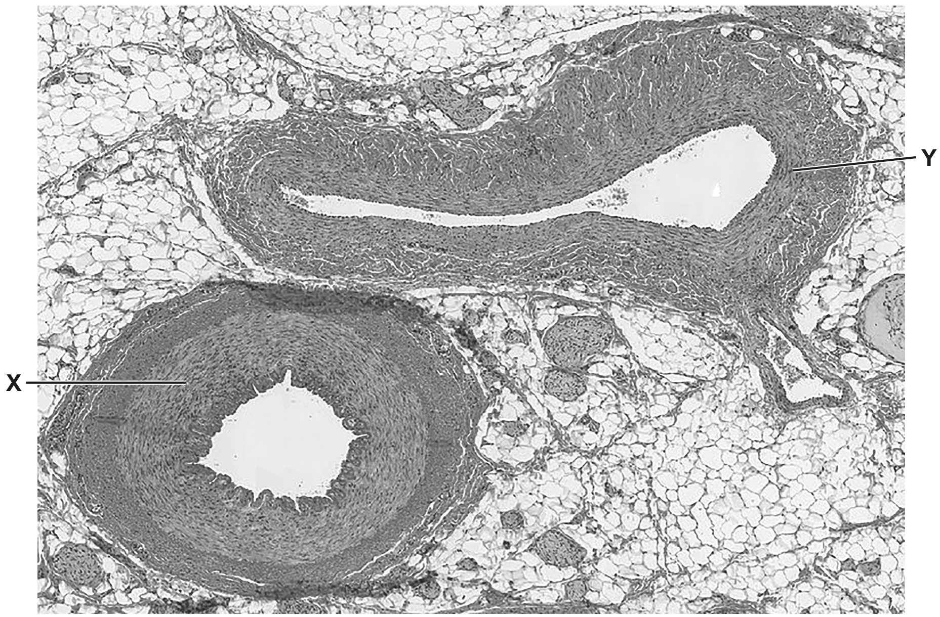
Fig. 3.1
State the reasons for your identification of the type of blood vessel shown by Y in Fig. 3.1.
In mammals, arteries branch to form smaller blood vessels called arterioles.
Arterioles branch to form capillaries that supply blood to tissues.
Explain the ways in which the structure of an artery is adapted to its function.
Fig. 4.1 shows transmission electron micrographs of cross-sections through an arteriole and a capillary.
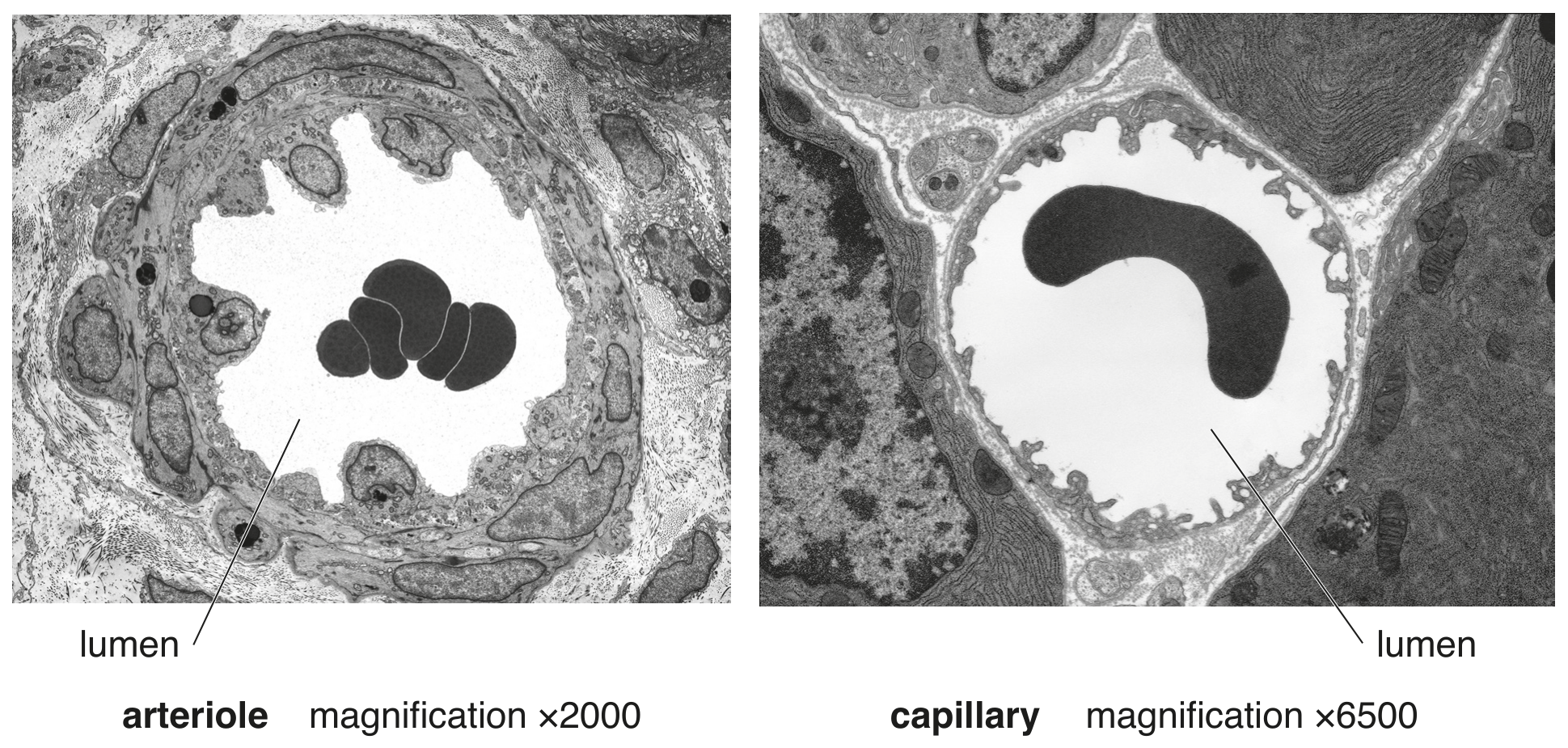
Fig. 4.1
Describe the differences between the arteriole and the capillary that are visible in Fig. 4.1.
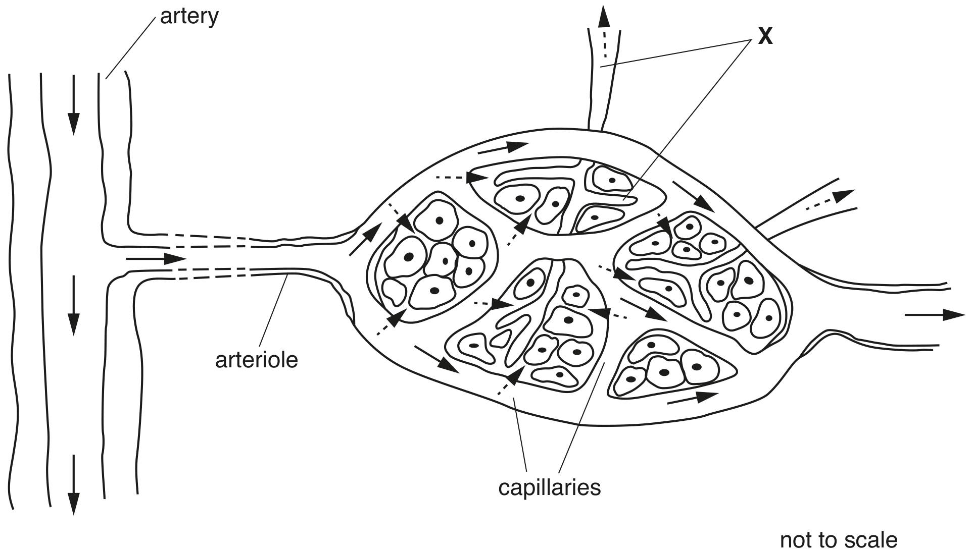
Fig. 4.2 shows a capillary network in a mammalian tissue. The arrows indicate the direction of flow of body fluids.
Which features enable the aorta to withstand high pressure at ventricular systole?
collagen fibres and elastin fibres
collagen fibres and endometrium
elastin fibres and large lumen
smooth muscle and small lumen