EduNinja
[Maximum number: 2]

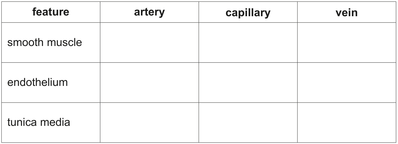
Smooth muscle is a tissue composed of smooth muscle cells. The cells contain cytoplasm packed with proteins that are involved in contraction and relaxation.

(a)

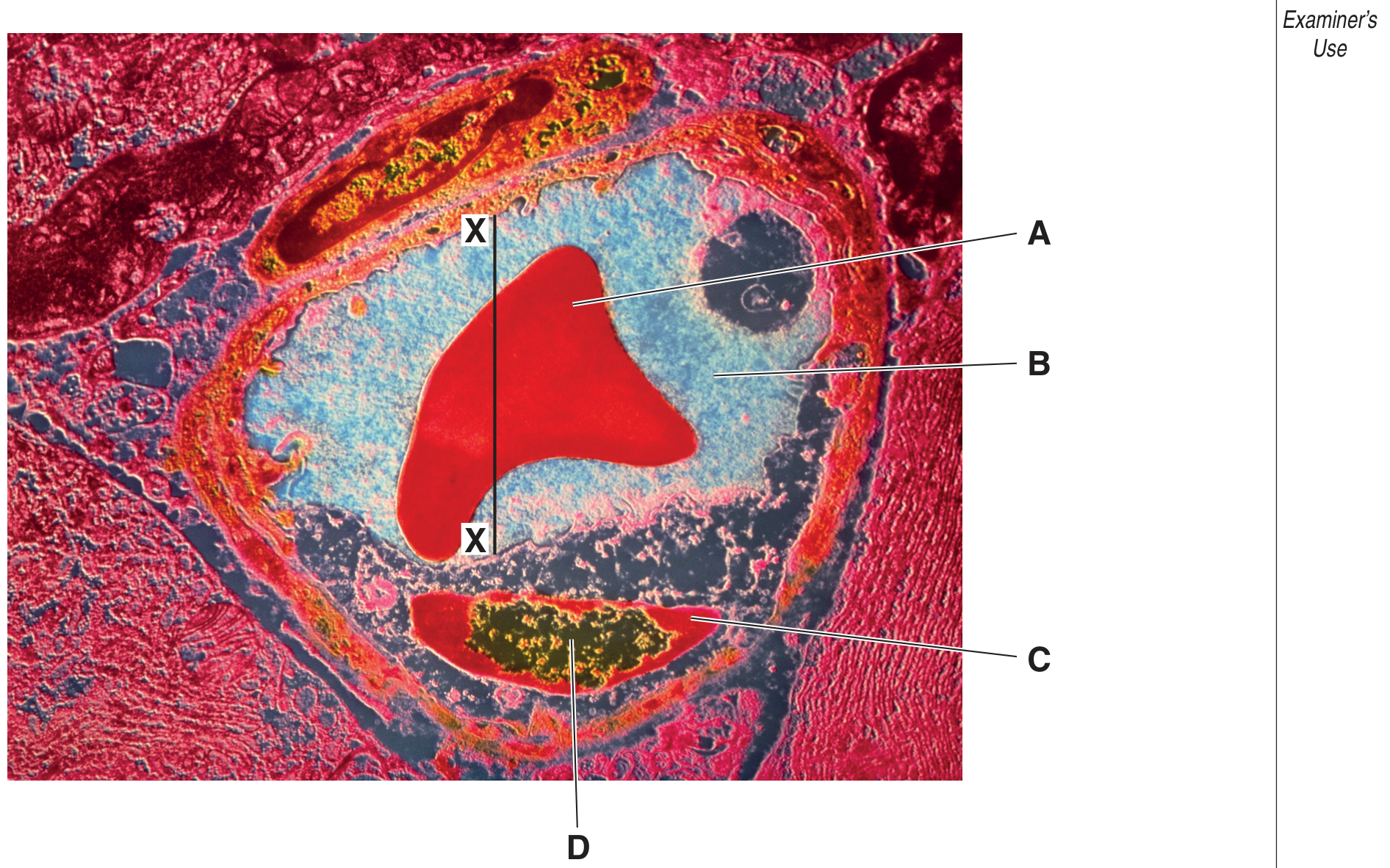
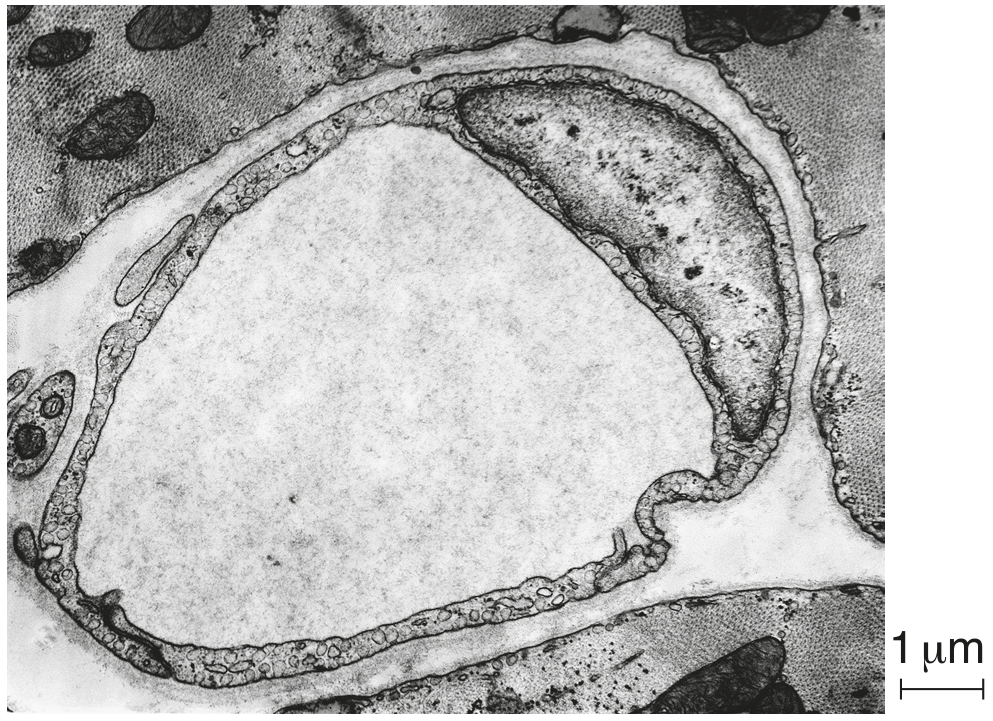
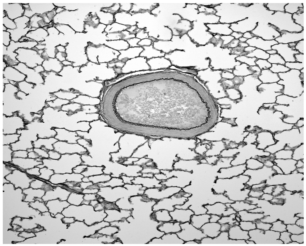
Fig. 1.2 is a photomicrograph of smooth muscle tissue in the wall of the intestines. A capillary is visible in addition to smooth muscle cells.
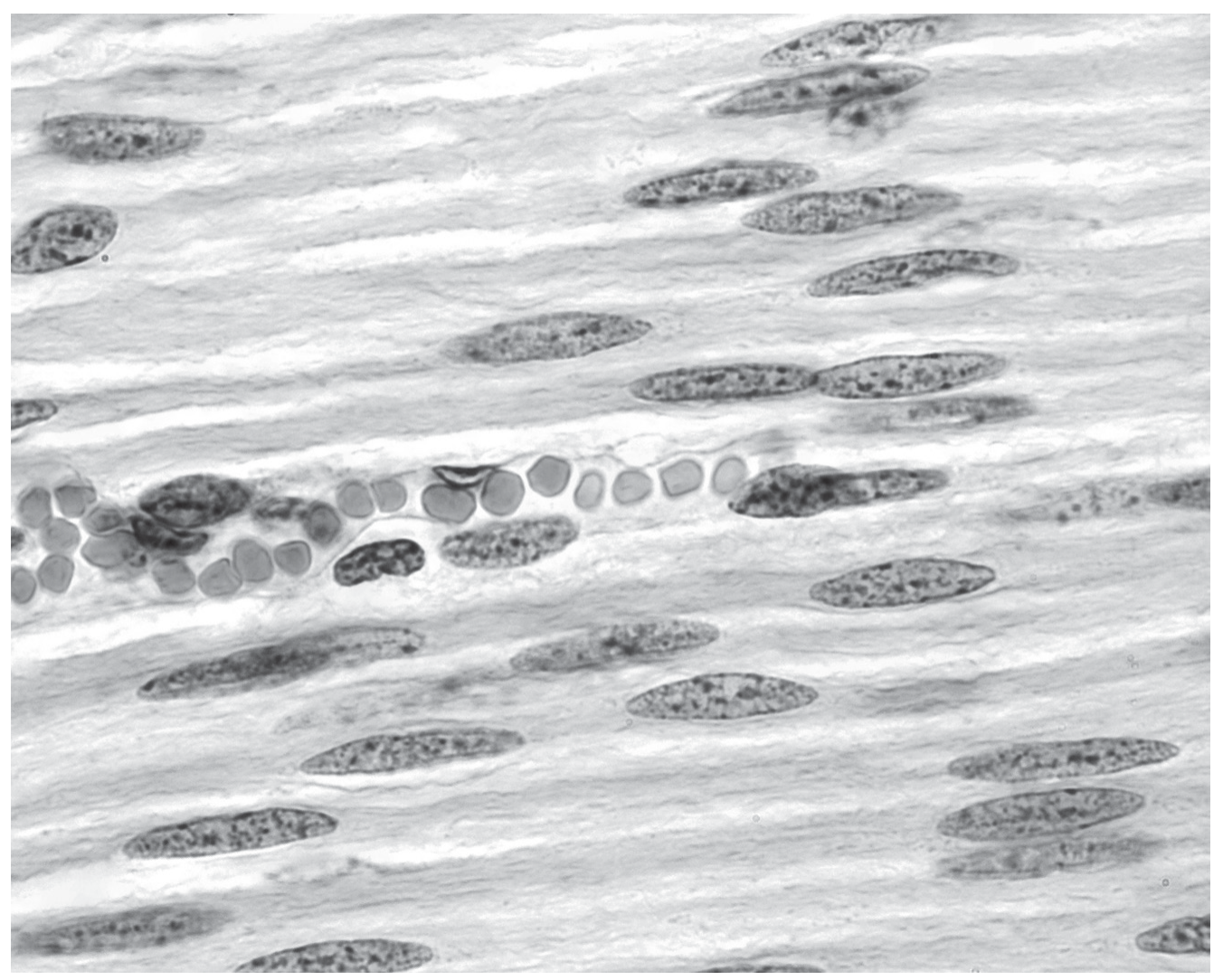
Fig. 1.2

Fig. 1.2

[ 2 ]
(i)

Outline the features that help to identify the blood vessel in Fig. 1.2 as a capillary.

[ 2 ]
[Maximum number: 2]

Fig. 1.1 is an electron micrograph of a cross section through a blood vessel.

Fig. 1.1

Fig. 1.1

(a)

Name the type of blood vessel shown in Fig. 1.1 and describe one visible feature which is characteristic of this type of vessel.
type of vessel
characteristic feature

[ 2 ]
(b)

Name:

(i)

structure A

[Maximum number: 3]

Fig. 2.1 is a transmission electron micrograph of a section through a blood vessel.

Fig. 2.1

Fig. 2.1

(a)

State the type of blood vessel shown in Fig. 2.1 and give two reasons for your choice. type of blood vessel
reason 1
reason 2

[ 3 ]
[Maximum number: 3]

Arteries, capillaries and veins are three types of blood vessel.

(a)

Table 3.1 shows some features of these three types of blood vessel.

Complete Table 3.1 by using a tick ()(\checkmark) if the feature is present in the blood vessel and a cross (x) if the feature is absent from the blood vessel.

Put a tick ()(\checkmark) or a cross (X) in every box.

Table 3.1

Table 3.1

[ 3 ]
[Maximum number: 1]

The photomicrograph shows a section through a structure found in mammals viewed using a light microscope.

Question image

×60\times 60

What are the main components of layer W?

A

collagen fibres only

B

elastic fibres and collagen fibres

C

smooth muscle and elastic fibres

D

squamous epithelial cells forming an endothelium

[Maximum number: 1]

Which structure is correctly identified?

Question image
A

aorta

B

pulmonary artery

C

pulmonary vein

D

vena cava

Plan diagrams of two blood vessels are shown.

Question image

Which labels are correct?

Table
[Maximum number: 1]

The photomicrograph shows a section through a tissue with an artery.

Question image

Which row correctly shows the type of artery and whether the blood inside the artery is oxygenated or deoxygenated?

type of artery

blood

muscular

deoxygenated

muscular

oxygenated

elastic

deoxygenated

elastic

oxygenated

[Maximum number: 1]

The photomicrograph shows a section through two structures found in mammals viewed using a light microscope.

\(\times 40\)

\(\times 40\)

Which row is correct?

W

X

feature

artery

vein

X contains cells with haemoglobinic acid

bronchiole

vein

X contains cells with oxyhaemoglobin

trachea

artery

the lumen of W allows air to pass through

vein

atrium

W contains deoxygenated blood

0