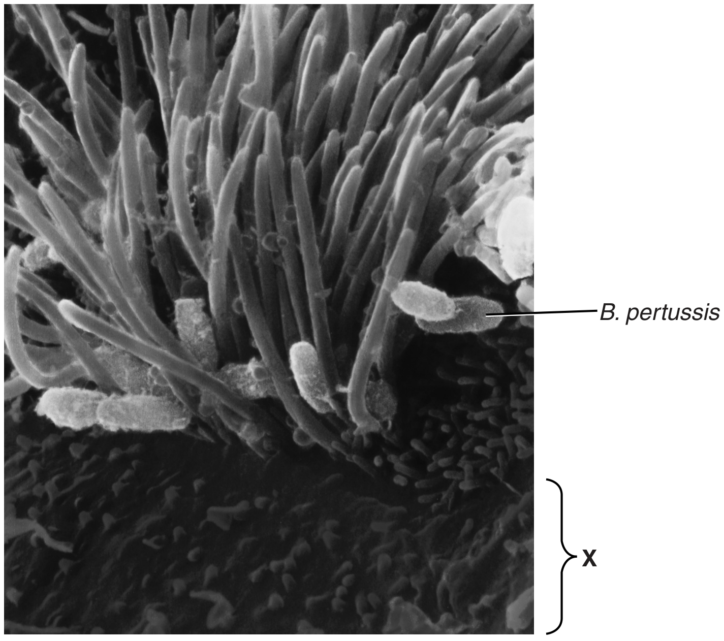
EduNinja
[Maximum number: 4]

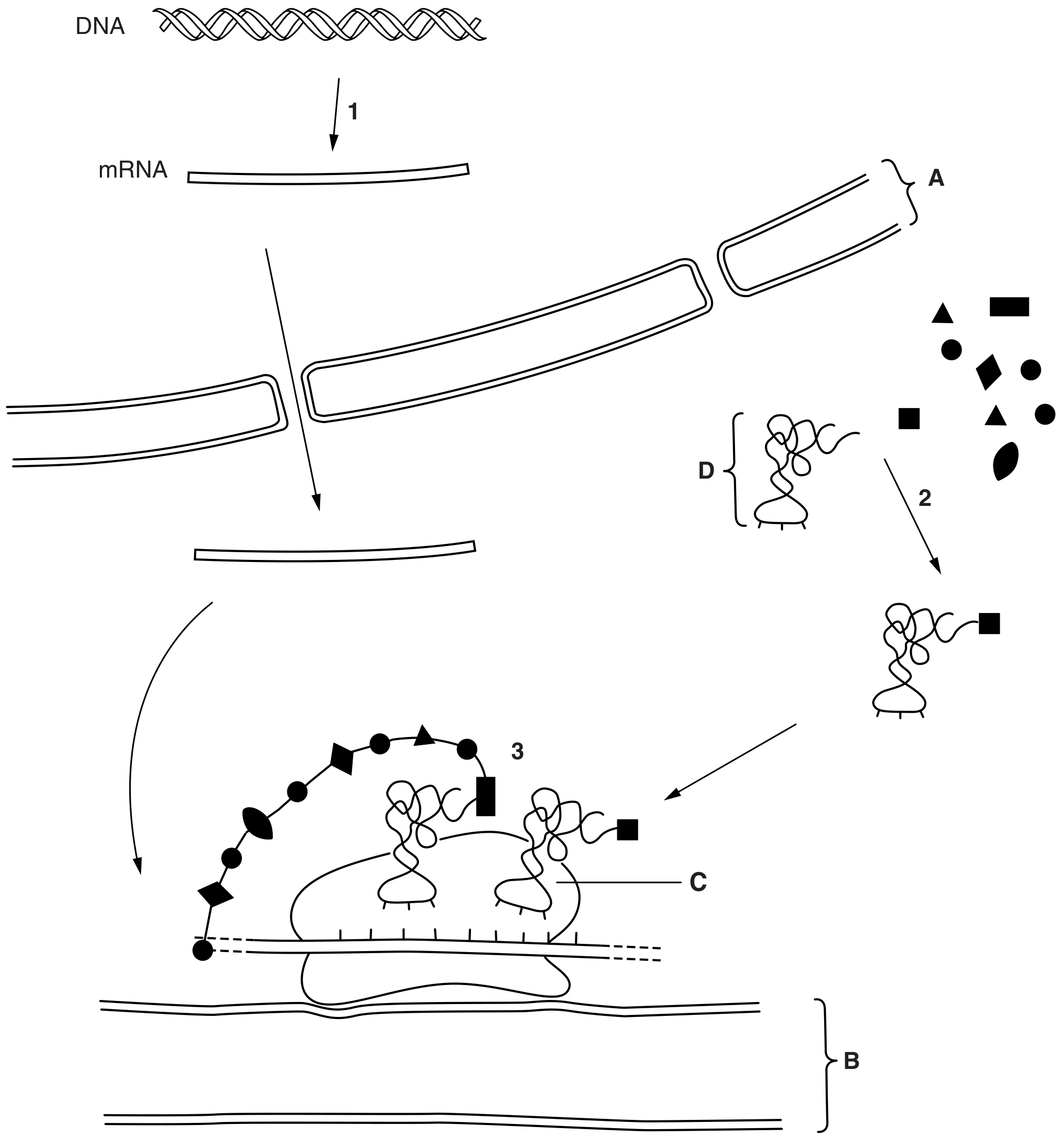
Fig. 1.1 is a diagram showing a stage in protein synthesis.
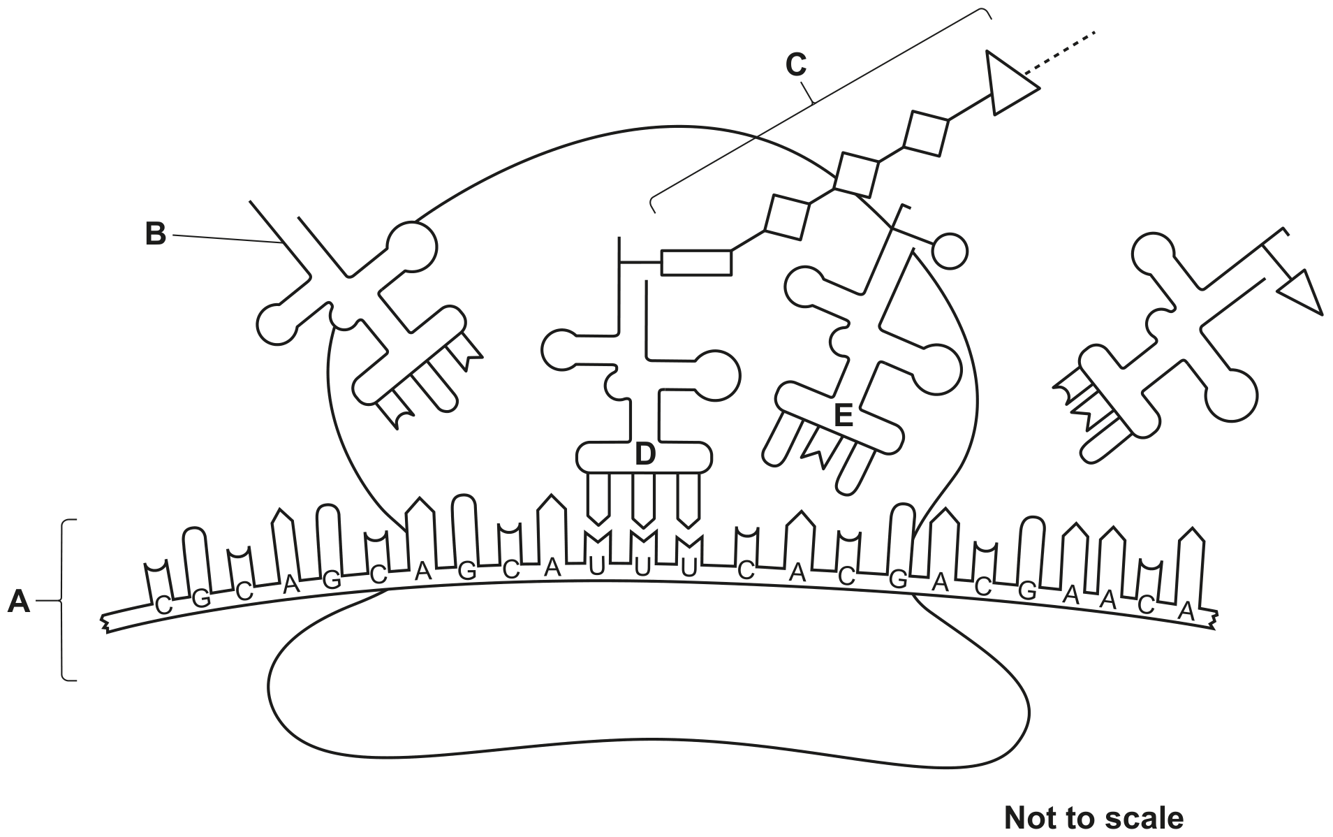
Fig. 1.1

Fig. 1.1

(a)

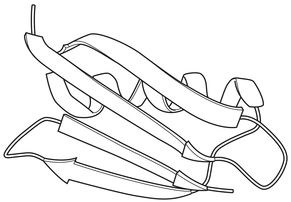
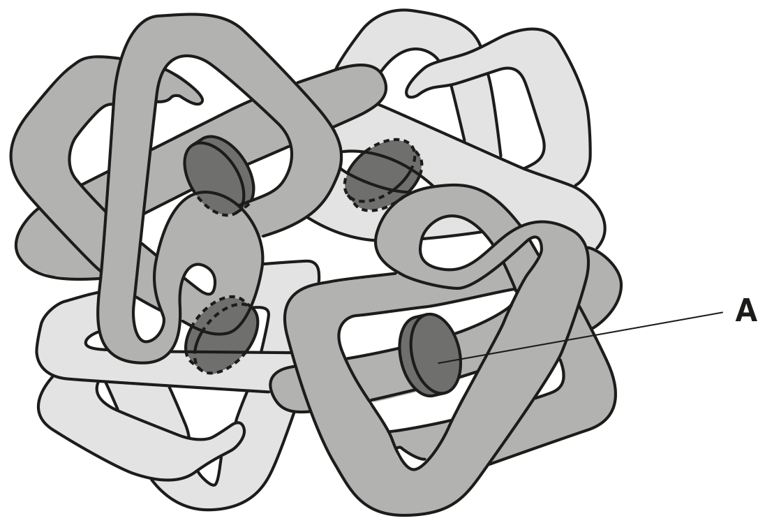
Mutagenesis is a process that leads to a change in the amino acid sequences of proteins. Scientists carry out mutagenesis to investigate the importance of particular amino acids in protein structure and function.

Outline how changing one amino acid in the β\beta-globin polypeptide of haemoglobin may change the structure and function of a molecule of haemoglobin.

[ 4 ]
[Maximum number: 1]

Smooth muscle is a tissue composed of smooth muscle cells. The cells contain cytoplasm packed with proteins that are involved in contraction and relaxation.

(a)

Caldesmon is a large protein with a number of binding sites to attach to other proteins.

Caldesmon exists in two different forms, H-caldesmon and L-caldesmon.
H-caldesmon helps to regulate contraction and relaxation in smooth muscle cells.
L -caldesmon is found in some non-muscle cells, where it also acts as a regulatory protein.
- Caldesmon is coded for by a gene known as CALD1.
- CALD1 has 17 exons.
- The primary structure of H-caldesmon has a repeating sequence in the middle of the amino acid chain that is not present in L-caldesmon.

[ 1 ]
(i)

Suggest how the two different forms of caldesmon can still have similar functions, even though they have a different primary structure.

[ 1 ]
(a)

Fig. 1.2 is a ribbon diagram showing the three-dimensional structure of a protein from the bacterium Streptococcus.

Fig. 1.2

Fig. 1.2

[ 4 ]
(i)

Describe the secondary structure of the protein shown in Fig. 1.2.

[ 2 ]
(ii)

Explain why the protein shown in Fig. 1.2 has tertiary structure, but not quaternary structure.

[ 2 ]
[Maximum number: 3]

Fig. 1.1 is a diagram of a molecule of haemoglobin.

Fig. 1.1

Fig. 1.1

(a)

A single base change in the DNA of the gene H B B results in a change to the amino acid sequence of β\beta-globin. In the sequence, a single glutamic acid is replaced by valine.

Outline the effects of this change in the amino acid sequence of β\beta-globin on the structure and function of a haemoglobin molecule.

[ 3 ]
[Maximum number: 4]

Antibodies are secreted by activated B-lymphocytes known as plasma cells.
Fig. 1.1 is a diagram showing the cellular processes involved in the production of a polypeptide of an antibody molecule (not drawn to scale).

Fig. 1.1

Fig. 1.1

(a)

Describe and explain how the structure of an antibody molecule is related to its functions.
Question 2 starts on page 6

[ 4 ]
[Maximum number: 1]

Fig. 1.1 is a diagram of an antibody molecule.

Fig. 1.1

Fig. 1.1

(a)
(i)

The antibody molecule in Fig. 1.1 has quaternary structure.

Explain the meaning of the term quaternary structure as applied to proteins.

[ 1 ]
[Maximum number: 2]

Fig. 2.1 is a scanning electron micrograph of an area of the trachea showing the presence of Bordetella pertussis bacteria.
B. pertussis is the causative organism of a respiratory disease in humans known as whooping cough. The disease is transmitted from person to person in a similar way to tuberculosis (TB).

A symptom that is common to TB and to whooping cough is the production of an excess of mucus.

Fig. 2.1

Fig. 2.1

(a)

The presence of B. pertussis stimulates the production of mucin, a gel-like glycoprotein that is the main component of mucus.

The mucin produced by the cell is packaged into vesicles ready for exocytosis.

[ 2 ]
(i)

Following translation, the polypeptide formed is modified by the addition of many short chains of monosaccharides in a process called glycosylation.

Suggest where glycosylation occurs in the cell and explain why mucin is packaged into vesicles.

[ 2 ]
[Maximum number: 1]

Most plants are C3 plants and are so-called because their first photosynthetic product is a three carbon compound.

The enzyme ribulose bisphosphate carboxylase/oxygenase (rubisco) catalyses the fixation of carbon dioxide in the Calvin cycle and is used by both C3 and C4 plants.

Each molecule is made up of eight large polypeptides and eight small polypeptides. Fig. 2.1 shows a side view of the molecule.

Fig. 2.1

Fig. 2.1

(a)
(i)

State why rubisco is said to have quaternary structure.

[ 1 ]
[Maximum number: 4]

In mammalian red blood cells, carbonic anhydrase has an important role in the transport of carbon dioxide.

Carbonic anhydrase is an enzyme.

(a)

A ribbon structure of a molecule of carbonic anhydrase is shown in Fig. 2.2. The zinc ion is associated with the active site of the enzyme and is essential for the enzyme to function.

Fig. 2.2

Fig. 2.2

The molecule of carbonic anhydrase has primary, secondary and tertiary structure.
Explain the extent to which Fig. 2.2 shows the primary, secondary and tertiary structure of carbonic anhydrase.

[ 4 ]
[Maximum number: 1]

Fig. 2.1 is a simplified diagram of the human circulatory system.

Fig. 2.1

Fig. 2.1

(a)

Blood plasma plays an important role in the transport of molecules such as antibodies.

Scientists discovered that some of the antibodies in the blood plasma of sharks have a different structure to the antibodies found in human blood plasma.

Fig. 2.2 shows the structure of an antibody molecule found in the blood plasma of a shark.

Fig. 2.2

Fig. 2.2

[ 1 ]
(i)

State how the quaternary structure of a human antibody molecule differs from the quaternary structure of the shark antibody molecule shown in Fig. 2.2.

[ 1 ]
0