EduNinja
[Maximum number: 3]

The malarial pathogen, Plasmodium falciparum, enters red blood cells after a person becomes infected. After some time, each cell of P. falciparum divides to form daughter cells.

Fig. 1.1 shows a cell of P. falciparum that is forming many daughter cells.
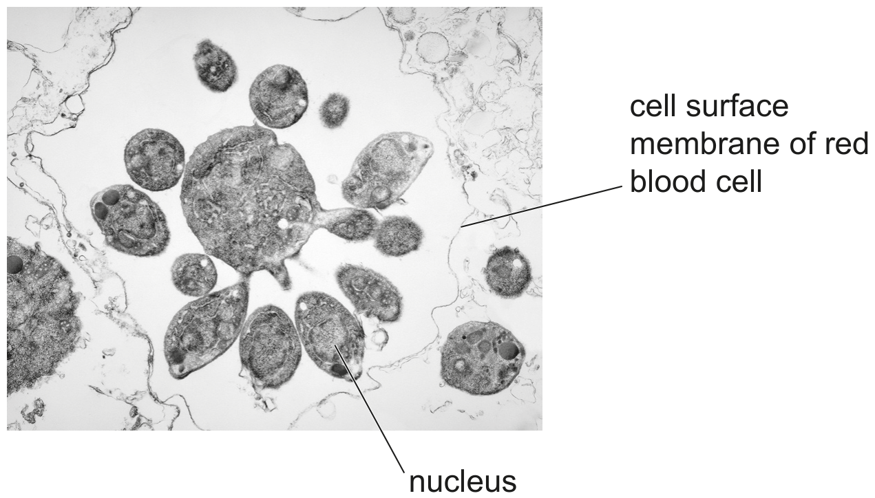
Fig. 1.1

Fig. 1.1

(a)

Explain how antibodies will reduce the spread of the malarial pathogen through the bloodstream.

[ 3 ]
[Maximum number: 1]

Name as precisely as you can the structure described in each of the following statements.

(a)

The cell that secretes antibodies.

[ 1 ]
[Maximum number: 2]

Fig. 2.1 is a simplified diagram of the human circulatory system.

Fig. 2.1

Fig. 2.1

(a)

Blood plasma plays an important role in the transport of molecules such as antibodies.

Scientists discovered that some of the antibodies in the blood plasma of sharks have a different structure to the antibodies found in human blood plasma.

Fig. 2.2 shows the structure of an antibody molecule found in the blood plasma of a shark.

Fig. 2.2

Fig. 2.2

[ 2 ]
(i)

Human antibodies are used in the treatment of some forms of cancer. However, the antibodies injected into the bloodstream can only reach a small percentage of the cancer cells that form the cancerous tumour.

Shark antibodies are smaller than human antibodies. Scientists are researching the possibility of injecting shark antibodies into the bloodstream to treat cancerous tumours in humans.

Suggest how using the smaller shark antibodies may be more effective in reaching a greater percentage of cancer cells than human antibodies and lead to greater success at treating cancer.

[ 2 ]
[Maximum number: 2]

Many tumours release a protein growth factor called VEGF. This is a chemical signal that causes nearby blood vessels to grow new branches into the tumour.

The monoclonal antibody, bevacizumab (Avastin ® { }^{\text {® }} ), specifically binds to VEGF.

(a)

A second monoclonal antibody, ranibizumab (Lucentis ® { }^{\text {® }} ) is used to treat eye diseases. Lucentis ®{ }^{\circledR} is a fragment of Avastin ®{ }^{\circledR} and is shown in Fig. 2.1.

Fig. 2.1

Fig. 2.1

Complete Fig. 2.1 to show a molecule of Avastin ®{ }^{\circledR}.
Labels are not required.

[ 2 ]
[Maximum number: 5]

Fig. 3.1 shows a method of producing monoclonal antibodies.

Fig. 3.1

Fig. 3.1

(a)
(i)

Describe what is meant by the term monoclonal antibody.

[ 2 ]
(ii)

Describe, in detail, the molecular structure of one of the antibodies produced in step 6. You may wish to use an annotated diagram to answer the question.

[ 3 ]
[Maximum number: 4]

Fig. 3.1 is a diagram that shows the structure of an antibody molecule.

Fig. 3.1

Fig. 3.1

(a)
(i)

Use Fig. 3.1 to explain how the structure of the variable region of an antibody molecule is related to its function.

[ 3 ]
(ii)

State the role of the constant region of an antibody.

[ 1 ]
[Maximum number: 5]

Cholera is a life-threatening and infectious disease caused by the bacterium Vibrio cholerae. One of the symptoms of the disease is severe diarrhoea.
V. cholerae O1 and V. cholerae O139 are the two forms of the pathogen that are associated with cholera epidemics (large outbreaks). These two forms have different antigens that can be detected.

(a)

Rapid diagnostic testing (RDT) can be used to diagnose cholera by detecting the presence of V. cholerae. One type of RDT involves using a dipstick that contains mobile and immobilised monoclonal antibodies.

Monoclonal antibodies (mAbs) are specific in their action.
Fig. 2.1 shows a simplified diagram of an RDT dipstick that can be used to distinguish between V. cholerae O1 and V. cholerae O139. A faecal sample from a person with suspected cholera is collected and added to a reagent solution to form the test mixture.

Fig. 2.1

Fig. 2.1

The test mixture moves up the dipstick through area 1. The mobile monoclonal antibodies are attached to tiny gold particles. If these antibodies collect in test area 2 or 3, a coloured band becomes visible.

A coloured band that becomes visible in area 4 confirms that the test strip is working and that the results are valid.

[ 5 ]
(i)

Explain how the structure of the monoclonal antibodies in the dipstick allows them to be specific in their action.

[ 2 ]
(ii)

Fig. 2.2 shows the results for samples taken from two different people, A and B, who are suspected of having cholera.

Fig. 2.2

Fig. 2.2

With reference to Fig. 2.2, state and explain the conclusions that can be drawn from the results of the RDT dipsticks for each person.

[ 3 ]
[Maximum number: 4]

In 2015, the World Health Organization (WHO) published the Global Technical Strategy for Malaria 2016-2030. The aim of this global strategy, which follows on from the 2008 Global Malaria Action Plan (GMAP), is to make progress in the control and elimination of malaria.

Both the global strategy and GMAP aim to reduce:
- the case incidence (number of new cases each year) of malaria
- the mortality rate (number of deaths each year) from malaria.

(a)

Another recommendation of the global strategy is to carry out rapid diagnostic testing (RDT) of individuals who may have malaria. This involves testing human blood samples for the presence of proteins specific to Plasmodium. Test sticks can be used.

Table 3.1 contains information about two RDT test sticks.

Table 3.1

Table 3.1

Some details of the design of these RDT test sticks are shown in Fig. 3.2.

Fig. 3.2

Fig. 3.2

The immobilised monoclonal antibodies in the test window are not visible.
If the blood sample contains a Plasmodium protein that can be detected by the RDT test stick:
- the mobile monoclonal antibodies bind to one part of the protein
- the immobilised monoclonal antibodies bind to another part of the protein
- a coloured line in the test window indicates a positive result for the protein.

[ 4 ]
(i)

With reference to Table 3.1 and Fig. 3.2, explain why test stick 1 and test stick 2 will contain different mobile monoclonal antibodies.

[ 2 ]
(ii)

Two blood samples were removed from a person. One sample was added to test stick 1 and the other sample was added to test stick 2.

With reference to Table 3.1 and Fig. 3.2, explain what can be diagnosed for this person from a positive result for test stick 1 and a negative result for test stick 2.

[ 2 ]
[Maximum number: 4]

Cells of the immune system function to protect the body against infectious diseases.

(a)
(i)

Name the type of cell that produces antibodies.

[ 1 ]
(ii)

The virus that causes the infectious disease influenza has two antigens, H and N. Antibodies are produced in response to an infection by this virus. The antibodies are specific for either antigen H or for antigen N.

Describe how the structure of an antibody molecule allows it to be specific for one antigen, such as H or N.

[ 3 ]
[Maximum number: 1]

Nerium oleander is a xerophytic plant. A photomicrograph of a section through the leaf of N. oleander is shown in Fig. 3.1.

Fig. 3.1

Fig. 3.1

(a)

Monoclonal antibody can be produced that is specific to cyFBPase. This antibody is used by investigators to check that the plants with the c y F B P mutation do not synthesise this enzyme.

[ 1 ]
(i)

Anti-cyFBPase monoclonal antibody is added to extracts taken from the leaves of the plants with the cyFBP mutation.

State the expected results following addition of the monoclonal antibody that would confirm the absence of cyFBPase in the leaf extracts.

[ 1 ]
0