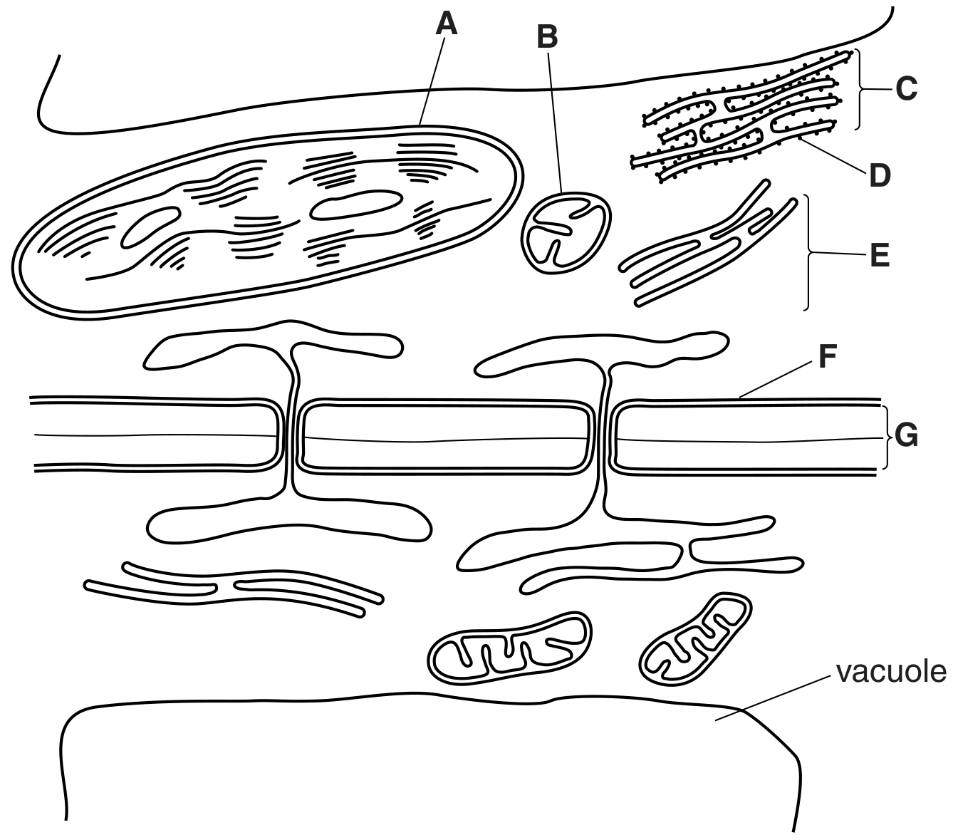
[Maximum number: 3]
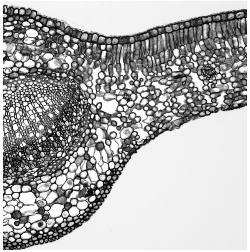
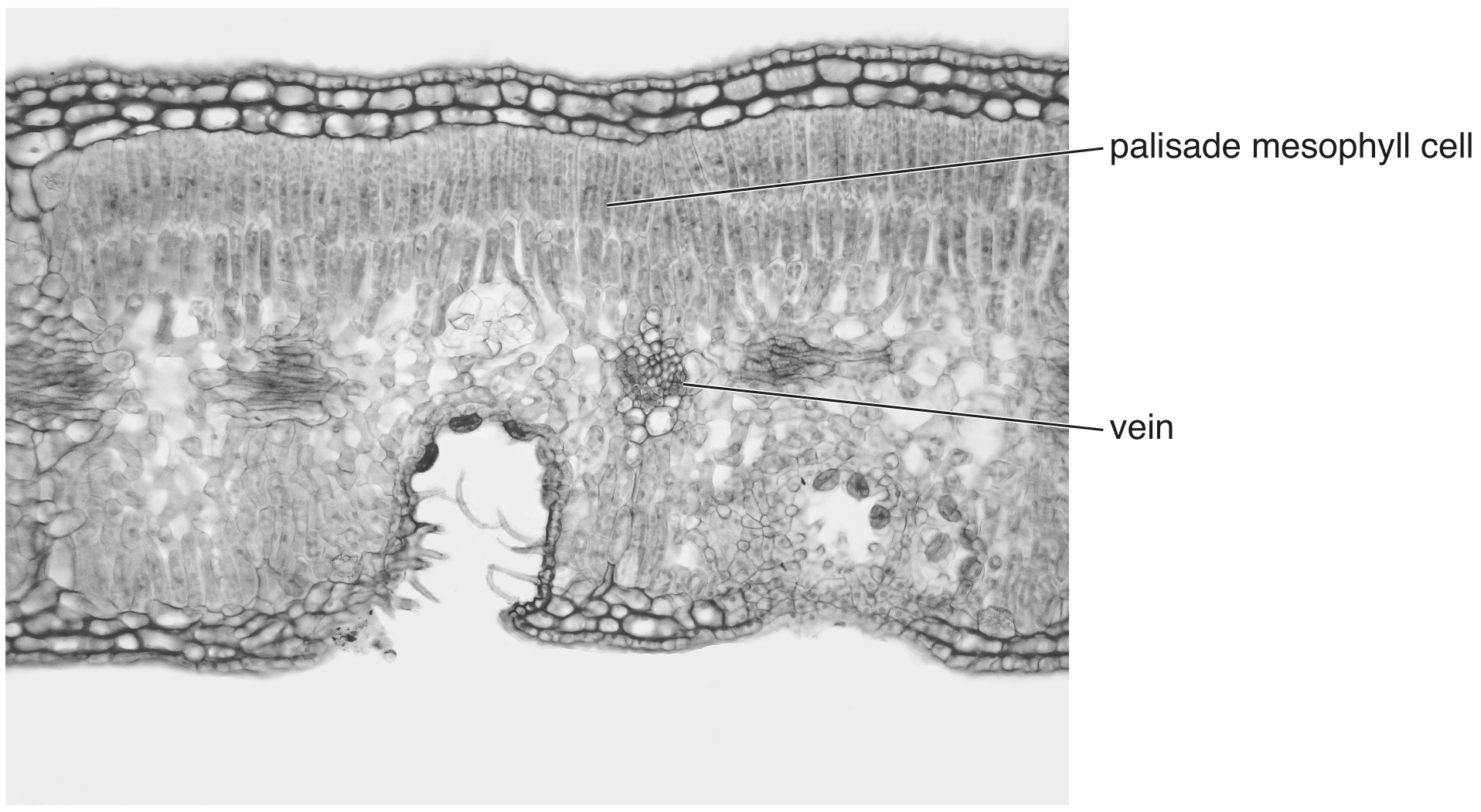
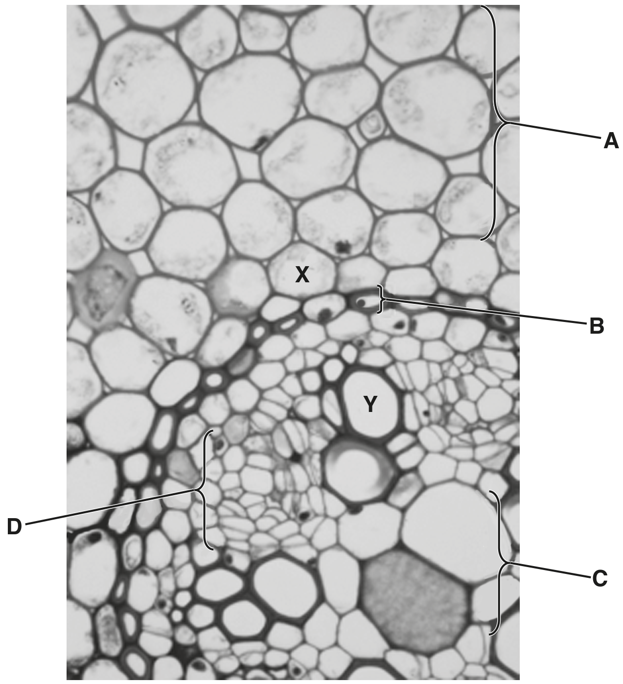
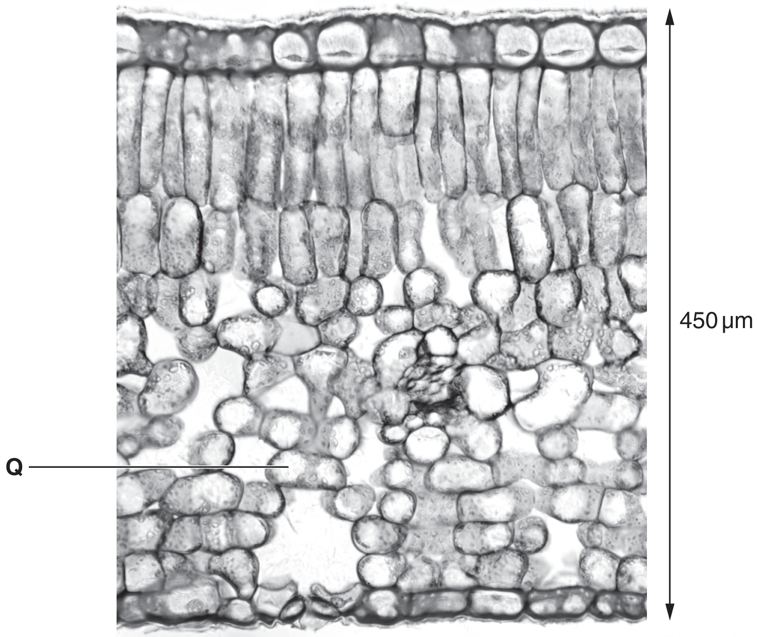
Fig. 1.1 and Fig. 1.2 are photomicrographs of sections through the leaves of two different plants.
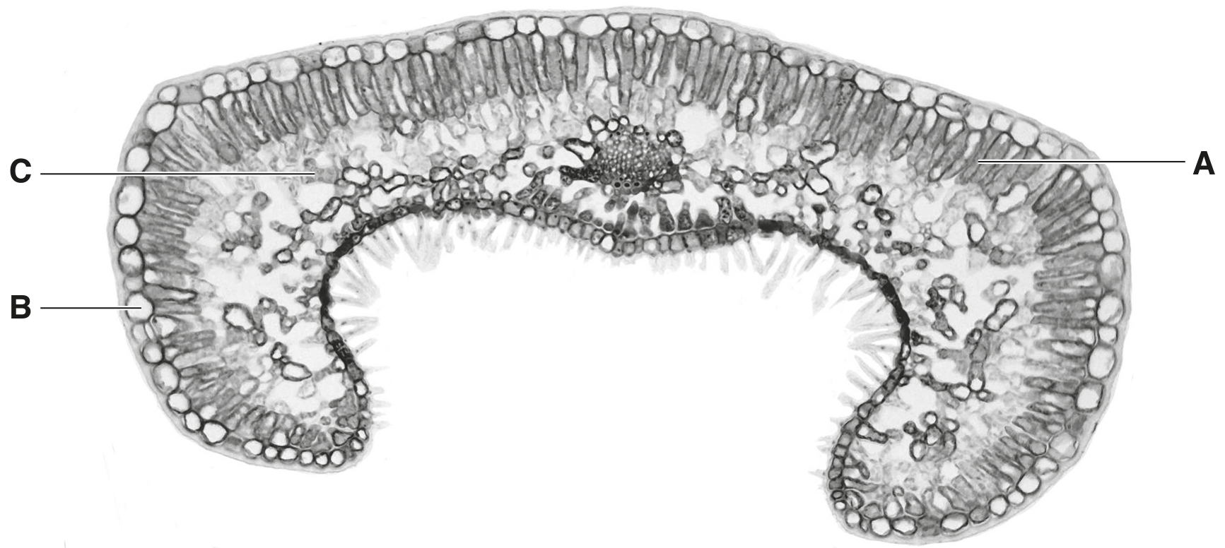
Fig. 1.1 Fig. 1.2 is a photomicrograph of a section through a leaf of Himalayan cedar, Cedrus deodara.
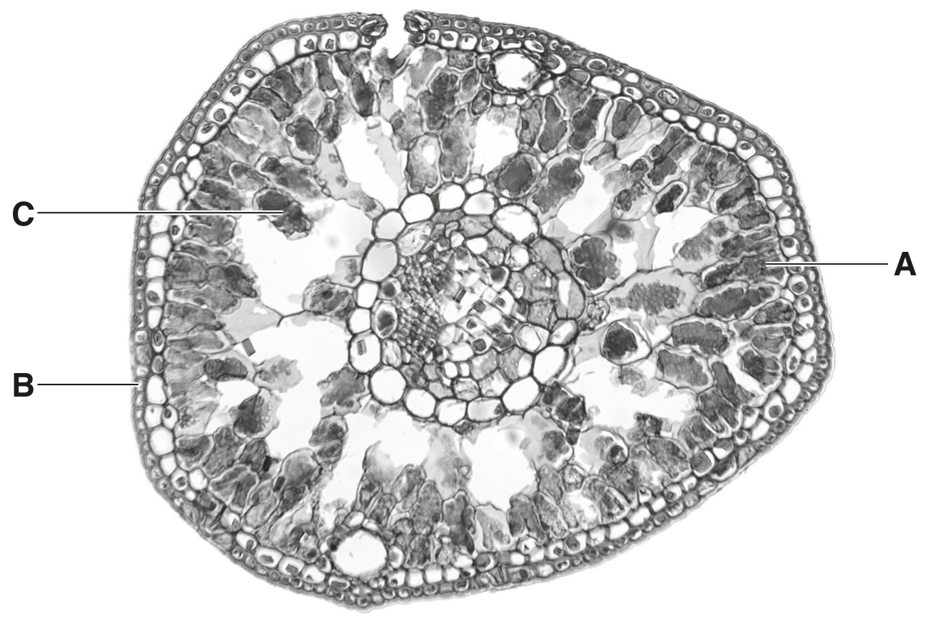
Fig. 1.2 Fig. 1.1 and Fig. 1.2 are not shown at the same magnification.
(a)
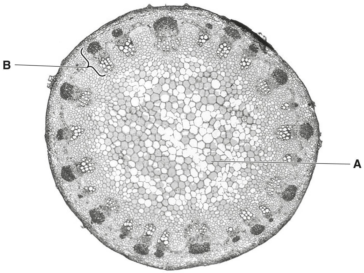
Cells labelled A, B and C in Fig. 1.1 and Fig. 1.2 each form a different tissue.
Name each tissue formed.
tissue formed from A
tissue formed from B
tissue formed from C
tissue formed from C
[ 3 ]