(a)
Fig. 1.1 is a diagram of a part of a sarcomere in striated muscle.
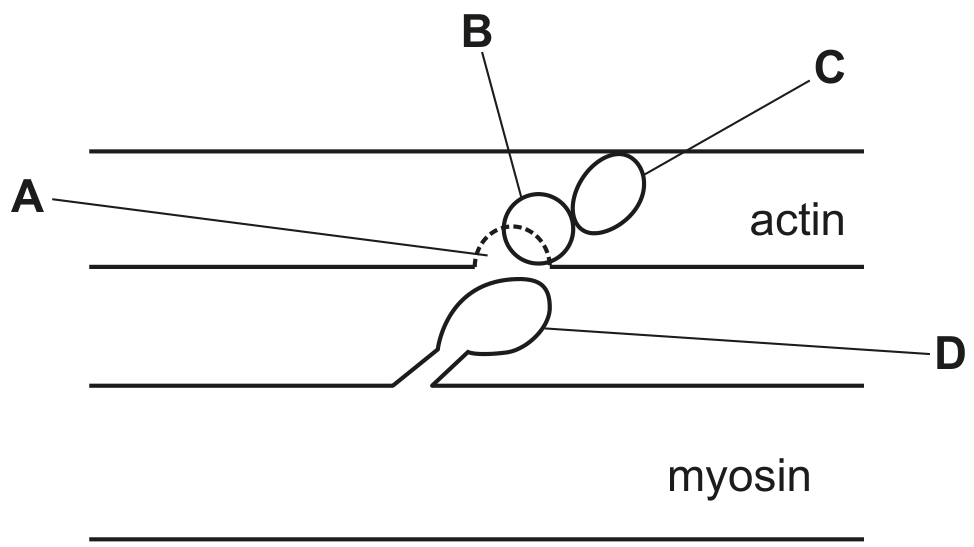
Fig. 1.1
With reference to Fig. 1.1, name A, B, C and D.
A
B
C
D
[ 4 ]
 EduNinja
Fig. 1.1 is a diagram of a part of a sarcomere in striated muscle.
Fig. 1.1
With reference to Fig. 1.1, name A, B, C and D.
A
B
C
D
Fig. 3.1 shows a diagram of two sarcomeres of relaxed striated muscle.
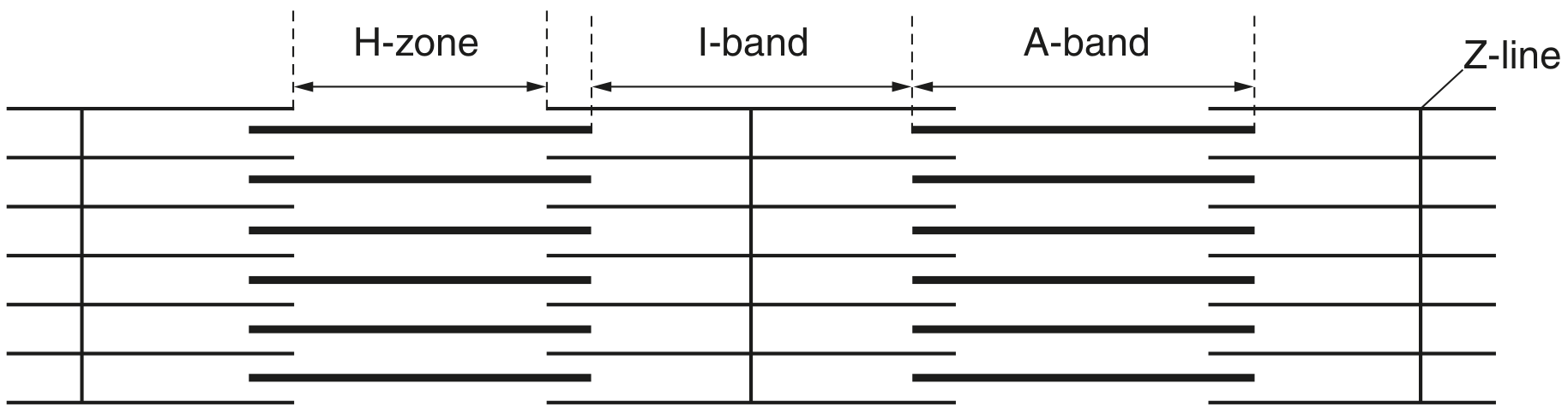
Fig. 3.1
When the striated muscle contracts, state what happens to the length of:
the I-band
the A-band.
Fig. 4.1 shows a transmission electron micrograph of a section through striated muscle.
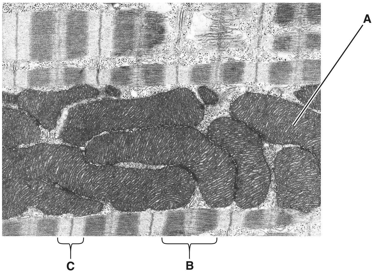
Fig. 4.1
Complete Table 4.1, using the letters A, B or C, to show the location of proteins associated with striated muscle structure.
You may use each letter once, more than once, or not at all.
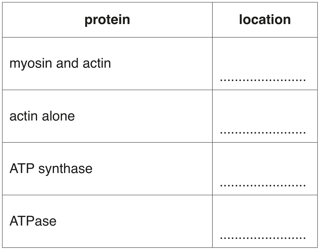
Table 4.1
Fig. 7.1 shows a mammalian neuromuscular junction.
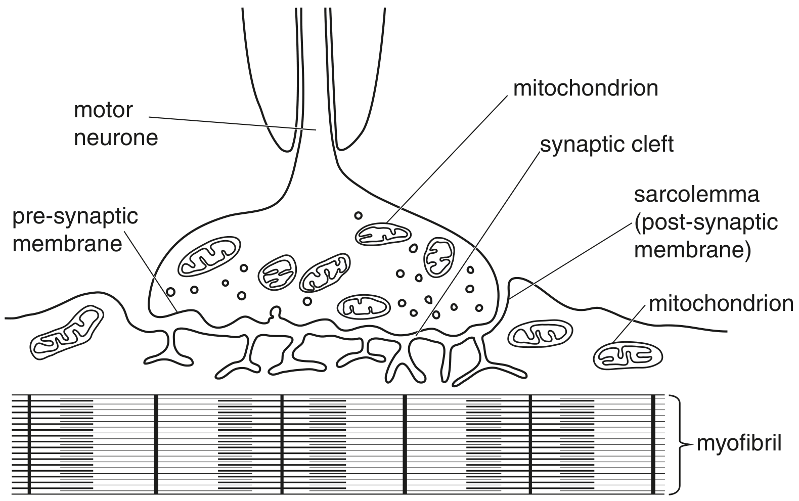
Fig. 7.1
On Fig. 7.1, use label lines and letters to label each of the following parts:
- a region containing only actin
- a region containing both actin and myosin.
Fig. 9.1 is a diagram of a relaxed sarcomere in striated muscle.
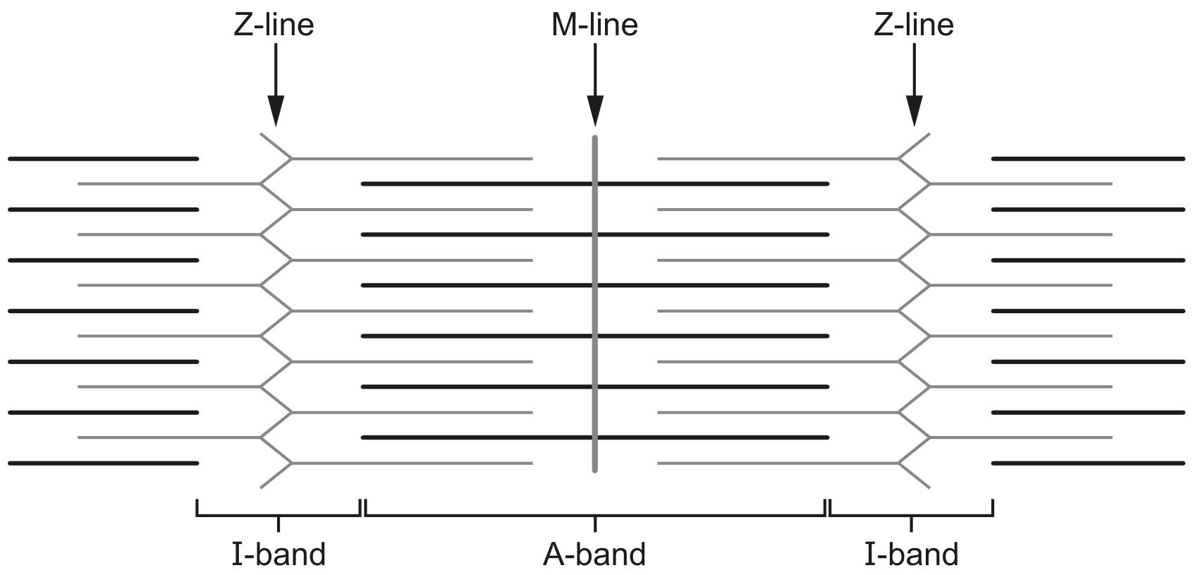
Fig. 9.1
On Fig. 9.1, use label lines and letters to label:
- an actin filament with the letter P
- a myosin filament with the letter R.
Fig. 7.1 is an electron micrograph of a section of striated muscle.
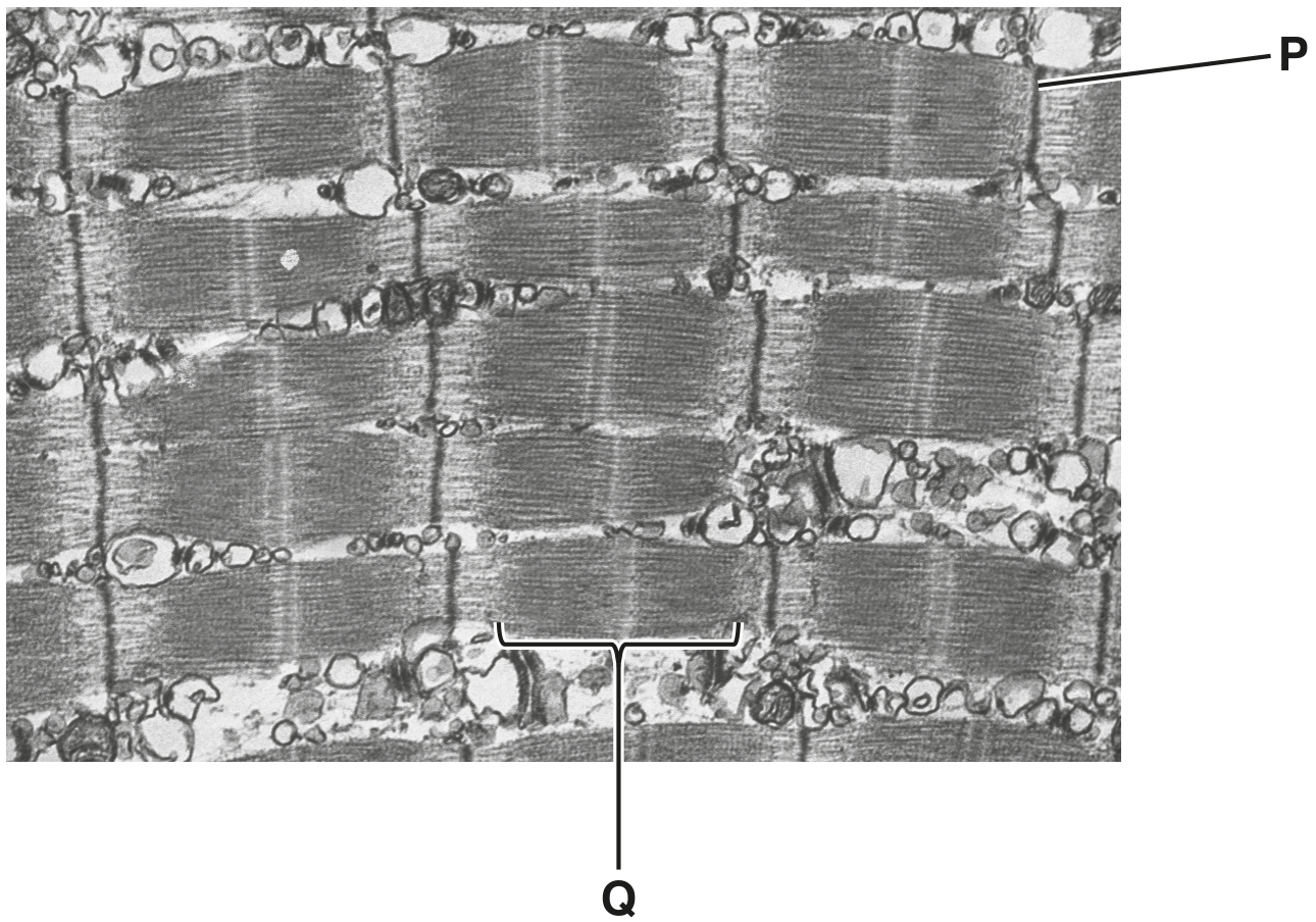
Fig. 7.1
Name structure P and the region represented by Q.
P
Q
Striated muscle is made of many fibres. Each fibre is composed of myofibrils.
The striated appearance of the muscle fibre is due to the arrangement of two types of protein filaments, thick filaments and thin filaments, within the sarcomeres of the myofibril.
Describe the main structural features of thick filaments and thin filaments in the sarcomere.
thick filaments
thin filaments
Describe the ultrastructure of a striated muscle fibre.