EduNinja
[Maximum number: 3]

Hypoxia is a condition in which tissues of the body are deprived of an adequate oxygen supply. A study was carried out in rats to examine the effects of continuing hypoxia on the structure of the diaphragm, and to determine whether nitric oxide is implicated in adaptation of the diaphragm to hypoxia. The diaphragm helps to supply oxygen to tissues and organs in the body by ventilating the lungs.

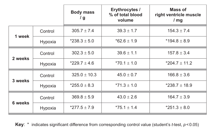
A group of 36 adult male rats were kept for 6 weeks in low oxygen while 36 adult male rats were kept in normal oxygen levels.

Question image
(a)

The graph shows the effect of hypoxia on the endurance of rats' diaphragm muscle after 6 weeks. Endurance is the change in force measured as a percentage of the initial force.

Question image

Using the data in the graph, deduce whether hypoxia increases or decreases the endurance of the rats' diaphragm muscle.

[ 2 ]
(b)

Skeletal muscle contractions can take two different forms: if they are stimulated by a single action potential they take the form of a twitch and if they are stimulated by a series of action potentials the contraction is longer lasting (tetanic). The table shows the effects of hypoxia on the force of twitch and peak tetanic contraction in the diaphragm.

Table
[ 1 ]
(i)

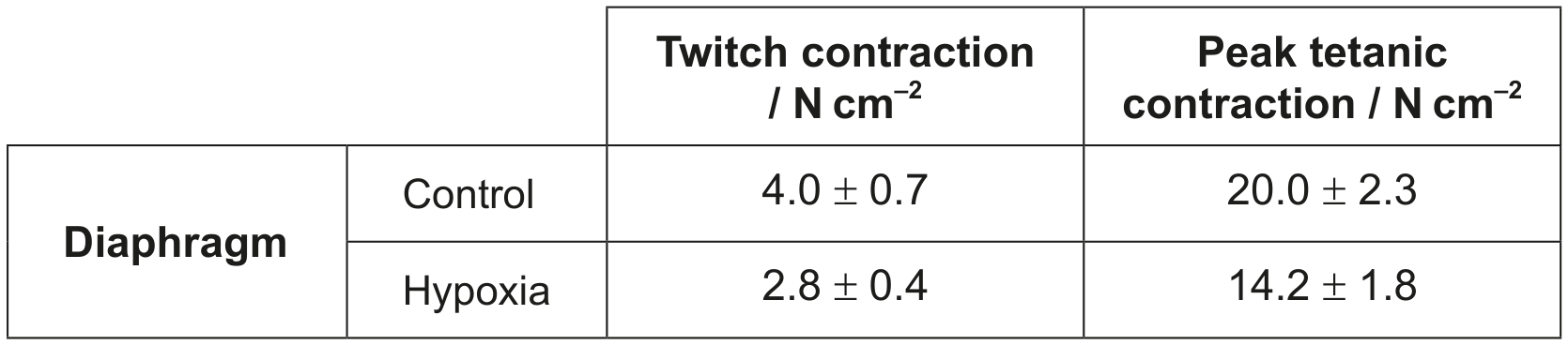
Outline the effect of hypoxia on the force of contraction of the diaphragm.

[ 1 ]
[Maximum number: 4]

A1. Elderly people lose bone mineral density (BMD) with age, and this is the source of many health issues, including higher risk of bone fractures. Researchers measured the change in BMD amongst elderly men considering many factors, over a period of four years. The results presented below show the difference between bone density change due solely to vitamin C intake and bone density loss considering a combination of the other factors represented by the baseline (zero). BMD was measured in the spine and at two femur (thigh bone) locations (femoral neck and trochanter) using scanner images. Daily intake of total vitamin C was categorized as high, medium or low.

Question image
(a)

Compare the changes in bone density of the femoral neck with those of the spine.

[ 2 ]
(b)

Evaluate the evidence provided by the data that the intake of vitamin C supplements may reduce bone density loss in elderly people.

[ 2 ]
[Maximum number: 1]

A student used two different methods to measure the left ring finger joint angles in 120 healthy people. The metacarpophalangeal (MCP), proximal interphalangeal (PIP) and distal interphalangeal (DIP) joint angles were measured while the finger was extended (straight) or flexed (bent).

The box-and-whisker plots show the angle measurements at the different joints obtained for both methods.

Question image
(a)
(i)

State the dependent variable in this experiment.

[ 1 ]
[Maximum number: 5]

Ehlers-Danlos syndrome (EDS) causes joint hypermobility, where joint movement extends well beyond the normal range. Researchers used two different methods to measure angles of movement at a joint of the smallest (fifth) finger.

(a)

Suggest in which direction the finger was moved to assess hypermobility.

The graph shows all angles of movement of the finger, measured in degrees ( { }^{\circ} ) using both methods.

Question image

Key:
- Individual with EDS
- Individual without EDS

[ 1 ]
(b)

Suggest a method which could have been used to measure the angle of movement of the finger.

[ 1 ]
(c)

Estimate the threshold (minimum) angle for a diagnosis of EDS.

[ 1 ]
(d)

Suggest which structure at the finger joint is affected by EDS.

[ 1 ]
(e)

Describe the level of correlation between the data from the two measurement methods.

[ 1 ]
(a)

Titin is the longest protein in the human body, consisting of a single strand of over 34000 amino acids.

Question image
[ 2 ]
(i)

Explain the role of titin in sarcomeres.

[ 2 ]
(a)

The electron micrographs show sections of muscle fibre in two different states.

I

Question image

II

Question image
[ 3 ]
(i)

Label a sarcomere on electron micrograph I.

[ 1 ]
(ii)

Deduce what events occur in muscle fibres to change them from the state shown in electron micrograph I to the state shown in electron micrograph II. provided.

[ 2 ]
[Maximum number: 1]

B1. In a study of athletes in Brazil, doctors examined the relationship between the type of injuries sustained by the athletes and the type of activity in which they were competing. The percentages of injuries to muscles, joints and tendons were compared across a range of sporting activities.

Question image
(a)
(i)

Hurdling is a sport that involves both sprinting and jumping. Using the data, suggest with a reason, which of these two activities contributes most of the injuries sustained by hurdlers.

[ 1 ]
[Maximum number: 5]

Strenuous short-duration exercise requires a lot of energy in different muscle fibre types. This causes a reduction in the mechanical power output.

The graph shows the decreases in ATP concentration in different human muscle fibres during brief strenuous exercise. It also shows muscle fatigue, measured as the percentage decrease in power.

Question image
(a)

Distinguish between power decline for the three types of muscle fibre.

[ 3 ]
(b)

Scientists have distinguished muscle fibres on the basis of their colour as red or white and their contractile properties as fast or slow. White fibres have a low myoglobin content, few mitochondria and fatigue quickly. Using the data, deduce with reasons, which muscle fibre type is most probably a white muscle fibre.

[ 2 ]
[Maximum number: 7]

In a study to investigate the effect of training on muscles, seven healthy males were subjected to training using the right leg only. They then exercised using both legs and the glycogen content was measured in both the untrained (left leg) and trained (right leg) muscle.

Question image
(a)

Calculate the percentage of type I muscle fibres containing moderate glycogen in the untrained leg.
\%

[ 1 ]
(b)

Distinguish between the results for type I and type IIA muscle fibres in untrained muscles.

[ 2 ]
(c)

Describe the impact of training on the glycogen content of the three types of muscle fibres.

[ 3 ]
(d)

Based on the data for the trained muscle, deduce with a reason, which type of fibre could be fast muscle fibre.

[ 1 ]
[Maximum number: 1]

(a) (i) Label the structures indicated on the X-ray of a human elbow.

(a)
(i)

State the role of tendons.

[ 1 ]
0