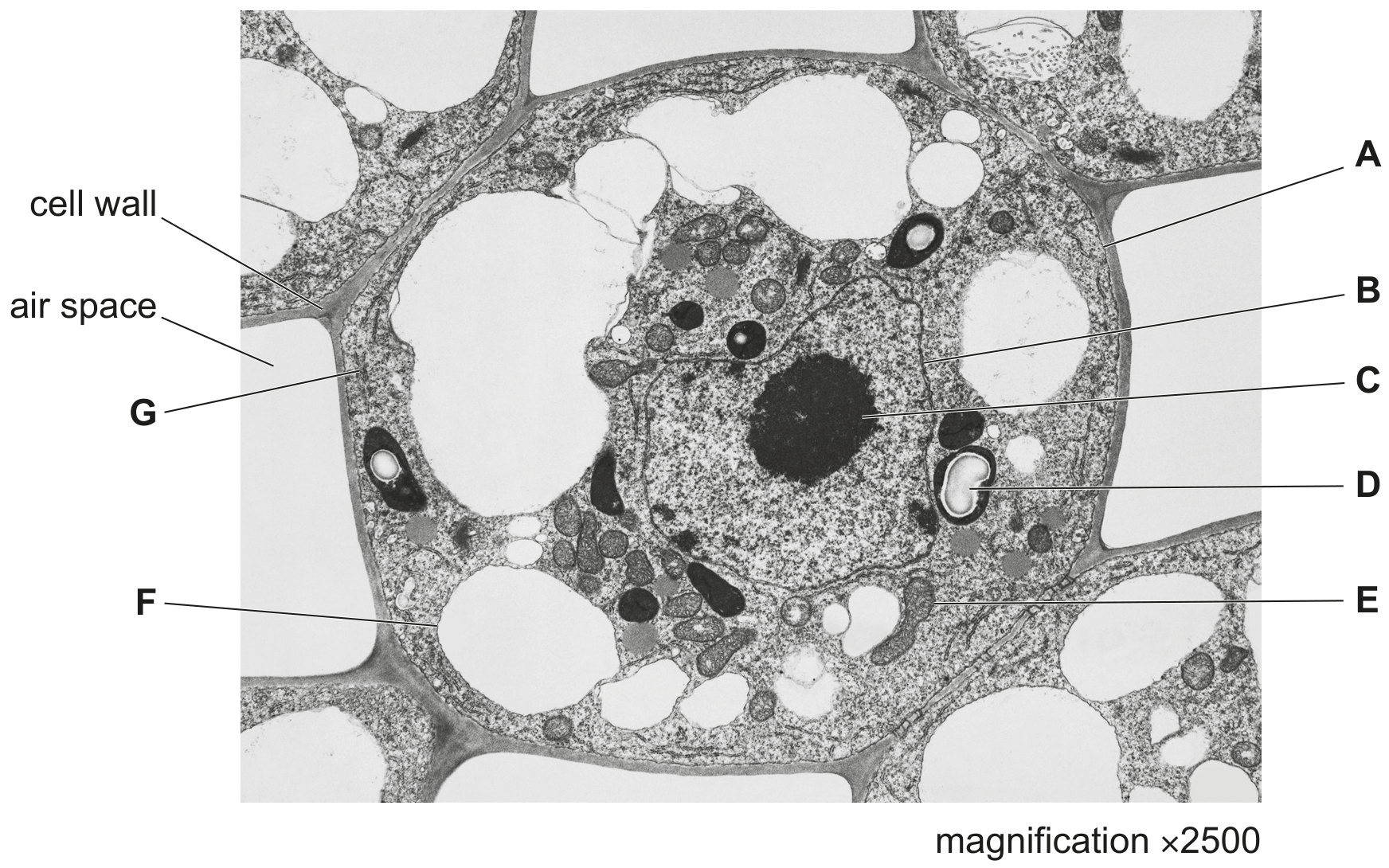
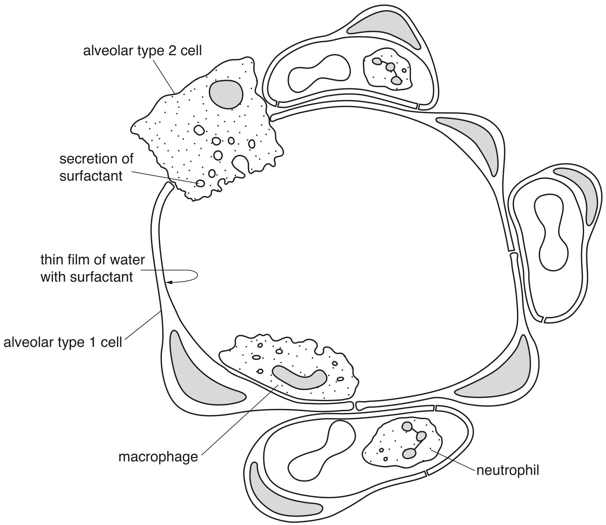
EduNinja
[Maximum number: 2]

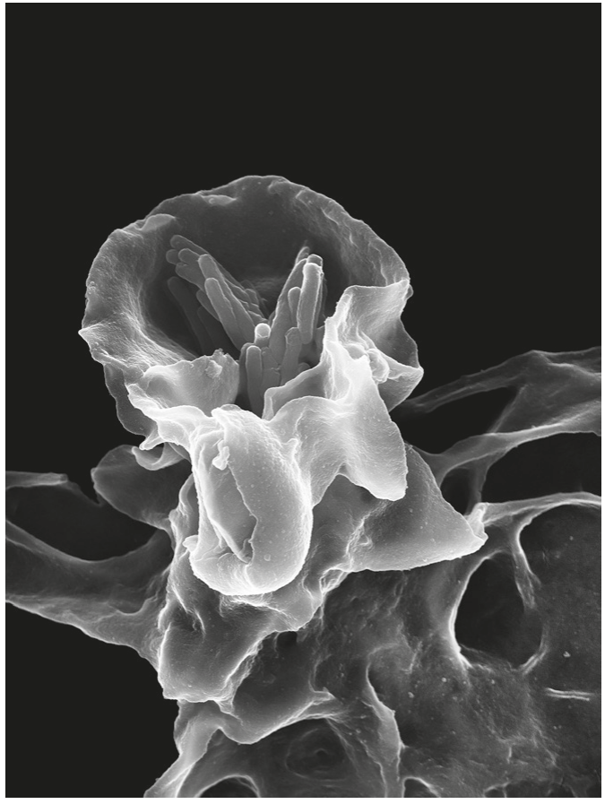
Fig. 1.1 is a scanning electron micrograph of part of the wall of the bronchus of a healthy human.
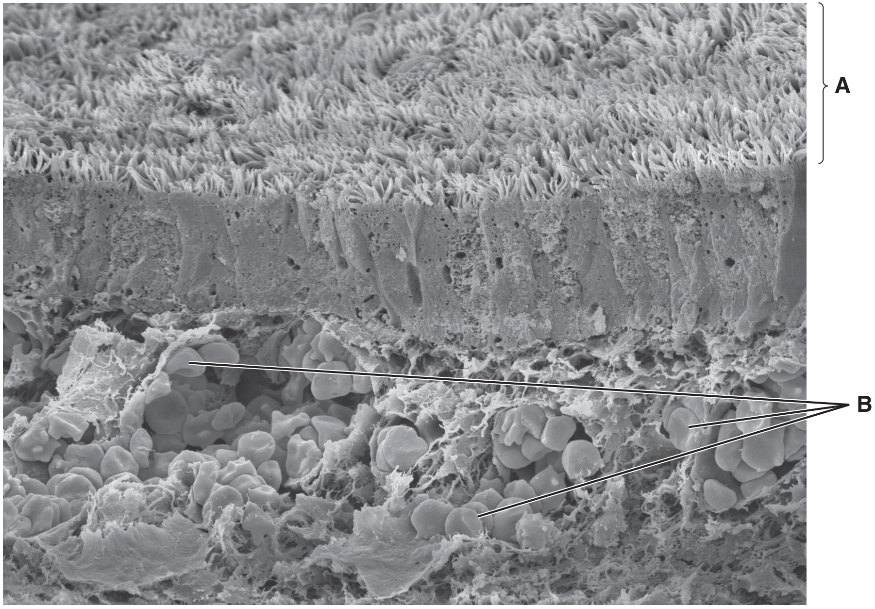
Fig. 1.1

Fig. 1.1

(a)

Suggest why a person with chronic bronchitis is more likely than a healthy person to suffer from infectious diseases of the gas exchange system.

[ 2 ]
[Maximum number: 1]

Name as precisely as you can the structure described in each of the following statements.

(a)

The cell that ingests and digests cell debris and bacteria in the lungs.

D

[ 1 ]
[Maximum number: 2]

Fig. 1.1 is a transmission electron micrograph of a cell from the stem of sago pondweed, Stuckenia pectinata.

Fig. 1.1

Fig. 1.1

(a)

Small vacuoles in S. pectinata may have roles similar to lysosomes in animal cells. Describe the role of lysosomes in animal cells in defence against pathogens.

Question 2 is on page 6.

[ 2 ]
[Maximum number: 1]

B-lymphocytes are activated to form plasma cells during immune responses.
Fig. 2.1 is a drawing of a plasma cell made from a transmission electron micrograph.

Fig. 2.1

Fig. 2.1

(a)
(i)

State the name of the process that is occurring at T.

[ 1 ]
[Maximum number: 4]

The main cause of tuberculosis (TB) in humans is the bacterium Mycobacterium tuberculosis. Most cases of the disease involve the lungs. The bacterium can enter cells and remain inactive in a latent (dormant) state. However, the bacterium can become active to produce symptoms of the disease.

In a person with active TB, the pathogen can be present in airborne droplets that are exhaled. Generally, a healthy person who inhales these droplets has effective defence mechanisms in the gas exchange system to prevent infection.

(a)

One example of a defence mechanism against pathogens in the gas exchange system involves the action of macrophages.

[ 4 ]
(i)

State the location in the body where macrophages have their origin.

[ 1 ]
(ii)

Describe the mode of action of a macrophage.

[ 3 ]
[Maximum number: 4]

The main cause of tuberculosis (TB) in humans is the bacterium Mycobacterium tuberculosis. Most cases of the disease involve the lungs. The bacterium can enter cells and remain inactive in a dormant state. However, the bacterium can become active to produce symptoms of the disease.

In a person with active TB , the pathogen can be present in airborne droplets that are exhaled. Generally, a healthy person who inhales these droplets has effective defence mechanisms in the gas exchange system to prevent infection.

(a)

One example of a defence mechanism against pathogens in the gas exchange system involves the action of macrophages.

[ 4 ]
(i)

Describe the mode of action of a macrophage against a bacterial cell.

[ 3 ]
(ii)

Sometimes M. tuberculosis survives within a macrophage instead of being destroyed by the macrophage.

Suggest one way in which M. tuberculosis may survive within a macrophage.

[ 1 ]
[Maximum number: 2]


Chronic bronchitis is one of the conditions that contributes to chronic obstructive pulmonary disease (COPD).

(a)

White blood cells play an important role in defence.
State precisely the type of white blood cell that fits each of the descriptions given in (i) to (iv).

[ 2 ]
(i)

It is formed in the bone marrow and matures from a monocyte. It contains many lysosomes with hydrolytic enzymes.

[ 1 ]
(ii)

It is formed, and matures in, the bone marrow. It contains a lobed nucleus and has the ability to ingest microorganisms by endocytosis.

[ 1 ]
[Maximum number: 3]

Fig. 2.1 is a scanning electron micrograph showing a macrophage engulfing some bacteria.

Fig. 2.1

Fig. 2.1

(a)
(i)

Describe how macrophages engulf bacteria.

[ 3 ]
[Maximum number: 1]

Tobacco smoking is known to be associated with atherosclerosis and emphysema.

(a)

Fig. 2.1 is a scan of the lungs of a person with emphysema. One common feature in the damaged areas labelled is a loss of the elastic fibres of the alveoli. Another feature is an increased number of macrophages and neutrophils.

Fig. 2.1

Fig. 2.1

[ 1 ]
(i)

State the general role shared by macrophages and neutrophils.

[ 1 ]
[Maximum number: 8]

Fig. 3.1 shows the structure of an alveolus and surrounding structures in a mammalian lung. The lining of each alveolus is formed by two types of epithelial cell, alveolar type 1 and alveolar type 2.

Fig. 3.1

Fig. 3.1

(a)

Describe the role of macrophages in the lungs.

[ 3 ]
(b)

Neutrophils leave the blood and secrete the extracellular enzyme, elastase.

[ 5 ]
(i)

Suggest why neutrophils secrete elastase.

[ 2 ]
(ii)

The protein alpha-1 antitrypsin is produced in cells in the liver and is transported to the lungs, where it inhibits the action of elastase.

Some people produce a different form of this protein that remains within liver cells. These people are at an increased risk of developing emphysema, in which alveolar walls break down. Emphysema is one of the conditions associated with chronic obstructive pulmonary disease (COPD).

Explain why these people are at increased risk of developing emphysema.

[ 3 ]
0