[Maximum number: 1]
Fig. 3.1 is a light micrograph of cells in the root tip of the garlic plant Allium sativum. It has a diploid number (2 n) of 16.
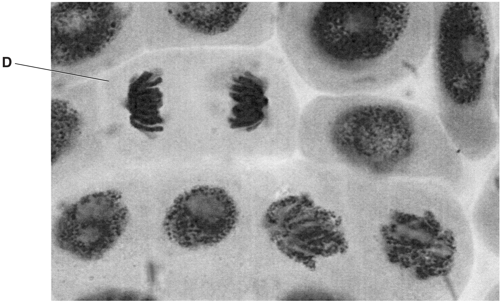
Fig. 3.1
(a)
Name the stage of mitosis shown in cell D.
[ 1 ]
 EduNinja
Fig. 3.1 is a light micrograph of cells in the root tip of the garlic plant Allium sativum. It has a diploid number (2 n) of 16.
Fig. 3.1
Name the stage of mitosis shown in cell D.
The photomicrographs show cells in various stages of the cell cycle.
Which stage of mitosis is not shown?
anaphase
prophase
metaphase
telophase
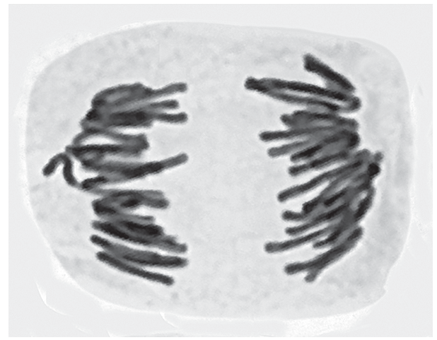
The transmission electron micrograph shows a cell in a stage of the mitotic cell cycle.
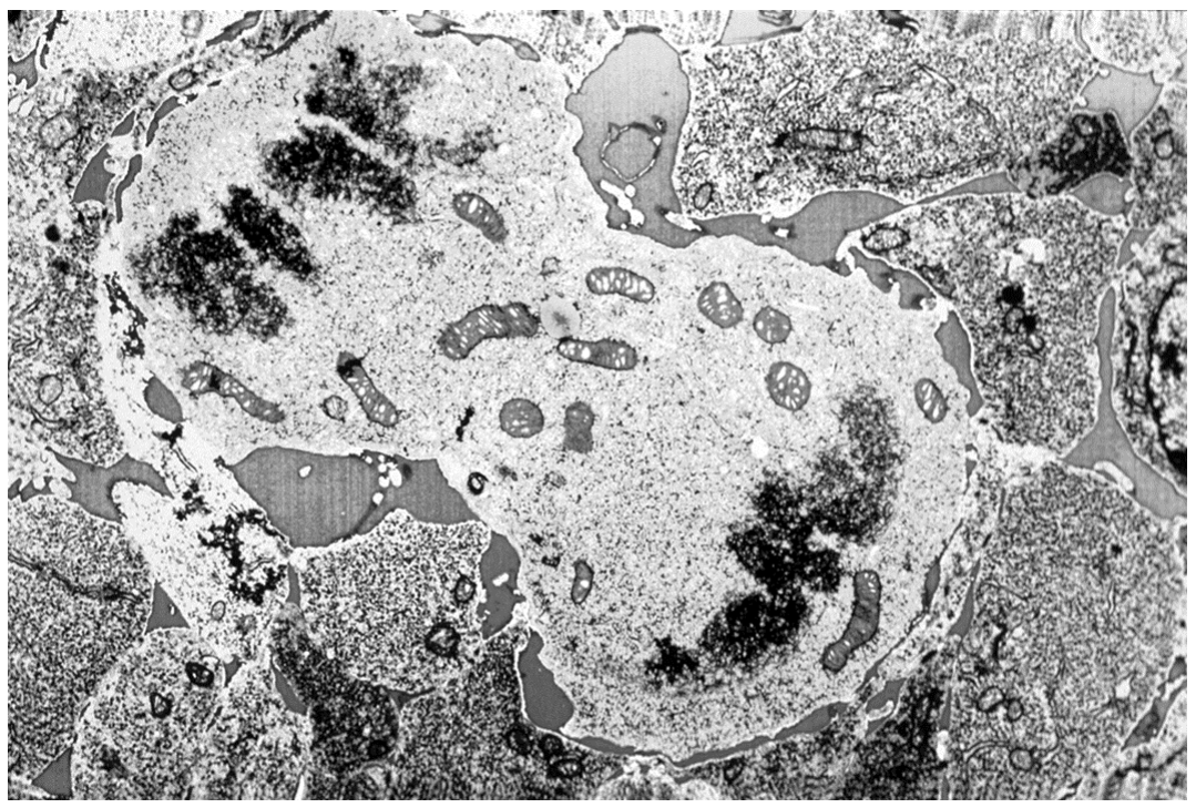
Which statement explains why it is difficult to identify the stage of the mitotic cell cycle shown?
Chromosomes have supercoiled and are visible, but centrioles are not visible.
Anaphase may be continuing, or telophase may be starting.
It is unclear whether the electron micrograph shows two cells in metaphase.
Some people may consider interphase to have started.
The photomicrograph shows cells at different stages of mitosis.
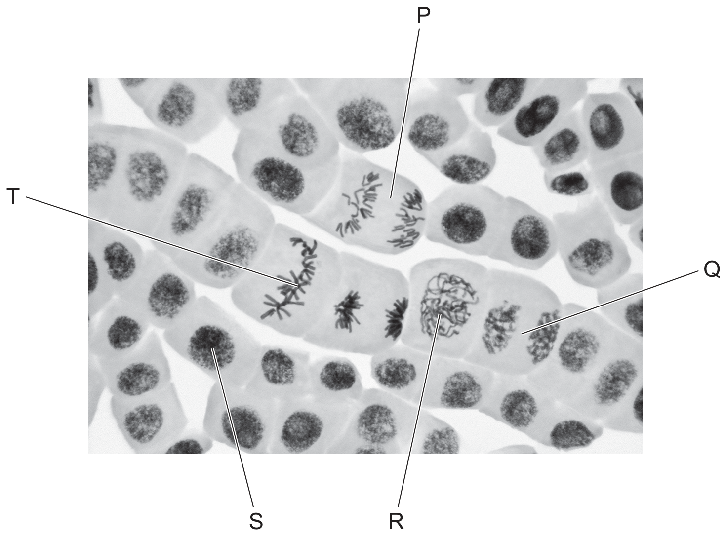
A student wrote four statements about the photomicrograph.
1 Cell P shows anaphase.
2 Spindle formation is occurring in cell Q.
3 The amount of DNA in cell R is the same as in cell T.
4 The correct order for the stages is .
Which statements are correct?
1,2 and 3
1, 2 and 4
1, 3 and 4
2, 3 and 4
Stained onion cells undergoing mitosis were observed using a microscope.
Which row is correct for mitosis in plant cells?
prophase
metaphase
anaphase
telophase
centrioles visible
chromosomes pair up at the equator
two telomeres are visible on each chromatid
two nuclear membranes form
centromeres present
chromosomes align at the equator
chromosomes replicate to form chromatids
centrioles disappear
each chromosome is visible as two chromatids
centromeres attach to spindle fibres
chromatids separate and migrate to opposite poles
chromosomes decondense
spindle fibres formed by centrioles
centromeres attach to spindle fibres
chromatids are pulled apart by centrioles
spindle fibres form new nuclear membranes
The diagram shows a cell during mitosis.
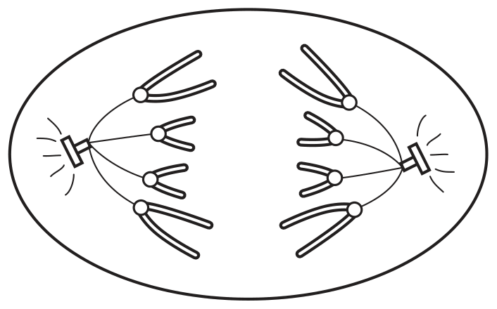
Which description correctly identifies the cell?
a plant cell during anaphase
a plant cell during metaphase
an animal cell during anaphase
an animal cell during metaphase
In a dividing cell, DNA replication occurs before mitosis.
Fig. 6.2 is a photomicrograph of root tip cells at different stages in the cell cycle.
A cell in interphase is labelled.
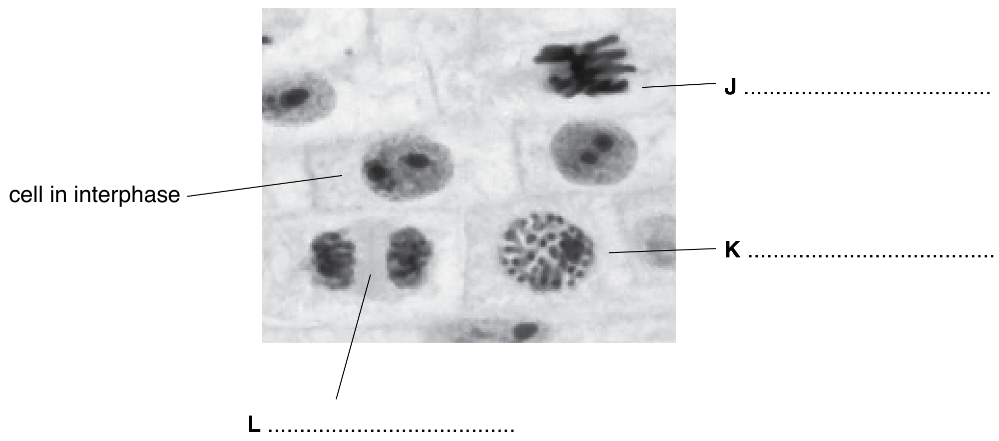
Fig. 6.2
Complete Fig. 6.2 by naming the stage of mitosis shown in each of cells J, K and L in Fig. 6.2.