[Maximum number: 2]
The root apical meristem is a region of undifferentiated cells in the root tips of plants. Mitosis occurs in this region.
(a)
(i)
Describe the behaviour of the nuclear envelope during mitosis.
[ 2 ]
 EduNinja
The root apical meristem is a region of undifferentiated cells in the root tips of plants. Mitosis occurs in this region.
Describe the behaviour of the nuclear envelope during mitosis.
Fig. 1.1 is a transmission electron micrograph of part of an animal cell.
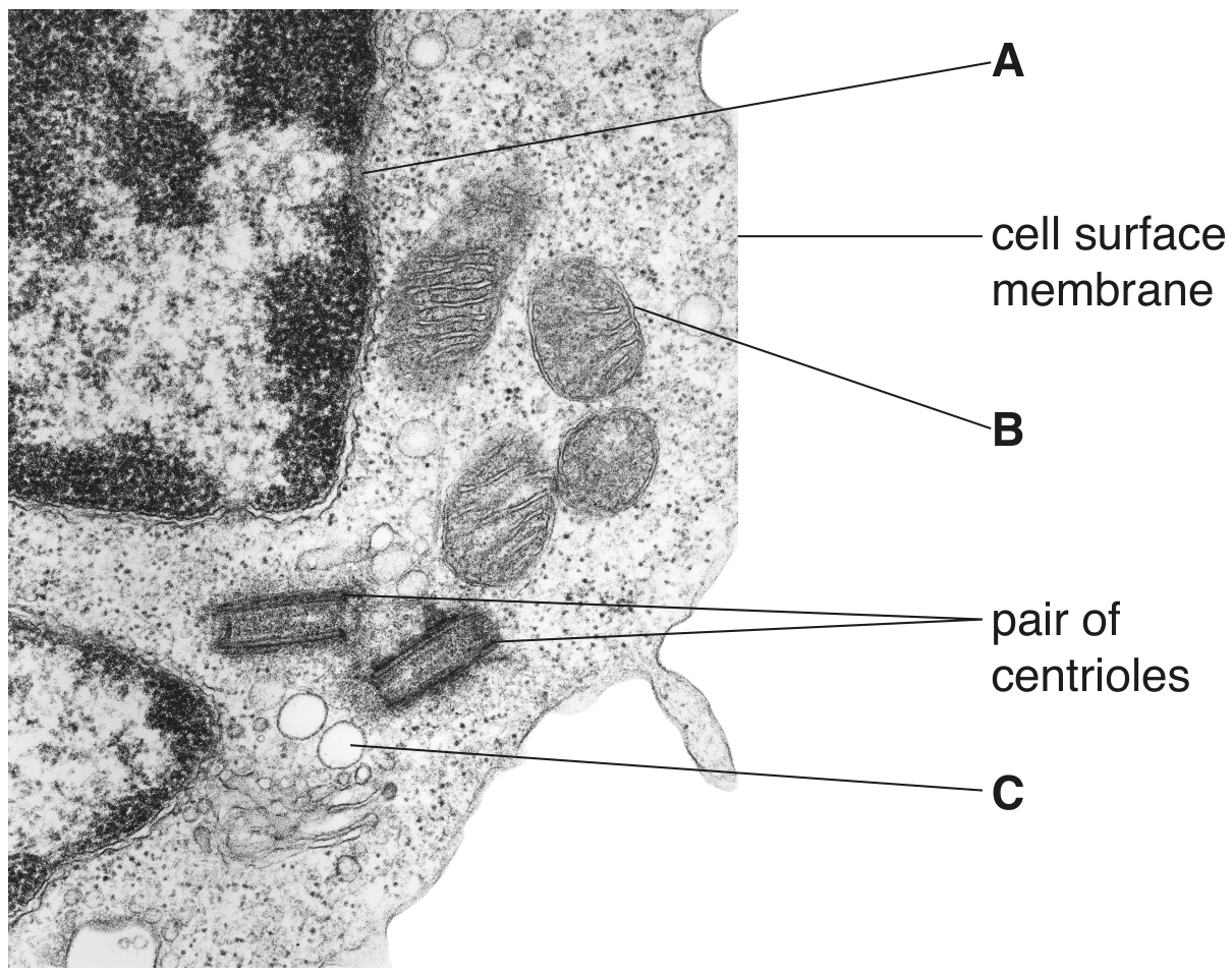
Fig. 1.1
Cells such as that in Fig. 1.1 can divide by mitosis.
Describe the role of centrioles in mitosis.
Fig. 2.1 shows some stages of the cell cycle in the meristematic tissue in the root tip of a plant. Three of these stages, P, Q and R, are identified in Table 2.1.
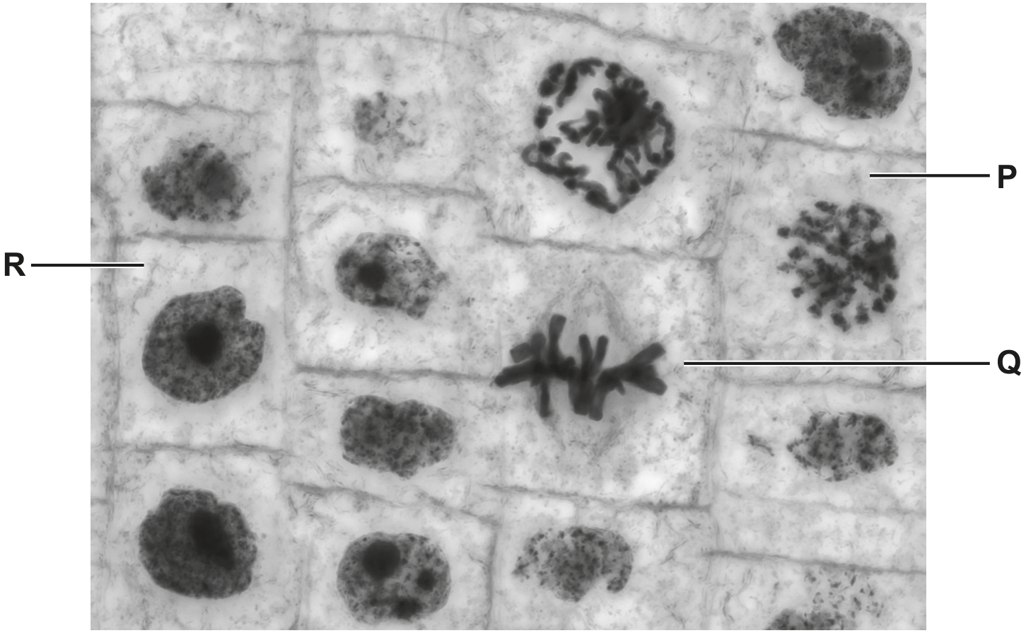
Fig. 2.1
Describe how the spindle is involved during the process of mitosis.
Meristematic tissue is found in the growing region of plants, such as root tips.
Fig. 2.1 shows a section through the meristematic region of a root tip of onion, Allium cepa.
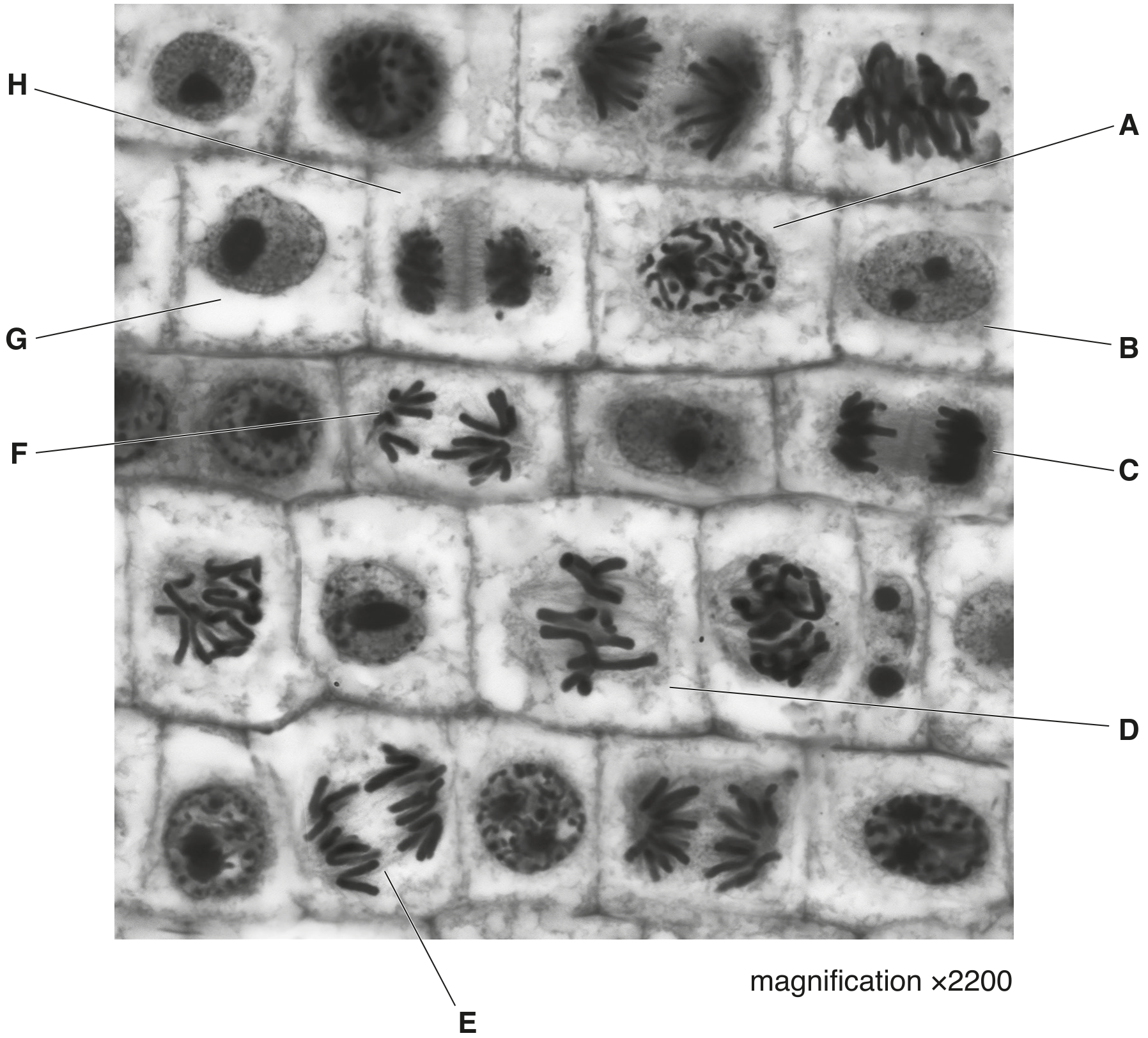
Fig. 2.1
Table 2.1 shows the numbers of cells in different stages of the cell cycle that were observed in sections of the meristematic regions of root tips of A. cepa.
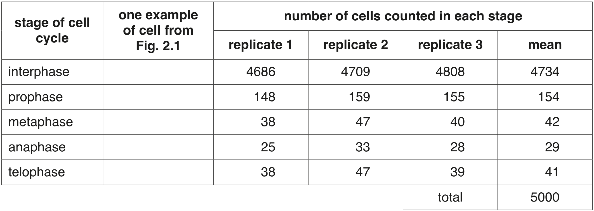
Table 2.1
State one event that occurs during cytokinesis in the cell cycle of plant cells, such as those shown in Fig. 2.1.
Which row correctly shows the behaviour of the nuclear envelope, the centrioles and the spindle during a stage of mitosis?
nuclear envelope
centrioles
spindle
disappears
replicate
spindle microtubules begin to form
not present
begin to move to poles of cell
spindle microtubules fully formed
begins to reform
at opposite poles of the cell
some spindle microtubules shorten
reforms
one beside each nucleus
spindle microtubules break down
The early development of an animal involves divisions of the zygote and daughter cells by mitosis to form an embryo consisting of genetically identical cells.
Fig. 4.1 shows several cells at various stages of the cell cycle in an embryo of whitefish, Coregonus artedi.
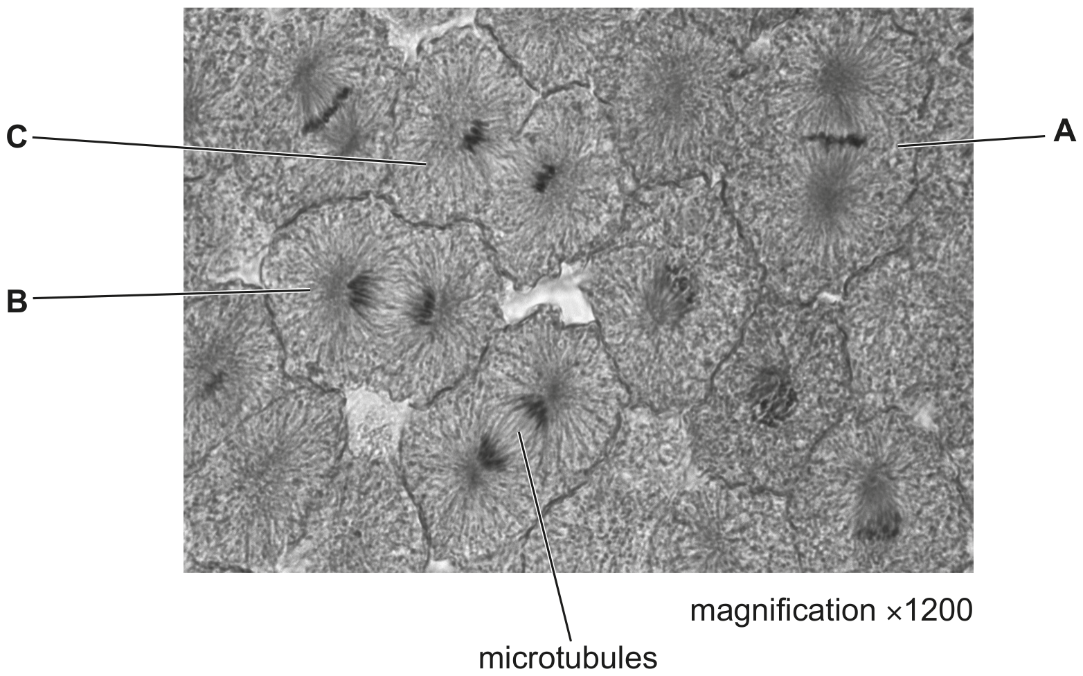
Fig. 4.1
Fig. 4.1 shows microtubules in the cells that are dividing.
Describe the role of microtubules in mitosis.
Plant and animal cells carry out mitosis to form two genetically identical cells from one original cell.
Plant cells require microtubules to form structures that are needed for mitosis.
Name one of these structures.