EduNinja
[Maximum number: 5]

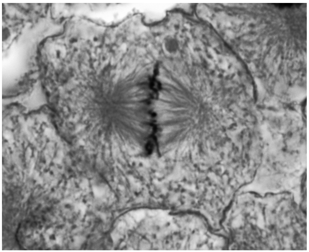
Fig. 1.1 is an electron micrograph of cells from the ciliated epithelium of the trachea.
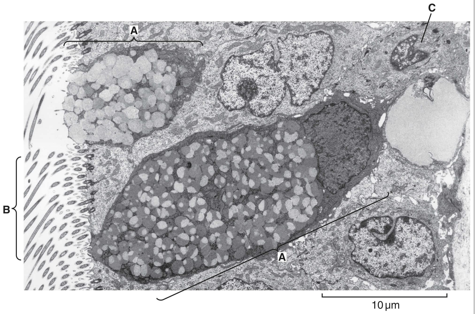
Fig. 1.1

Fig. 1.1

(a)

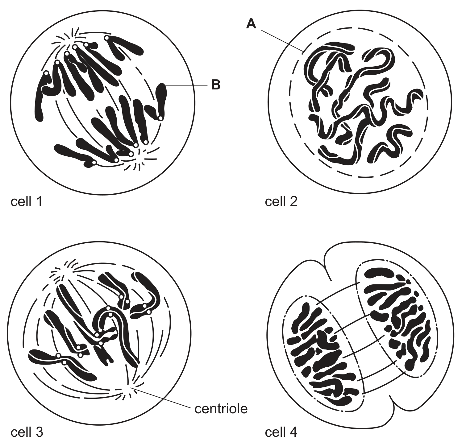
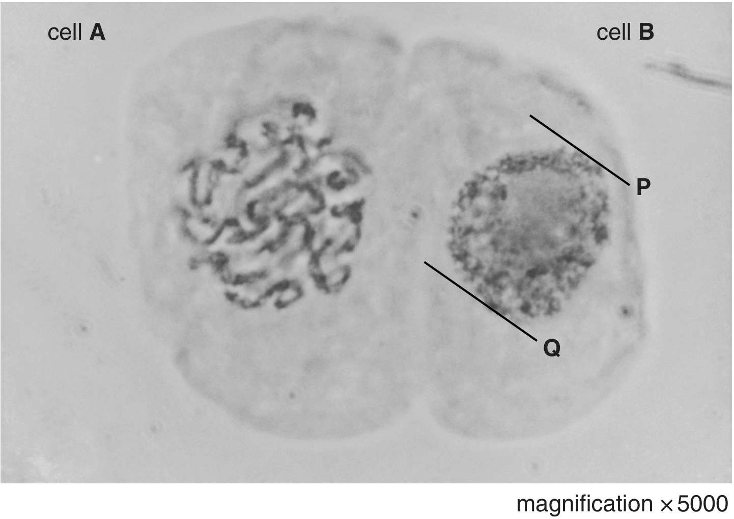
Cells, such as C, at the base of the epithelium of the trachea divide by mitosis.

Describe the changes that occur within the cell between the beginning of prophase and the end of metaphase.

[ 5 ]
[Maximum number: 3]

Fig. 1.1 shows a stage in the mitotic cell cycle in an animal cell.

Fig. 1.1

Fig. 1.1

(a)
(i)

State three features which are characteristic of the stage of mitosis shown in Fig. 1.1.
1.

[ 3 ]
[Maximum number: 2]

Fig. 1.1 is a diagram of a transverse section through a vein.

Fig. 1.1

Fig. 1.1

(a)

Fig. 1.2 is a diagram of a cell in late prophase of mitosis.

Fig. 1.2

Fig. 1.2

Complete Fig. 1.3 to show the same cell in the anaphase stage of mitosis.

Fig. 1.3

Fig. 1.3

[ 2 ]
[Maximum number: 2]

Fig. 1.1 is a photomicrograph of plant root cells near the growing tip. Some of the cells are undergoing mitosis.

Fig. 1.1

Fig. 1.1

(a)

Describe two events occurring in cell B.
1.
2.

[ 2 ]
[Maximum number: 1]

Fig. 1.1 shows four animal cells in different stages of the mitotic cell cycle.

Fig 1.1

Fig 1.1

(a) Using the number given to each cell in Fig. 1.1, arrange the stages as they occur in the mitotic cell cycle.

(a)
(i)

State what is occurring at A in cell 2 .
(ii) Label B is pointing to a region of the chromatid that contains repetitive nucleotide sequences.
State the name given to this region.

[ 1 ]
(a)

Fig. 1.1 is a diagram of a human chromosome at a stage in mitosis.

Fig. 1.1

Fig. 1.1

[ 1 ]
(i)

State one stage during mitosis when the chromosome would appear as shown in Fig. 1.1.

[ 1 ]
[Maximum number: 1]

A diagram of a chromosome from a dividing cell is shown in Fig. 1.1.

Fig. 1.1

Fig. 1.1

(a)

The chromosome shown in Fig. 1.1 consists of one long DNA molecule associated with histone proteins.

Name one stage of mitosis in which a chromosome would have the same general structure as the chromosome shown in Fig. 1.1.

[ 1 ]
[Maximum number: 4]

Fig. 3.1 is a photomicrograph of two animal cells, A and B, at different stages of the mitotic cell cycle.

Fig. 3.1

Fig. 3.1

(a)
(i)

Describe the events that occur during the stage of the cell cycle named for cell A in (a)(i).

[ 4 ]
(a)

Vinblastine disrupts the formation of the spindle apparatus during mitosis.

Explain how vinblastine has its effect as an anti-cancer drug.

[ 3 ]
[Maximum number: 1]

Which process occurs during prophase of the mitotic cell cycle in an animal cell?

A

division of centromeres

B

formation of chromosomes

C

replication of DNA

D

separation of centrioles

0