[Maximum number: 3]
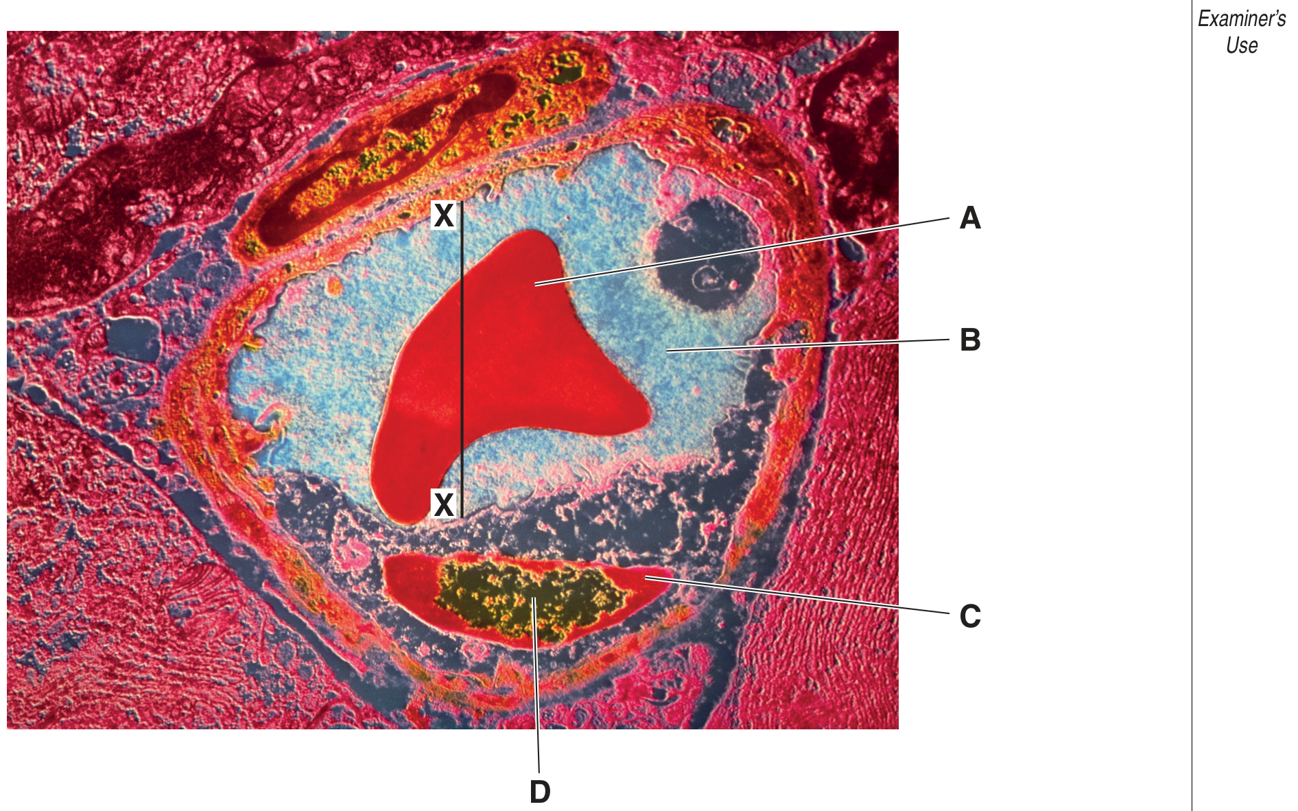
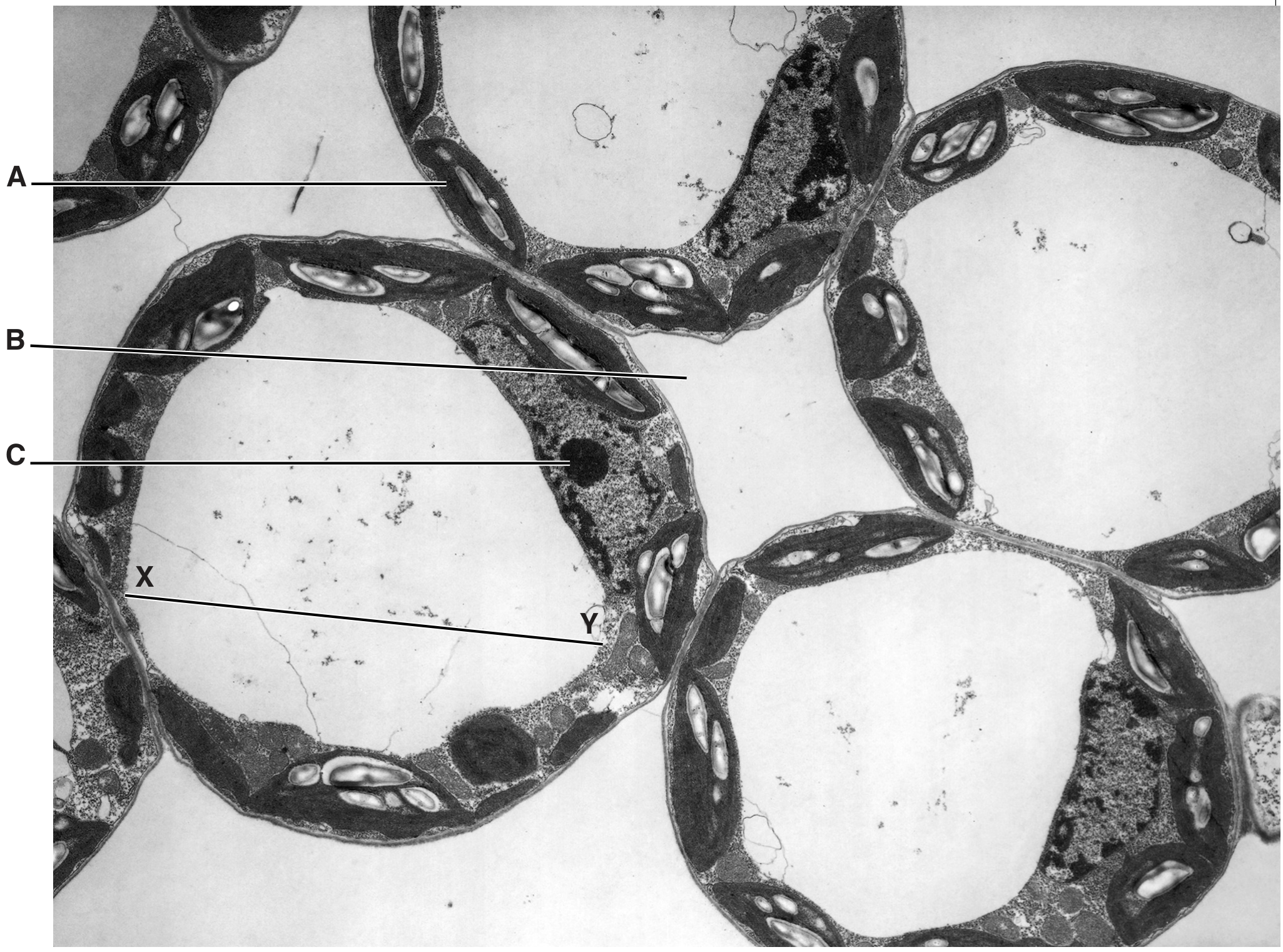
Fig. 1.1 is an electron micrograph of part of a eukaryotic cell.
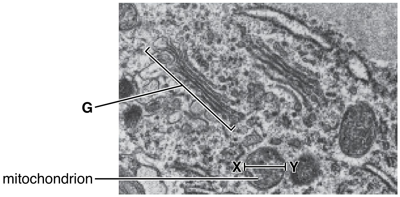
Fig. 1.1
×47000
(a)
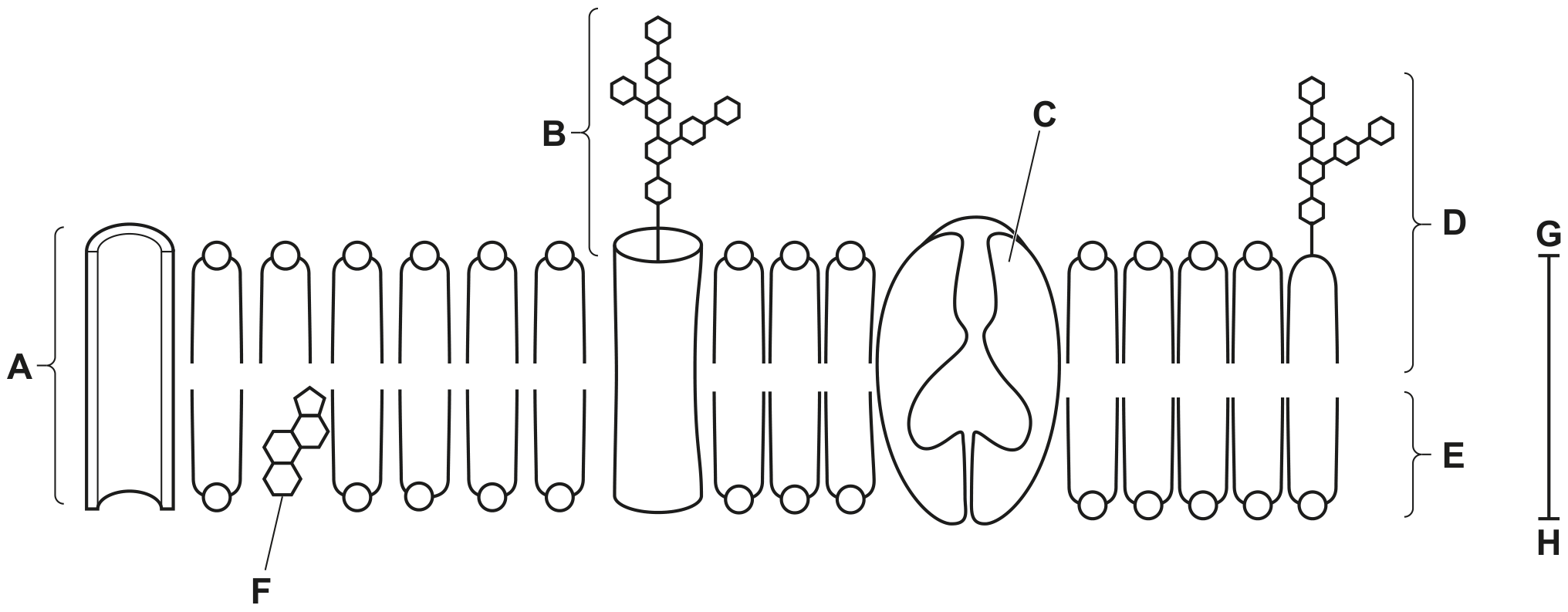
Many of the cell structures in Fig. 1.1 are surrounded by membranes.
Membranes are approximately 6 nm to 7 nm wide.
[ 3 ]
(i)
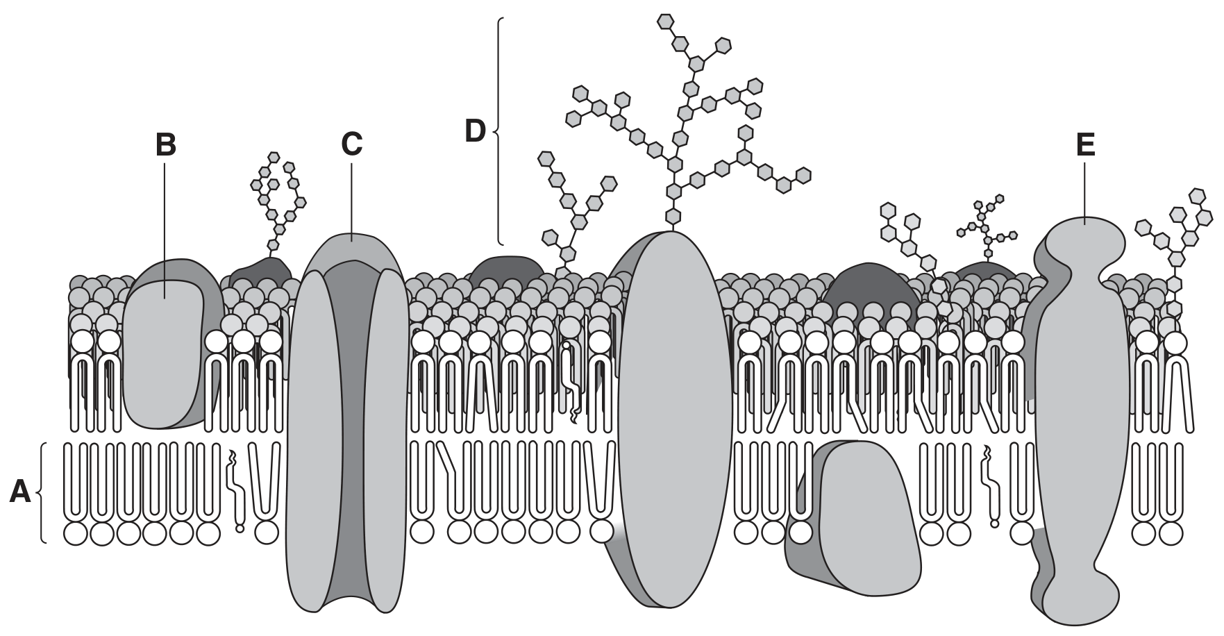
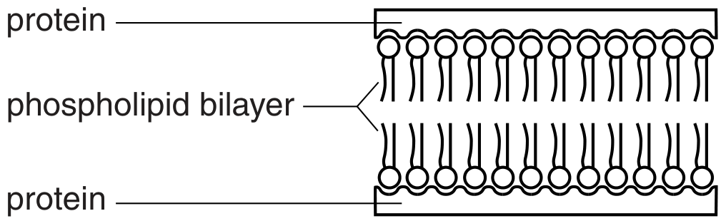
Describe the fluid mosaic model of membrane structure.
There is space below for a diagram.
[ 3 ]