EduNinja
[Maximum number: 2]

A number of diseases, such as dengue fever, are spread by mosquitoes. The incidence of this disease has increased dramatically in recent years and this has been linked with the spread of the mosquito, Aedes aegypti.

In an attempt to reduce the numbers of A. aegypti, genetically modified (GM) male mosquitoes were produced. One of the genes added to these mosquitoes, when switched on, results in the production of a protein which is toxic to mosquitoes.

In 2010, in the Cayman Islands and in Malaysia, GM male mosquitoes were released into the wild to mate with females. All the resulting offspring died in the larval stage.

(a)

GM mosquitoes carrying the tTA gene can live and reproduce normally when fed on a diet containing an added chemical, A.

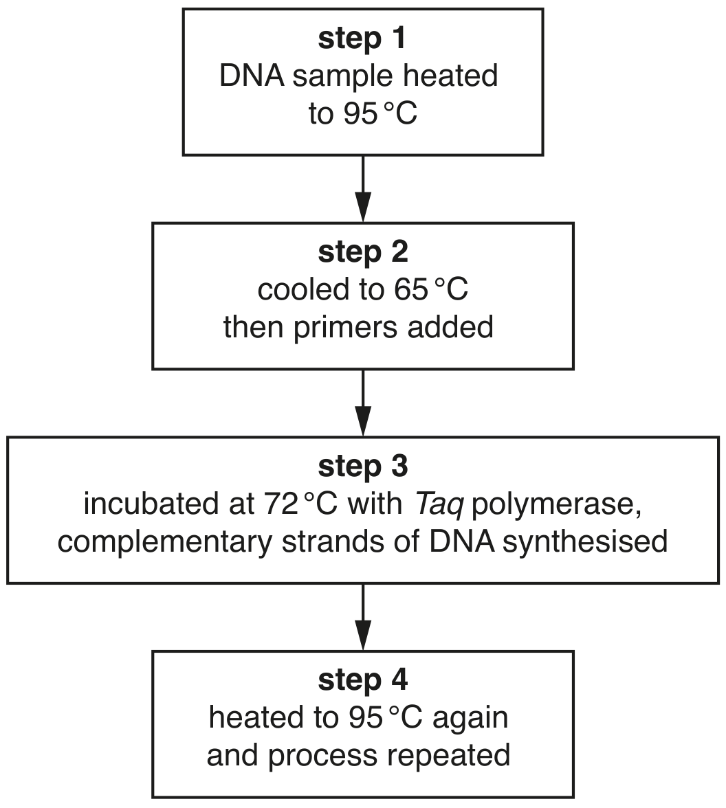
With reference to Fig. 2.1:

[ 2 ]
(i)

suggest how large numbers of adult GM male mosquitoes can be produced for release into the wild, from an original stock of GM males

[ 2 ]
[Maximum number: 6]

The polymerase chain reaction (PCR) is used to produce large amounts of DNA from a very small original sample. The main stages of a PCR are shown in Fig. 3.1.

Fig. 3.1

Fig. 3.1

(a)
(i)

Explain why the DNA sample is heated to 95C95^{\circ} \mathrm{C} in step 1 .

[ 2 ]
(ii)

Explain why primers are added in step 2.

[ 2 ]
(iii)

Explain why the enzyme Taq polymerase is used in step 3.

[ 2 ]
(a)

In 1984, the geneticist Alec Jeffreys invented a DNA testing technique, known as DNA profiling, that produces a DNA banding pattern on a gel. The DNA banding pattern (profile) is unique to each individual.

DNA profiling can be used in police forensic work to catch criminals.
Since 1987, police in many countries have collected and stored DNA from crime scenes to create DNA profiles, which they try to match with the DNA profiles of criminal suspects.

[ 2 ]
(i)

DNA at a crime scene may be obtained from hairs and traces of blood, semen and saliva.

Explain why PCR may be needed before DNA from a crime scene can be profiled.

[ 2 ]
[Maximum number: 2]

The β\beta-globin gene codes for the β\beta-globin polypeptide of haemoglobin. It has two alleles, HbA\mathbf{H b}^{\mathbf{A}} (normal) and HbS\mathbf{H b}^{\mathbf{S}} (sickle cell). The sickle cell allele differs from the normal allele due to a base substitution mutation and this mutation results in a single amino acid change to the β\beta-globin polypeptide.

There are three possible genotypes and phenotypes.
- HbSHbS\quad \mathbf{H b}^{\mathbf{S}} \mathbf{H b}^{\mathbf{S}}, sickle cell anaemia, a severe disease
- HbAHbS\quad \mathbf{H b}{ }^{\mathbf{A}} \mathbf{H b}^{\mathbf{S}}, sickle cell trait with mild or no symptoms of sickle cell anaemia
- HbAHbA\quad \mathbf{H b}{ }^{\mathbf{A}} \mathbf{H b}^{\mathbf{A}}, normal (healthy)

A man and woman who both have sickle cell trait may choose to have children by IVF. This allows the genotype of embryos to be determined by gene testing before the embryos are implanted. Embryos with the normal genotype can then be selected and implanted into the mother.

One technique that can be used in gene testing an embryo for the HbS\mathbf{H b}^{\mathbf{S}} allele is restriction fragment length polymorphism (RFLP) analysis. This involves digesting a DNA sample from an embryo with a restriction endonuclease and then separating the DNA fragments by gel electrophoresis. The position of the DNA fragments on the gel can show if the embryo has the HbS\mathbf{H b}^{\mathbf{S}} allele.

(a)

The first step in testing an embryo for the HbS\mathbf{H b}^{\mathbf{S}} allele by RFLP analysis requires many copies of the part of the β\beta-globin gene in which the mutation causing sickle cell anaemia occurs.

[ 2 ]
(i)

Name the technique used to produce many copies of a DNA sequence from a very small quantity of DNA.

[ 1 ]
(ii)

Explain why it is necessary to copy this DNA sequence many times in order to test embryos for HbS\mathbf{H b}^{\mathbf{S}} alleles by RFLP analysis.

In the next step of RFLP analysis, the copies of the part of the β\beta-globin gene from the first step are incubated with a restriction endonuclease, MstII. This enzyme cuts at a specific sequence of DNA (the restriction site).

The restriction site for MstII is shown in Fig. 3.1.

5CC TNAGG33GGANTCC5N= any nucleotide (A,T,C or G)\begin{aligned} & 5^{\prime}-\mathrm{CC} \mathrm{~T} N A G G-3^{\prime} \\ & 3^{\prime}-\mathrm{G} G A N T C C-5^{\prime} \quad \mathrm{N}=\text { any nucleotide }(A, T, C \text { or } G) \end{aligned}

Fig. 3.2 shows the part of an HbA\mathbf{H b}^{\mathbf{A}} allele obtained from the first step. All the MstII restriction sites and the number of DNA base pairs separating these restriction sites are shown.

Fig. 3.2

Fig. 3.2

Fig. 3.3 shows the same part of an HbS\mathbf{H b}^{\mathbf{S}} allele. The single base substitution in the HbS\mathbf{H b}^{\mathbf{S}} allele that causes sickle cell anaemia is indicated.

Fig. 3.3

Fig. 3.3

[ 1 ]
[Maximum number: 6]

White-clawed crayfish, Austropotamobius pallipes, live in rivers and lakes in Europe.
In the 1850s, the North American signal crayfish, Pacifastacus leniusculus, was introduced to Europe. The introduced species carried a pathogen that causes a disease known as crayfish plague. This disease kills A. pallipes.
Since 1850, the population size of A. pallipes has reduced in many areas of Europe due to the spread of crayfish plague.

(a)

It is difficult to locate crayfish because they live underwater.

After an outbreak of crayfish plague in one country in Europe, researchers used an environmental DNA technique to find the locations in a river where populations of A. pallipes were still surviving.
Water samples were taken from locations along the river. Each water sample was filtered to obtain any cells or DNA that had been released by organisms into the water. The polymerase chain reaction (PCR) was carried out on this DNA using primers specific to A. pallipes DNA.

[ 4 ]
(i)

Outline and explain the steps that occur in PCR.

[ 4 ]
(b)

The PCR technique used in this research is quantitative. A fluorescent dye binds to the DNA and the fluorescence is monitored throughout the process, showing the quantity of DNA present.

Fig. 4.1 shows standard curves for four known concentrations of DNA, A, B, Cand D, and the result obtained for A. pallipes DNA at one location along the river (dotted line).

Fig. 4.1

Fig. 4.1

[ 2 ]
(i)

State the letter of the curve with the highest starting concentration of DNA.

[ 1 ]
(ii)

The quantity of A. pallipes DNA reaches a threshold (marked as a horizontal line on Fig. 4.1) at 28 cycles. The DNA in curve C reaches the threshold after 25 cycles. In one cycle of PCR the concentration of DNA doubles.

Calculate the relative difference in the starting concentrations of A. pallipes DNA and the DNA in curve C.

[ 1 ]
[Maximum number: 6]

The polymerase chain reaction (PCR) is used to produce large quantities of DNA from a very small original sample. The main steps of one PCR method are shown in Fig. 4.1.

Fig. 4.1

Fig. 4.1

(a)
(i)

Explain why it is necessary to heat the mixture to 95C95^{\circ} \mathrm{C} (step 1).

[ 2 ]
(ii)

Explain why primers are included in the mixture.

[ 2 ]
(iii)

Explain why the enzyme Taq polymerase, rather than any other type of DNA polymerase, is used in PCR.

[ 2 ]
[Maximum number: 3]

Sickle cell anaemia is a non-infectious chronic disease. If not treated, sickle cell anaemia can be painful and life-threatening.

Sickle cell anaemia is caused by a base substitution mutation in the gene coding for the β\beta-globin polypeptide of haemoglobin. This leads to a change in the primary structure of the polypeptide, as valine is present instead of glutamine. This results in abnormal sickle-shaped red blood cells, which stick together in blood vessels.

Symptoms of sickle cell anaemia include painful attacks when red blood cells block capillaries in tissues and organs.

(a)

Sickle cell anaemia is an autosomal recessive inherited disorder:
- allele HbA\mathrm{Hb}^{\mathrm{A}} codes for the normal β\beta-globin polypeptide
- allele HbS\mathrm{Hb}^{\mathrm{S}} codes for the sickle-cell polypeptide.

People who are heterozygous ( HbAHbS\mathrm{Hb}^{A} \mathrm{Hb}^{S} ) have sickle cell trait (SCT). For a child to inherit sickle cell anaemia (HbSHbS)\left(\mathrm{Hb}^{\mathrm{S}} \mathrm{Hb}^{\mathrm{S}}\right), both parents must have SCT.

A genetic screening program is available for sickle cell anaemia and SCT:
- when a mother is screened and found to have SCT, the father is then screened
- if the mother becomes pregnant, the fetus is screened for both sickle cell anaemia and for SCT
- the test is done either by amniocentesis or by chorionic villus sampling, both of which carry a small risk of the pregnancy failing.

[ 3 ]
(i)

To test for the presence of HbS\mathrm{Hb}^{\mathrm{S}}, DNA is extracted and the polymerase chain reaction (PCR) is carried out with two specific primers.

One mutation to produce HbS\mathrm{Hb}^{S} is a base substitution in the sixth codon of the β\beta-globin gene. The normal codon GAG changes to GTG. The normal-specific primer detects GAG whereas the mutant-specific primer detects GTG.

Explain:
- why primers are used in PCR
- how the use of two specific primers allows the amplification of the normal, sickle cell anaemia and SCT genotypes.

[ 3 ]
[Maximum number: 2]

The gene BRCA1 is expressed in breast tissue and in several other tissues of the body. BRCA1 codes for a tumour suppressor protein. This protein is involved in either repairing damaged DNA, or in triggering the death of a cell if DNA cannot be repaired.

Some mutations in BRCA1 are associated with an increased risk of developing breast cancer.

(a)

DNA sequencing is one method used to test for mutations in BRCA1. Before DNA sequencing occurs, the DNA sample goes through the polymerase chain reaction (PCR).

Suggest why PCR is used for testing for mutations in BRCA1.

[ 2 ]
[Maximum number: 3]

Myosotis is a genus of small flowering plants. Many different Myosotis species grow on the islands of New Zealand, which are an important site of Myosotis evolution. Lowland Myosotis species grow at low altitude while alpine Myosotis species grow at high altitude at the tops of mountains.

(a)

Scientists wanted to obtain molecular data to determine the evolutionary relationships of New Zealand's Myosotis species. They extracted DNA from individuals of Myosotis species collected from three different islands in New Zealand.

To carry out a polymerase chain reaction before DNA sequencing, the DNA samples were mixed with primers, deoxynucleotides and Taq polymerase and put through 35 cycles of treatment. Each treatment cycle involved one minute at 95C95^{\circ} \mathrm{C}, followed by one minute at 50C50^{\circ} \mathrm{C} and then four minutes at 72C72^{\circ} \mathrm{C}.

Describe what happened to the DNA at each temperature.

[ 3 ]
0