EduNinja
[Maximum number: 4]

In a healthy mammalian heart, contraction of the four chambers is coordinated by the action of the sinoatrial node (SAN) and atrioventricular node (AVN).

(a)

State and explain how the structure of the heart allows the atria to contract before the ventricles.

[ 2 ]
(b)

Fig. 2.1 shows blood pressure changes that occur in the left ventricle, left atrium and aorta during one cardiac cycle.
E, F, G and H are the points at which a valve opens or closes as a result of blood pressure changes.
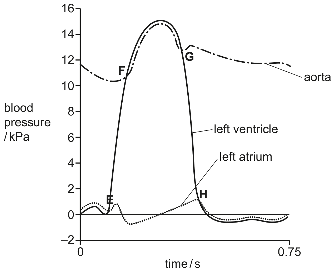
Fig. 2.1

Fig. 2.1

[ 2 ]
(i)

Explain how Fig. 2.1 provides evidence that the wall of the left atrium has a different thickness to the wall of the left ventricle.

[ 2 ]
(a)

Explain why the wall of the left ventricle in the heart is thicker than the wall of the right ventricle.

[ 2 ]
[Maximum number: 3]

Fig. 2.1 shows a diagram of a section through a human heart.

Fig. 2.1

Fig. 2.1

(a)

Explain why the region labelled Y is thicker than the region labelled Z.

[ 3 ]
[Maximum number: 3]

In a double circulatory system, blood passes through the heart twice in one complete circuit of the body. The complete circuit consists of the pulmonary circulation and the systemic circulation.

(a)

Fig. 4.1 is a diagram of a vertical section through the mammalian heart. The differences in the thickness of cardiac muscle in the walls of the four chambers of the heart are shown.

Fig. 4.1

Fig. 4.1

Explain, with reference to their functions, the difference in the thickness of the walls of the left ventricle and right ventricle of the heart.

[ 3 ]
(a)

Fig. 5.1 shows a drawing of an external view of a mammalian heart.

Two cross-sections were made of the heart:
- section 1 was made across the line A-B.
- section 2 was made across the line C-D.

Drawings of the two sections were viewed from above as shown by the arrow on Fig. 5.1. Fig. 5.2 is a drawing of section A-B. Fig. 5.3 is a drawing of section C-D.

Fig. 5.1

Fig. 5.1

Fig. 5.2

Fig. 5.2

Fig. 5.3

Fig. 5.3

[ 3 ]
(i)

Explain why the wall of chamber Y is thicker than the wall of chamber X, as shown in Fig. 5.3.

[ 3 ]
[Maximum number: 1]

Cardiac muscle is made up of many fibres that form the walls of the chambers of the heart.
When the heart contracts, these fibres shorten in length so the muscle creates a force that exerts a pressure on the blood, causing it to move.

Which statement explains the difference in thickness of the walls of ventricles of the heart?

A

There is more muscle in the wall of the right ventricle than that of the left ventricle because more pressure is needed to push blood into the aorta than into the pulmonary artery.

B

The number of muscle fibres in the left ventricle is greater than the number in the right ventricle so their contraction has more force, exerting more pressure on blood.

C

The space available to fill with blood inside the left ventricle is smaller than that of the right ventricle so more pressure is needed to force blood out.

D

The wall of the right ventricle is thicker than that of the left ventricle because it has to resist more pressure when the muscle of the right ventricle contracts.

[Maximum number: 2]

Fig. 5.1 shows a vertical section of the left side of the heart of a mammal.

Fig. 5.1

Fig. 5.1

(a)

Explain the difference in the thickness of the left ventricle and the left atrium.

[ 2 ]
[Maximum number: 1]

The diagram shows a cross-section through a mammalian heart.

Question image

Which chambers of the heart are represented by G and H ?

G

H

left ventricle

right ventricle

right atrium

left atrium

right atrium

right ventricle

right ventricle

left ventricle

[Maximum number: 1]

Aortic stenosis is a heart valve disorder in which the aortic semilunar valve opening is narrow.
Which effect could aortic stenosis have on heart structure and function?

A

The cardiac muscle of the left ventricle wall is thinned by blood leaking out of the left ventricle during ventricular diastole.

B

There is less cardiac muscle in the left ventricle and reduced diastolic blood pressure, caused by the smaller blood volume entering the left atrium.

C

The tendons of the heart valves are weakened by blood being forced back through the left atrioventricular valve into the left atrium.

D

The wall of the left ventricle thickens, leading to an enlarged heart and inability to relax and fill completely during diastole.

[Maximum number: 1]

Aortic stenosis is a heart valve disorder in which the aortic semi-lunar valve opening is narrow.
Which effect could aortic stenosis have on heart structure and function?

A

The cardiac muscle of the left ventricle wall is thinned by blood leaking out of the left ventricle during ventricular diastole.

B

There is less cardiac muscle in the left ventricle and reduced diastolic blood pressure, caused by the smaller blood volume entering the left atrium.

C

The tendons of the heart valves are weakened by blood being forced back through the left atrio-ventricular (bicuspid) valve into the left atrium.

D

The wall of the left ventricle thickens, leading to an enlarged heart and inability to relax and fill completely during diastole.

0