(a)
Prokaryotes and plant cells have cell walls.
Outline the composition of the cell wall of a prokaryote and the composition of the cell wall of a plant cell to show how they differ.
[ 2 ]
 EduNinja
Prokaryotes and plant cells have cell walls.
Outline the composition of the cell wall of a prokaryote and the composition of the cell wall of a plant cell to show how they differ.
Which size of ribosome is found in mitochondria?
60 S
70S
80 S
90 S
Which size of ribosome is found in chloroplasts and typical prokaryotic cells?
60 S
70 S
80 S
90 S
What are found in both mitochondria and typical prokaryotic cells?
70 S ribosomes and circular DNA
70S ribosomes only
80 S ribosomes and circular DNA
circular DNA only
Which size of ribosome is found in chloroplasts?
60 S
70S
80 S
90 S
Which feature is found in both prokaryotic and plant cells?
cell wall
DNA bound to protein
endoplasmic reticulum
Golgi apparatus
Fig. 1.1 is a transmission electron micrograph of a cell from the root of thale cress, Arabidopsis thaliana.
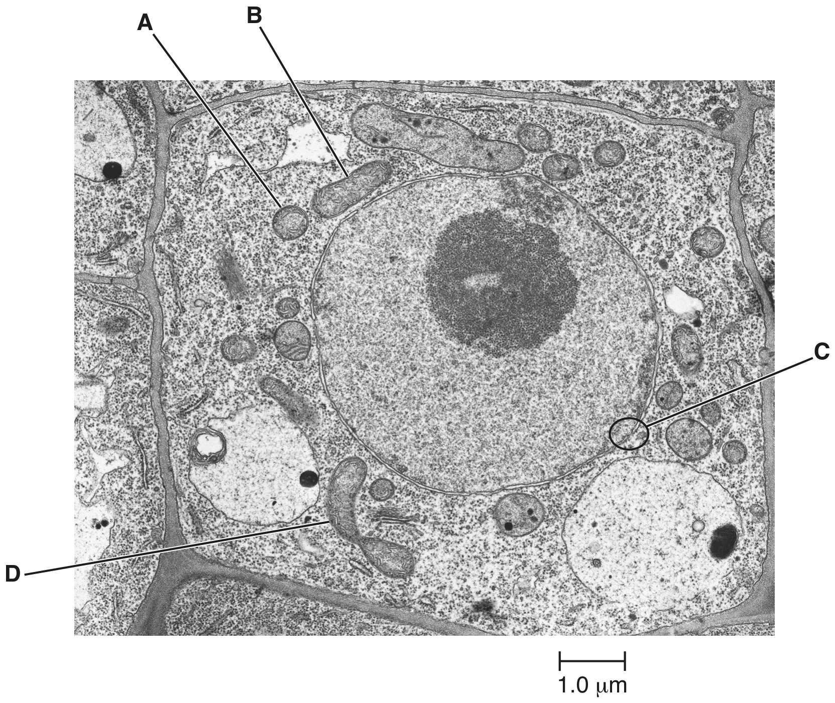
Fig. 1.1
The structures labelled A and B on Fig. 1.1 are sections of two mitochondria.
Suggest why A and B are different shapes.
Fig. 1.1 is a transmission electron micrograph of part of an animal cell.
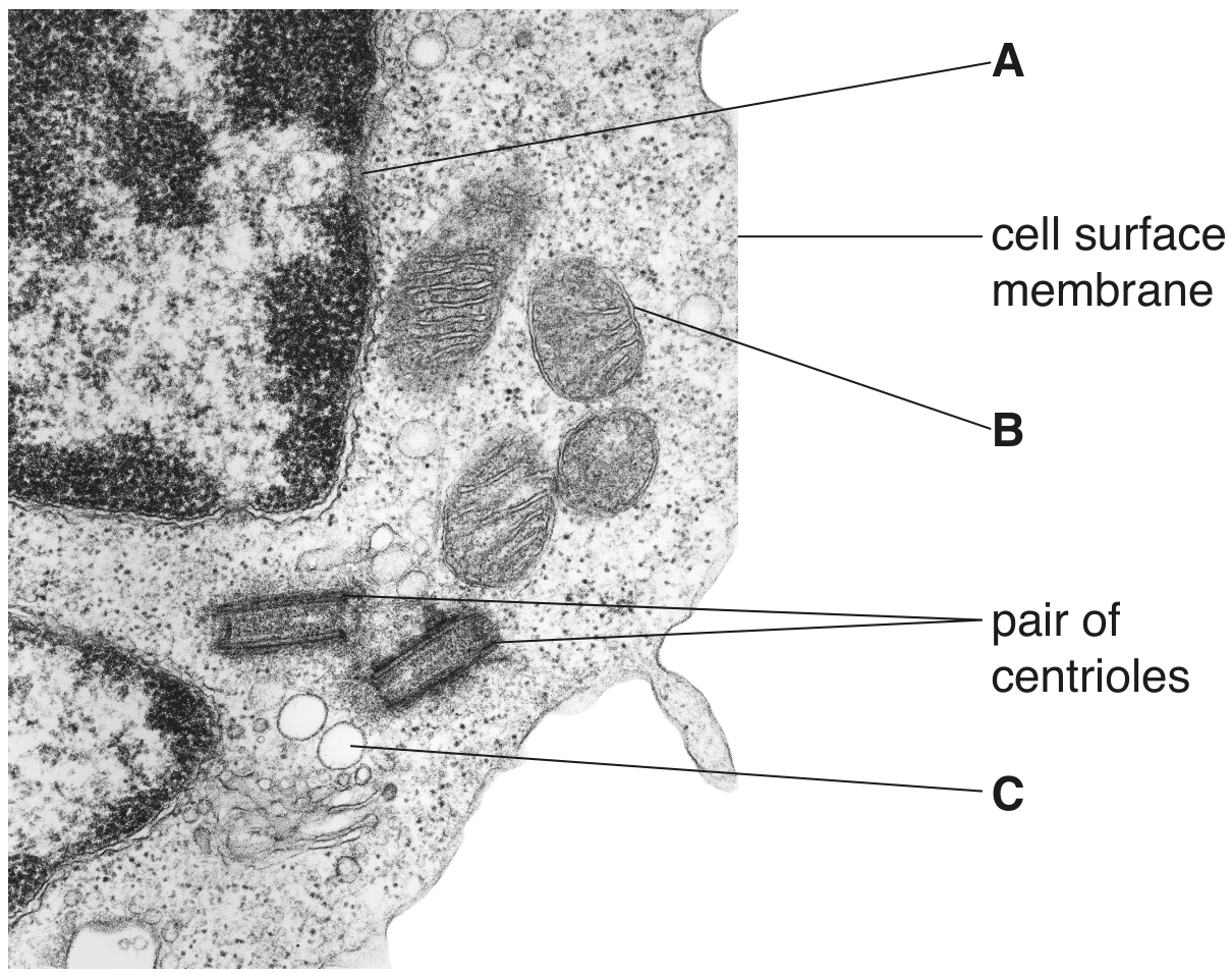
Fig. 1.1
Name one structure, visible in Fig. 1.1, that would also be present in a prokaryotic cell.
Fig. 1.1 is an electron micrograph of a mitochondrion.
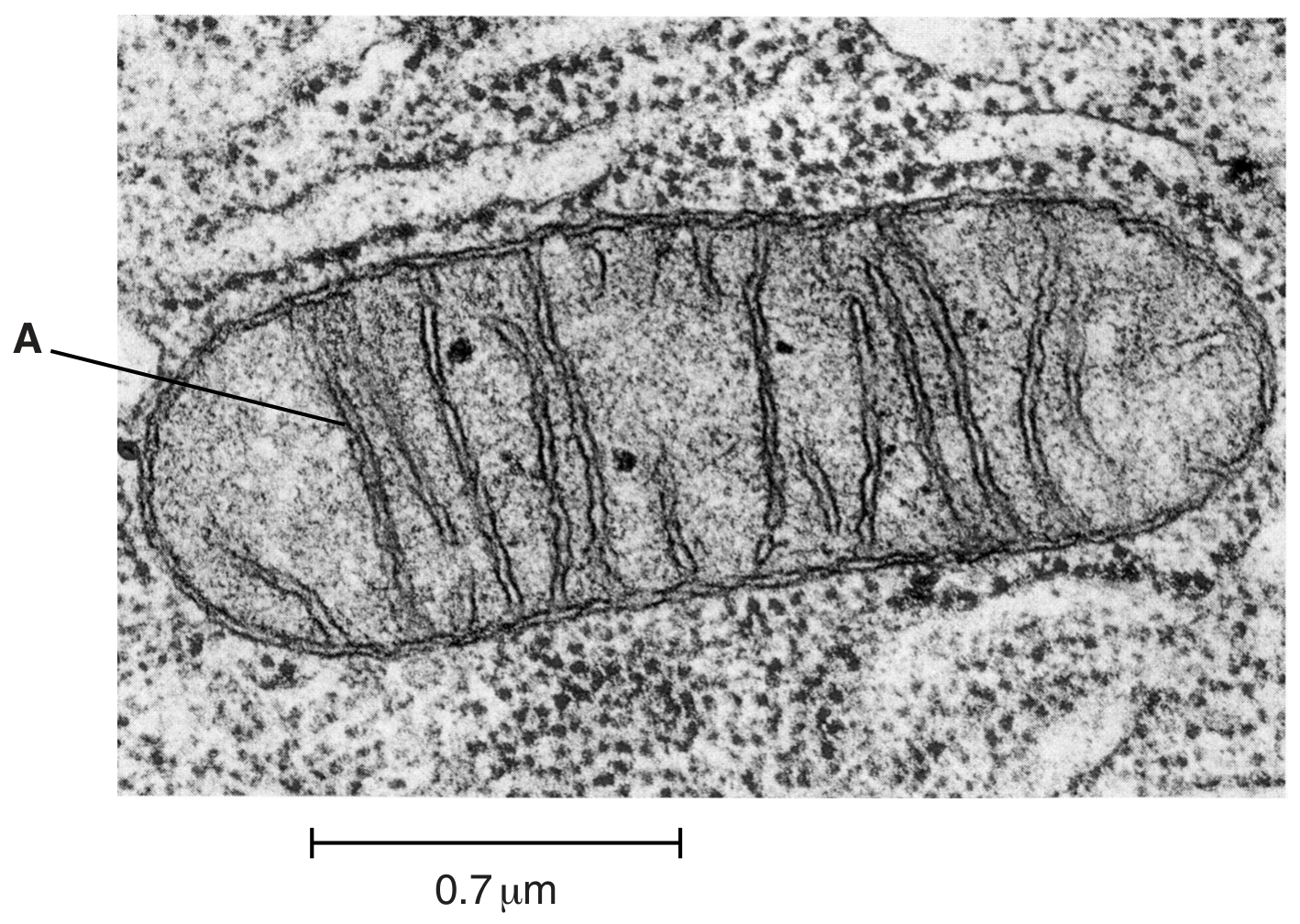
Fig. 1.1
Scientists think that mitochondria were once prokaryotes. The evidence for this is that mitochondria have features in common with prokaryotes.
State two features that mitochondria have in common with prokaryotes.
1.
2.
Vibrio cholerae is a prokaryotic organism.
Fig. 1.1 shows the structure of a cell of V. cholerae.
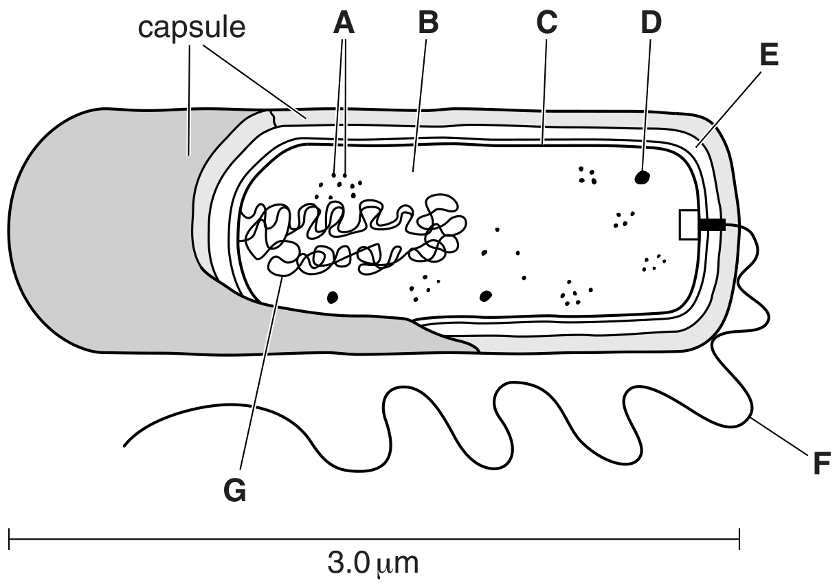
Fig. 1.1
Locate the structures in Fig. 1.1 that apply to each of the features shown in Table 1.1. Complete Table 1.1 by writing the appropriate letter and the name of the structure. You must only give one letter in each case. You may use each letter once, more than once or not at all. The first answer has been completed for you.
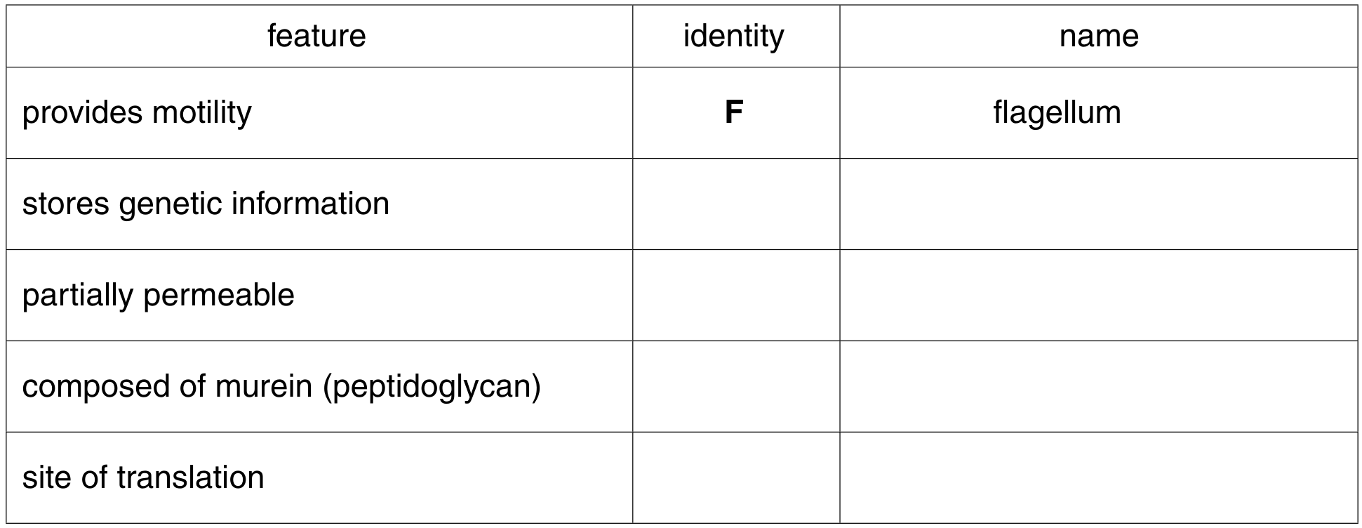
Table 1.1