[Maximum number: 1]
What is the diameter of a typical plant cell?
A
B
C
D
 EduNinja
What is the diameter of a typical plant cell?
Fig. 1.1 is a photomicrograph of plant root cells near the growing tip. Some of the cells are undergoing mitosis.
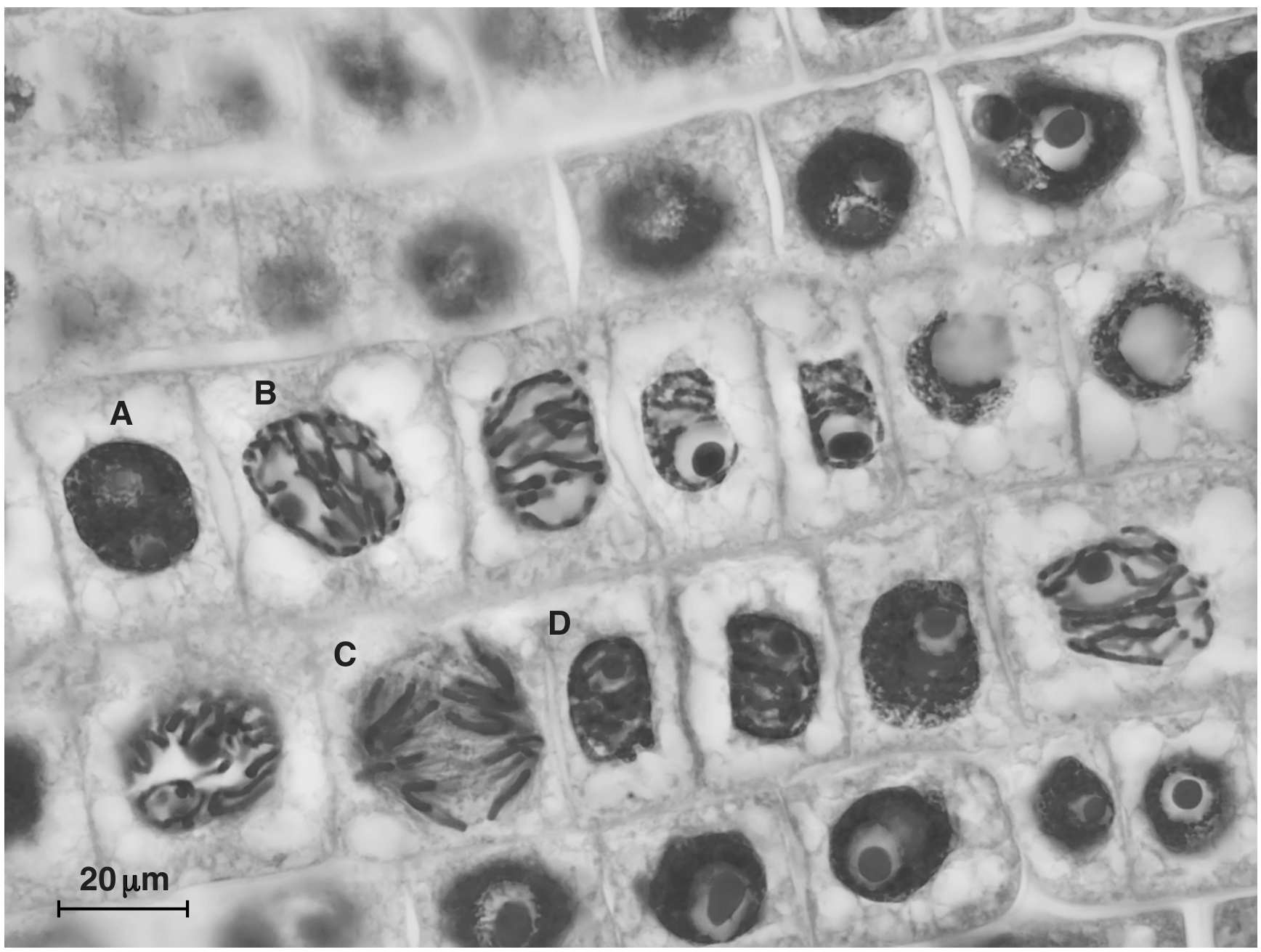
Fig. 1.1
State one feature, visible in Fig. 1.1, which indicates that the section is taken from plant tissue and not animal tissue.
A student compared an image of a plant cell with an image of an animal cell. Both images were at the same magnification.
Parts (a) to (c) are four correct comparative statements about these images.
There are strands of cytoplasm passing through channels in the cell wall of the plant cell. These are not visible in the animal cell.
Explain one advantage to the plant cell of having these structures.
A tissue is a collection of one or more types of cell, specialised to carry out a particular function.
An organ can be considered a structural unit within an organism that:
- consists of more than one type of tissue
- performs a particular function.
The aorta is the main artery of the body.
Explain, with reference to its structure and function, whether the aorta may be described as a tissue or an organ.
Vibrio cholerae is a prokaryotic organism.
Fig. 1.1 shows the structure of a cell of V. cholerae.
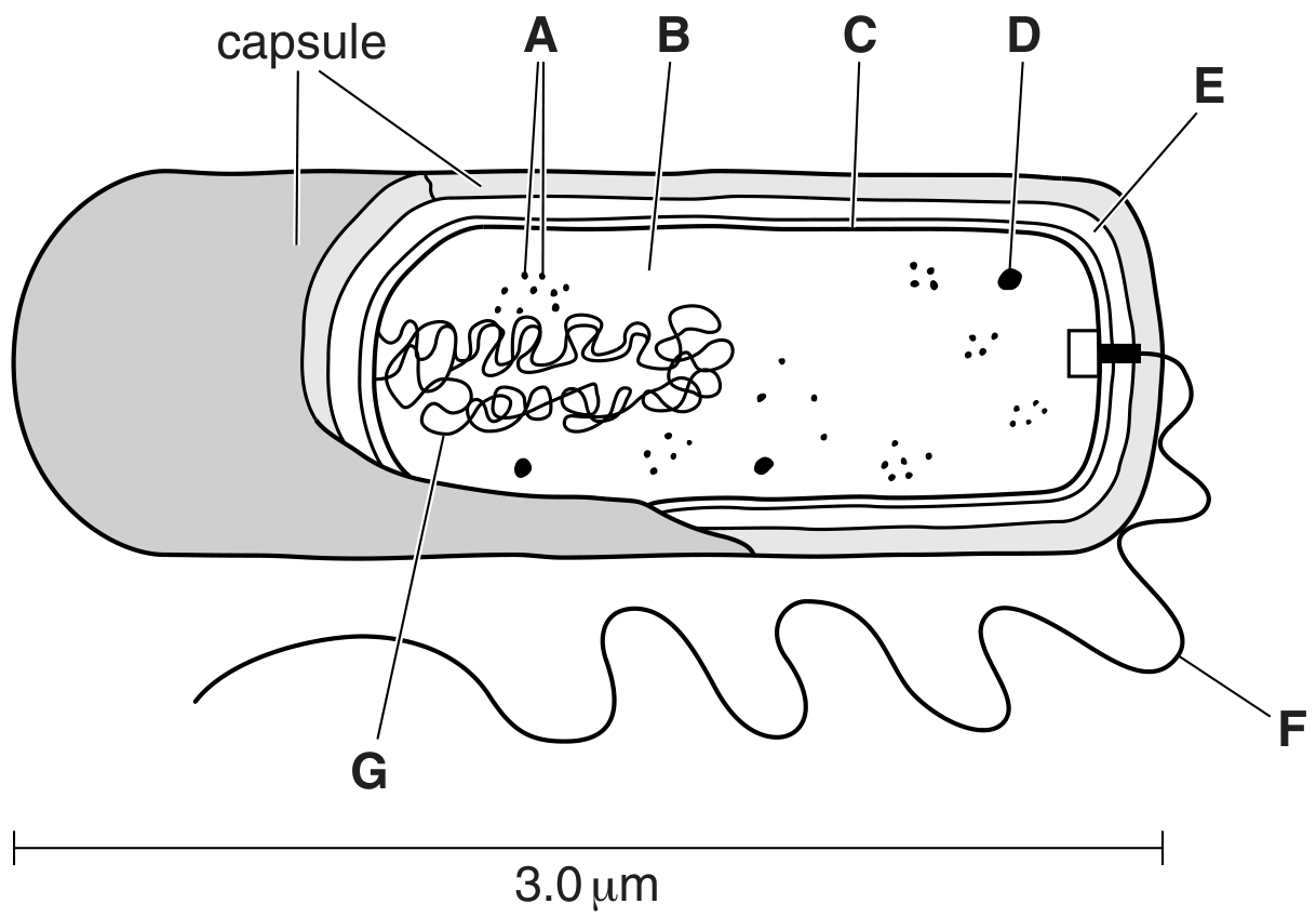
Fig. 1.1
State three structural features that are present in a mesophyll cell in a leaf that are not present in a prokaryotic cell such as that of V. cholerae.
1.
2.
3.
Water and mineral ions are transported up the stem of a plant to the leaves within xylem vessels.
Some water and mineral ions can pass out of xylem vessel elements to supply parenchyma tissue in the stem.
Fig. 1.2 is a diagram of a photomicrograph showing three adjacent parenchyma cells in the stem. These parenchyma cells can be described as typical plant cells.
The arrows show the direction of movement of water between the cells.
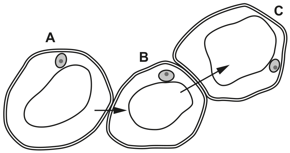
Fig. 1.2
Only some of the structures visible using the light microscope have been included in Fig. 1.2.
List the features that can be seen using the high power of a light microscope that help identify a parenchyma cell as a plant cell and not as an animal cell.
Fig. 1.1 is a transmission electron micrograph of cells from the leaf of a plant.
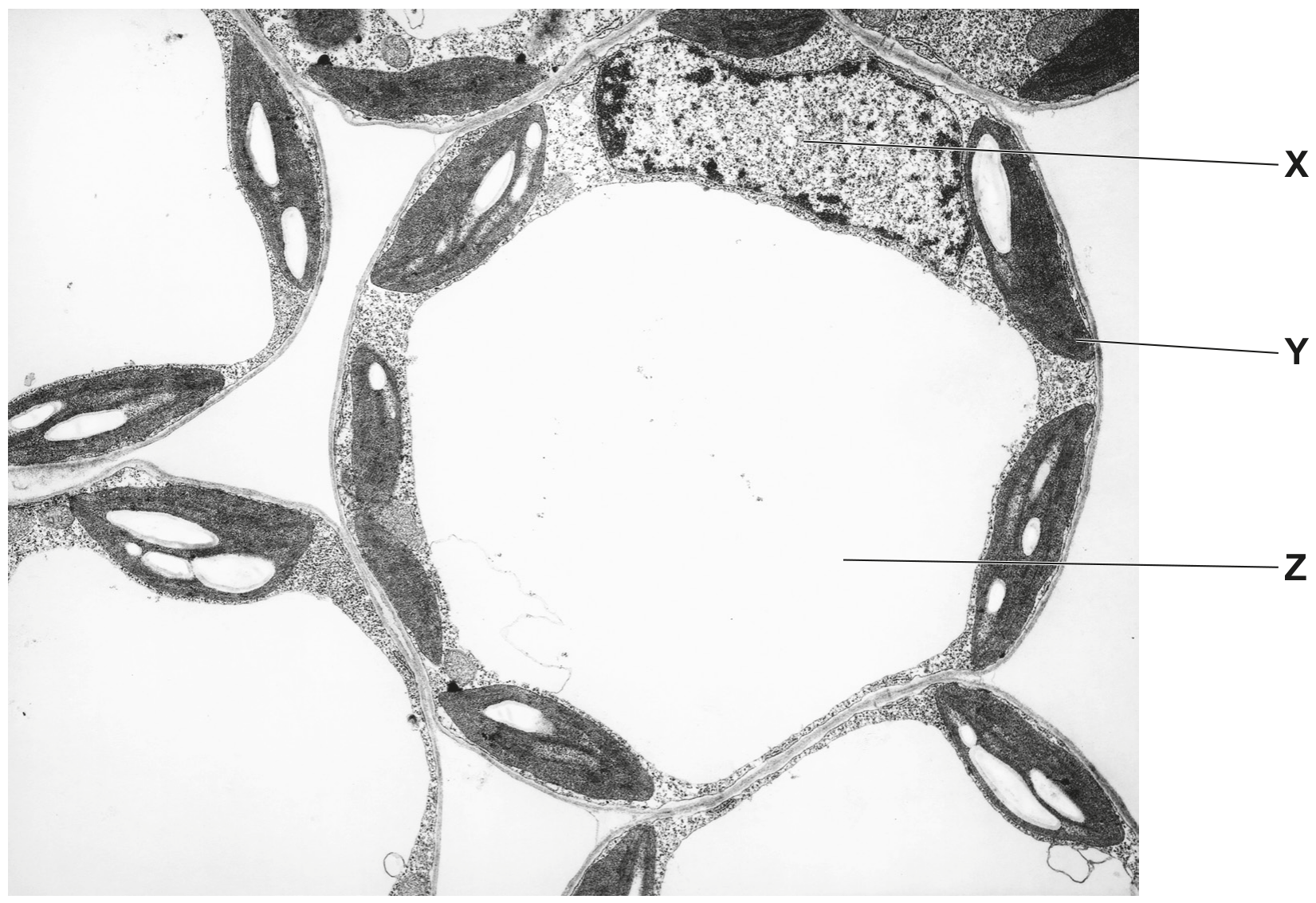
Fig. 1.1
State two ways in which the structure of an animal cell differs from plant cells such as those shown in Fig. 1.1.
1
2
Fig. 1.1 is a transmission electron micrograph of a cell from the stem of sago pondweed, Stuckenia pectinata.
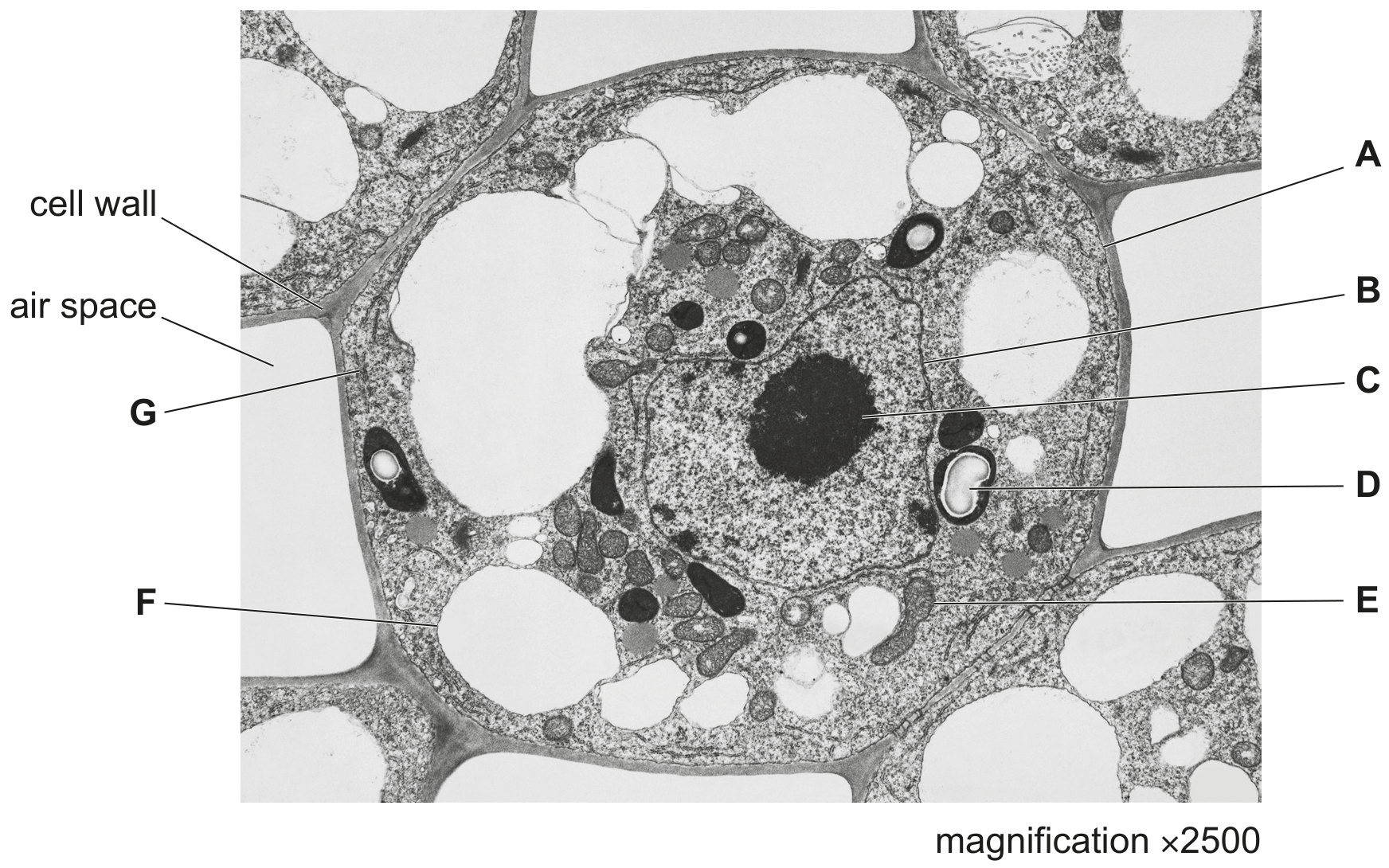
Fig. 1.1
State the evidence from Fig. 1.1 that shows that the cell is from the stem of S. pectinata and not from the mesophyll of a leaf.
Fig. 1.1 is a diagram of an electron micrograph of a plant cell.
Fig. 1.2 is a diagram of an electron micrograph of an animal cell. Both diagrams are incomplete.
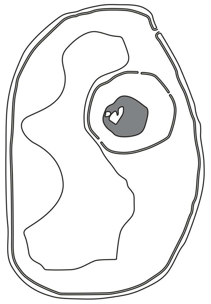
Fig. 1.1
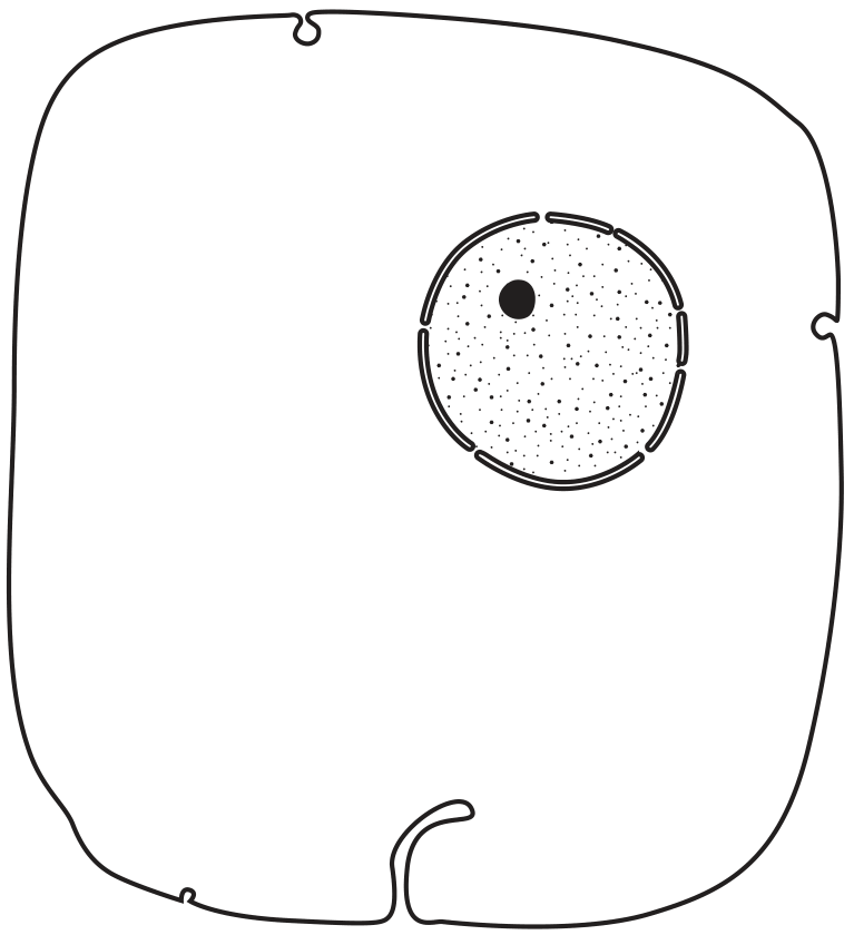
Fig. 1.2
Explain how Fig. 1.1 can be identified as a plant cell.
What is the diameter of a typical plant cell?