[Maximum number: 3]
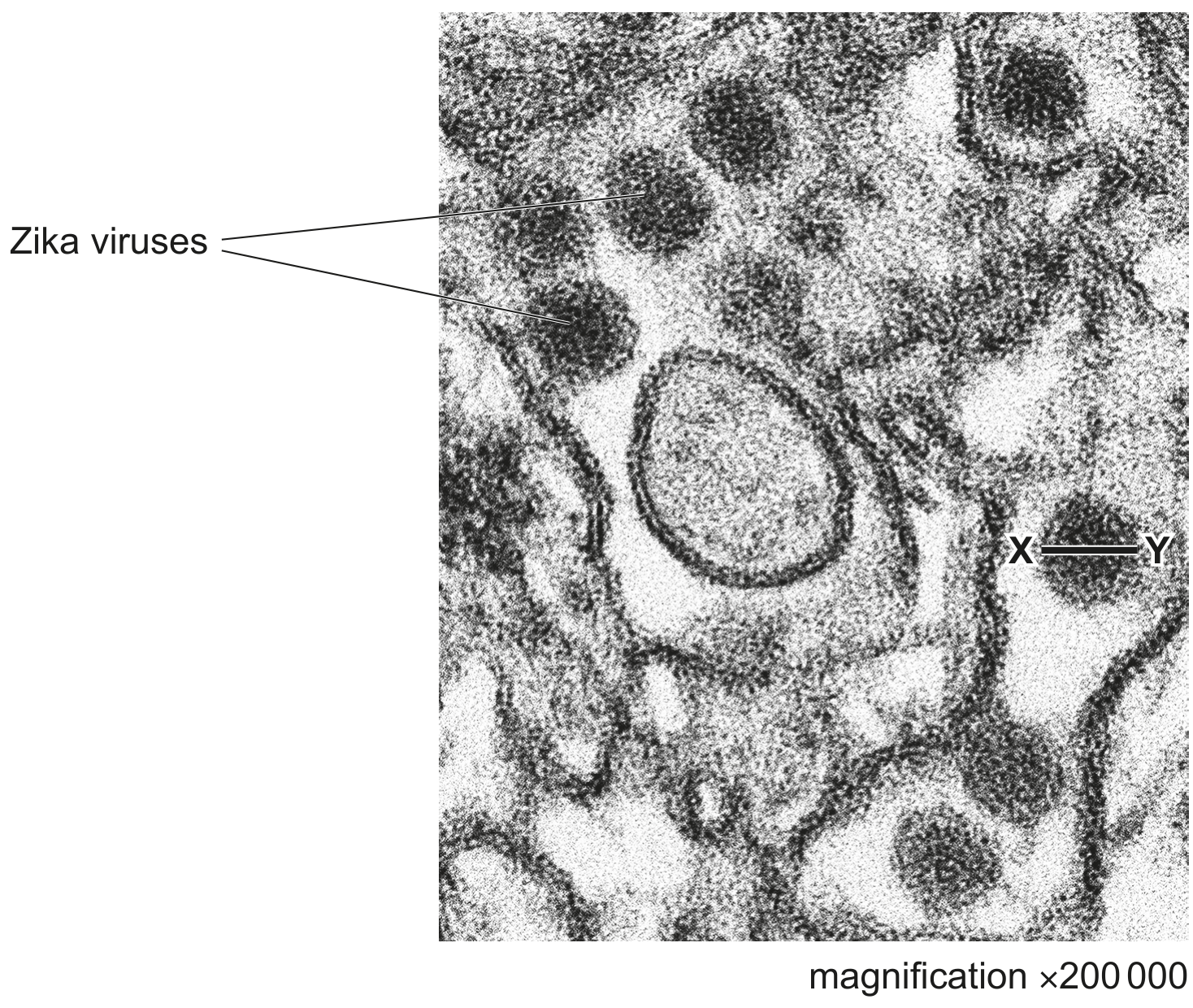
Picornaviruses are small viruses that are 30 nm in diameter. Picornaviruses are able to enter the cells of mammals and birds and can replicate within these cells.
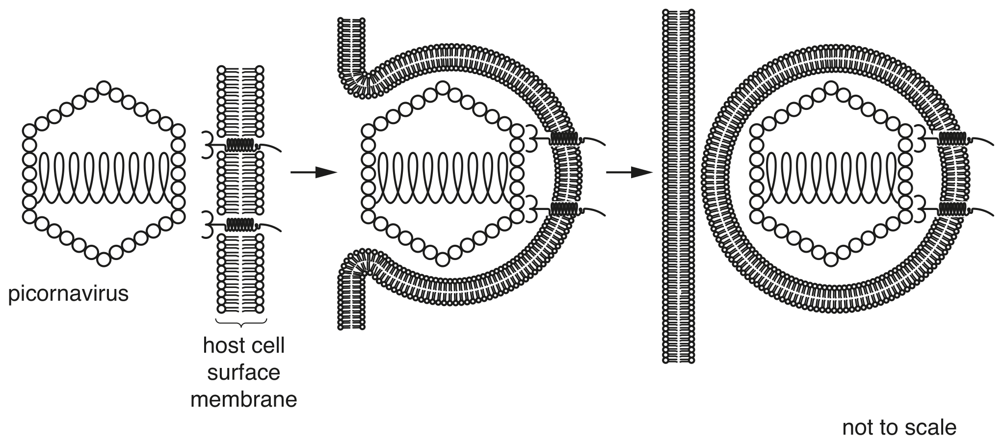
Fig. 1.1 shows the entry of a picornavirus into its host cell.
(a)
State, with reasons, whether a picornavirus can be seen using the light microscope.
[ 3 ]