[Maximum number: 1]
The diagram below was drawn from an electron micrograph of an animal cell.
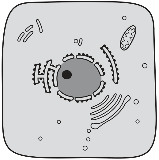
Which diagram would represent the same cell seen under a simple light microscope, using daylight as the only light source?
A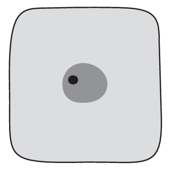
B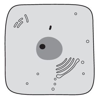
C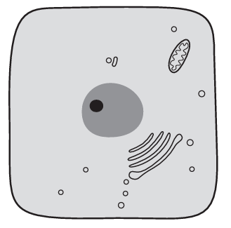
D
 EduNinja
The diagram below was drawn from an electron micrograph of an animal cell.
Which diagram would represent the same cell seen under a simple light microscope, using daylight as the only light source?
All cells have a cell surface membrane. Fig. 1.1 shows a transmission electron micrograph of part of two adjacent animal cells, cell 1 and cell 2 .
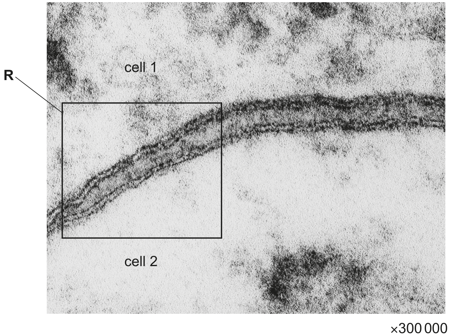
Fig. 1.1
In the space provided, draw a diagram of the region in the box labelled R on Fig. 1.1. Your diagram should show the four dark lines.
Label the diagram to identify what is shown by the dark lines and each of the three spaces between them.
space for diagram:
T-helper lymphocytes and Leydig cells are two types of mammalian cells. The main role of T-helper lymphocytes and Leydig cells is to synthesise and secrete cell-signalling molecules.
- T-helper lymphocytes synthesise proteins known as cytokines.
- Leydig cells synthesise the steroid (lipid) hormone testosterone from cholesterol. Leydig cells also synthesise cholesterol.
Fig. 3.1 shows part of a mammalian cell.
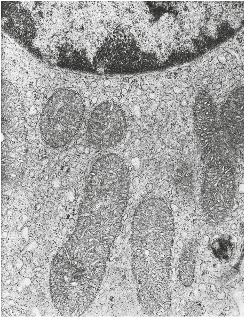
Fig. 3.1
Underline the correct name for the type of image shown in Fig. 3.1 and explain your choice.
photomicrograph
scanning electron micrograph
transmission electron micrograph
explanation
A group of students were asked to look at the photomicrograph of a cross-section of unfamiliar material and make observations.
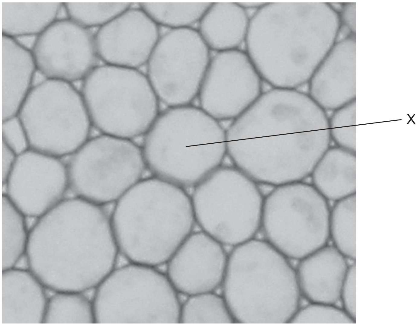
They described X as
1 circular
2 a hollow tube
3 a spherical structure
Which description(s) are correct?
1, 2 and 3
1 and 2 only
1 only
3 only