Protein synthesis links DNA information to functional proteins through transcription, RNA processing, translation, genetic-code reading and post-translational modification in cells.
mRNA Carries a Working Copy of Selected DNA Information
Chromosomal DNA is the long-term information store. Protein synthesis instead uses a short-lived messenger RNA (mRNA) copy of a selected gene.
selected gene in DNA → complementary mRNA copy → mRNA reaches a ribosome → nucleotide sequence directs amino-acid order
A working copy solves two problems:
The original DNA sequence remains protected and reusable.
Many mRNA copies can be made, so one gene can support repeated protein production.
mRNA carries information; it is not converted into protein matter. The ribosome reads its sequence while amino acids are joined into a separate molecule.
RNA Polymerase Builds mRNA from One DNA Template Strand
1
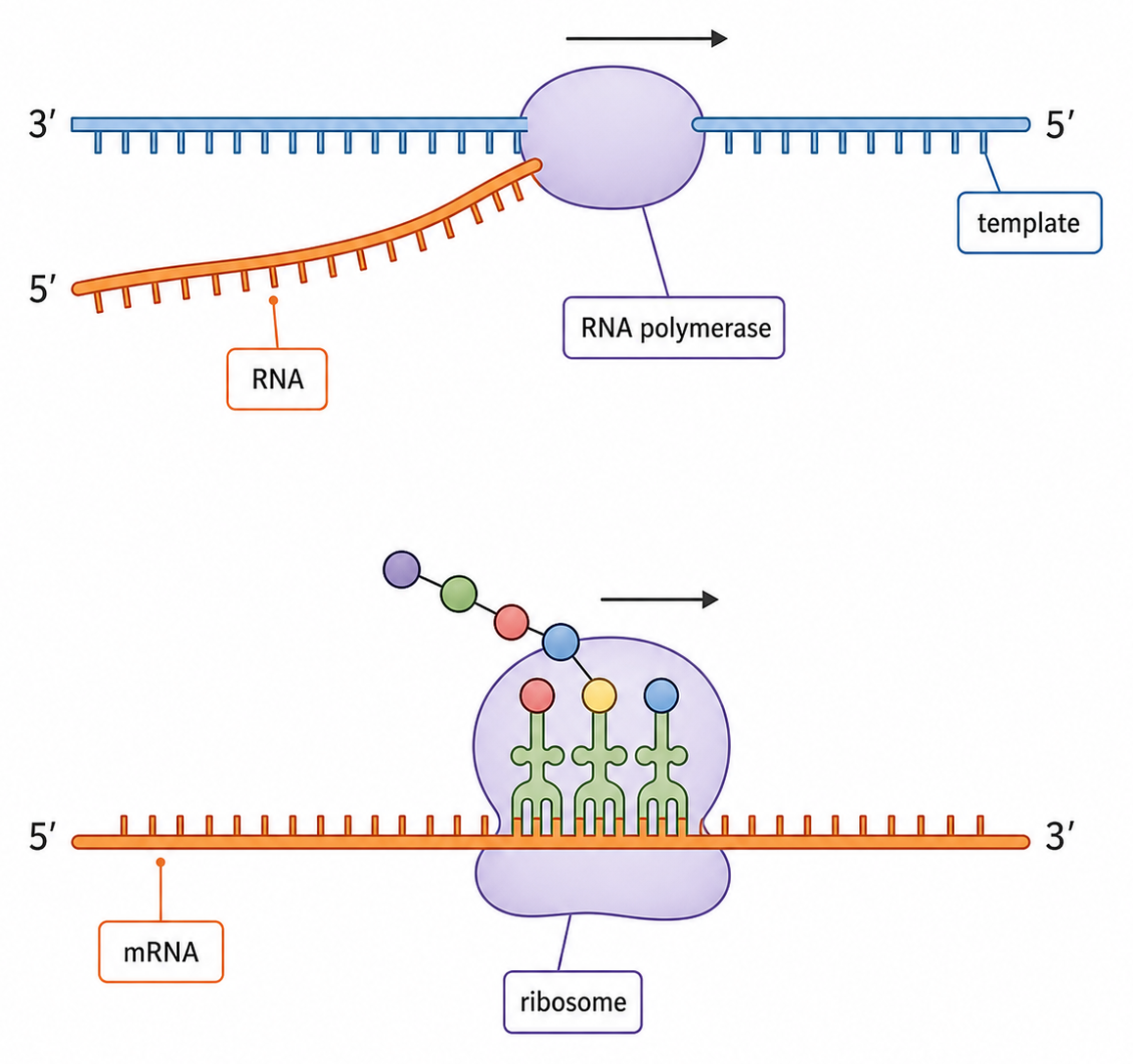
RNA polymerase binds near the gene and opens a short region of the DNA double helix, exposing the template strand.
2
Free RNA nucleotides align by complementary base pairing: DNA A pairs with RNA U, DNA T with RNA A, and C with G.
3
RNA polymerase joins the RNA nucleotides into a sugar–phosphate backbone. The mRNA separates, and the DNA strands pair again.
Template DNA Determines the mRNA Sequence
Strand
Relationship to the new mRNA
DNA template
antiparallel and complementary
DNA coding strand
same base order as mRNA, except DNA has T where RNA has U
DNA template: 3′–TAC CTT GCG–5′
mRNA: 5′–AUG GAA CGC–3′
Codon tables are read from mRNA 5′→3′. Do not look up a DNA template triplet directly.
Transcription Opens DNA Locally and Leaves Its Sequence Unchanged
Hydrogen bonds between complementary bases can break and re-form. This lets a short DNA region open temporarily without breaking the covalent sugar–phosphate backbones.
After the mRNA leaves, the original complementary DNA strands pair again. The gene's base sequence is conserved and can be transcribed repeatedly.
Transcription copies one gene region into RNA; it does not duplicate the whole DNA molecule and does not consume the template strand.
Selective Transcription Controls Which Proteins a Cell Can Make
Gene expression is the use of gene information to produce a functional RNA or polypeptide. Transcription is a major control point because an untranslated gene cannot supply mRNA to ribosomes.
Transcription state
Immediate consequence
Possible protein outcome
gene active
mRNA is produced
translation can occur
gene inactive
little or no mRNA is produced
little or no corresponding polypeptide is made
Most cells in one organism contain the same genome, but different cell types transcribe different gene sets. Their different protein mixtures produce different structures and functions.
Turning transcription on permits expression; it does not guarantee a fixed protein amount. RNA processing, translation and protein breakdown can also be regulated.
Reconstruct the Route from a Selected Gene to mRNA
gene selected → DNA opens locally → RNA polymerase reads one template strand → complementary RNA nucleotides are joined → mRNA leaves → DNA re-forms unchanged
Question
Correct check
Which DNA strand determines mRNA?
the template strand
Which base replaces thymine in RNA?
uracil
Which direction is mRNA written?
5′→3′
Why can the gene be reused?
DNA sequence and covalent backbones remain intact
The mRNA now carries codons in an order that a ribosome can translate into an amino-acid sequence.
Translation Converts Codon Order into Amino-Acid Order
Translation is the synthesis of a polypeptide whose amino-acid sequence is determined by the codon sequence of an mRNA.
mRNA codons read 5′→3′ → matching tRNAs deliver amino acids → ribosome forms peptide bonds → polypeptide grows from its amino end toward its carboxyl end
The immediate product is a polypeptide: a specific linear amino-acid sequence. Folding and later processing are needed before many polypeptides become functional proteins.
The genetic code determines amino-acid order, not the final three-dimensional shape directly. Shape emerges from interactions within the amino-acid sequence and its environment.
mRNA, tRNA and the Ribosome Divide Translation Work
Component
Information or material carried
Translation job
mRNA
ordered codons
supplies the sequence to be read
tRNA
one anticodon and its attached amino acid
matches a codon and delivers the corresponding amino acid
small ribosomal subunit
mRNA-binding site
positions the message for decoding
large ribosomal subunit
A, P and E sites
positions tRNAs and catalyses peptide-bond formation
Translation is accurate only when all three information links agree: the mRNA codon, the tRNA anticodon and the amino acid attached to that tRNA.
The ribosome does not choose amino acids by recognizing their chemical identity. It accepts charged tRNAs whose anticodons pair with the exposed codons.
Two Recognition Events Protect Amino-Acid Identity
At the ribosome, a tRNA anticodon pairs antiparallel with a complementary mRNA codon. This selects which tRNA can occupy the decoding site.
Before translation, a specific aminoacyl-tRNA synthetase attaches the correct amino acid to its matching tRNA. This charging step links anticodon identity to amino-acid identity.
Recognition
Example
What it secures
codon–anticodon pairing
mRNA 5′–AUG–3′ pairs with tRNA 3′–UAC–5′
the correct tRNA is selected
enzyme–tRNA/amino-acid recognition
methionine is attached to the initiator tRNA
the selected tRNA carries the correct amino acid
Complementary base pairing alone cannot check which amino acid is attached. Translation fidelity depends on both correct pairing and correct tRNA charging.
The Genetic Code Is Triplet, Degenerate and Nearly Universal
The genetic code has several linked properties:
Triplet: three mRNA bases form one codon.
Degenerate: most amino acids are specified by more than one codon.
Unambiguous: each codon specifies only one amino acid or stop signal.
Nearly universal: the same codons usually have the same meanings across organisms, with limited exceptions.
Punctuated: a start codon establishes the reading frame; stop codons end translation.
Degeneracy means some base substitutions are silent, but it does not mean one codon can represent several different amino acids.
Codon type
Function
AUG
usually codes for methionine and can establish the start of translation
UAA, UAG, UGA
stop signals recognized by release factors; they do not add an amino acid
Read the mRNA 5′→3′ before Using a Codon Table
1
If a DNA template is supplied, write the antiparallel complementary mRNA, replacing DNA A with RNA U.
2
Write the mRNA 5′→3′, locate the stated or biologically relevant start, and divide the sequence into consecutive triplets from that frame.
3
Look up each mRNA codon, record amino acids in order and stop when a stop codon is reached.
4
Template 3′–TAC CTT GCG ACT–5′ gives mRNA 5′–AUG GAA CGC UGA–3′, which translates as Met–Glu–Arg–Stop.
Elongation Repeats Until a Stop Codon Ends Translation
1
A charged tRNA with a complementary anticodon enters the A site beside the tRNA carrying the growing chain in the P site.
2
The ribosome catalyses a peptide bond, transferring the growing polypeptide to the amino acid on the A-site tRNA.
3
The ribosome moves one codon along the mRNA. The peptidyl-tRNA shifts to P, the empty tRNA shifts to E and exits, and the A site becomes available again.
4
The entry–bond–movement cycle repeats for successive codons. When a stop codon reaches the A site, no tRNA enters; a release factor releases the completed polypeptide and the ribosome dissociates.
A Polysome Makes Several Copies from One mRNA at Once
A polysome is one mRNA being translated simultaneously by several ribosomes. Each ribosome starts near the 5′ end and moves toward the 3′ end.
All ribosomes read the same codon sequence, so they produce polypeptides with the same amino-acid order. Simultaneous translation increases the rate of protein production from that mRNA.
The ribosomes do not cooperate to make one long polypeptide. Each ribosome makes a separate copy.
A DNA Mutation Does Not Always Change the Protein
A mutation can alter a DNA base sequence. If the changed region is transcribed, it can alter an mRNA codon and therefore the amino-acid sequence or the timing of translation.
Codon outcome
Possible protein consequence
codon still specifies the same amino acid
no change to primary structure
codon specifies a different amino acid
altered side-chain interactions, folding or function
codon becomes a stop signal
shortened polypeptide
insertion or deletion shifts the reading frame
many downstream codons may change
Effect size depends on where the mutation occurs and what the affected amino acid does. A substitution at an active site or interaction surface can matter more than one in a tolerant region.
Mutation creates a possibility of changed protein structure; it does not guarantee harm. Some mutations are silent, neutral or beneficial in a particular environment.
One β-Globin Substitution Can Reshape a Red Blood Cell
1
In the sickle-cell allele, a β-globin DNA substitution changes an mRNA codon from GAG to GUG, replacing glutamate with valine in the polypeptide.
2
Glutamate is charged and hydrophilic; valine is non-polar. The replacement creates a hydrophobic patch on deoxygenated haemoglobin S.
3
Under low oxygen conditions, haemoglobin S molecules associate into long fibres. The fibres distort the red blood cell into a rigid sickle shape.
4
Rigid cells can block small blood vessels and are removed rapidly from circulation, reducing oxygen delivery and causing anaemia and painful crises.
The mutation does not directly bend the whole cell. It first changes one amino acid, which changes intermolecular interactions, which then changes cell shape under particular oxygen conditions.
Trace Information—and the Places Where It Can Change
Is the mRNA complementary to the template and written 5′→3′?
code table
Are mRNA codons grouped from the correct start?
tRNA selection
Does the anticodon pair and is the tRNA correctly charged?
elongation
Has the ribosome advanced one codon while the chain remains on the peptidyl-tRNA?
mutation
Did the codon, amino acid or reading frame actually change?
A sequence change matters biologically only through a causal path: altered primary structure → altered interactions or folding → altered protein activity → possible cellular or organism effect.
Information Is Read in Opposite Strand Directions but Built 5′→3′
HL only
Process
Molecule being read
Reading direction
Product growth or movement
transcription
DNA template
3′→5′
RNA is synthesized 5′→3′
translation
mRNA
5′→3′
ribosome moves toward the mRNA 3′ end; polypeptide grows N-terminus→C-terminus
Nucleotides are added to a free 3′ hydroxyl group, so every new nucleic-acid strand grows 5′→3′. The template must therefore be read antiparallel to the new strand.
A diagram arrow showing polymerase movement and an arrow showing new-strand growth refer to opposite strands; identify which molecule each arrow belongs to.
Promoters and Transcription Factors Assemble the Start Complex
HL only
A promoter is a DNA region that positions the transcription machinery near a gene's transcription start site. General transcription factors bind first and help recruit RNA polymerase.
Regulatory input
Binding relationship
Typical effect on initiation
activator transcription factor
binds a regulatory DNA sequence such as an enhancer
stabilizes or recruits the initiation machinery
repressor transcription factor
binds a regulatory sequence or interferes with the complex
reduces assembly or activity of the initiation machinery
Regulatory DNA may be far from the promoter in the linear sequence. DNA looping can bring bound factors into contact with promoter-associated proteins.
The combined transcription-factor state changes how often RNA polymerase initiates, altering the amount of pre-mRNA produced from the gene.
Non-Coding DNA Includes Several Functional Categories
HL only
Non-coding DNA is DNA that is not translated into a polypeptide sequence. Non-coding does not mean untranscribed, unused or without function.
Important non-coding categories include:
Regulatory sequences such as promoters and enhancers that affect transcription.
Introns that are transcribed into pre-mRNA and removed during splicing.
Genes for functional RNAs, including rRNA and tRNA, whose products are not translated.
Structural regions such as telomeres and centromeric DNA.
Repeated sequences, some functional and some with no known function.
An exon can include untranslated regions, so exon is not a synonym for protein-coding sequence. Likewise, an intron can influence regulation or splicing even though it is removed from mature mRNA.
Capping, Polyadenylation and Splicing Produce Mature mRNA
HL only
Processing change
What happens
Main consequence
5′ cap
a modified nucleotide is added to the 5′ end
protects RNA and supports export and translation initiation
poly-A tail
many adenine nucleotides are added to the 3′ end
increases stability and supports export and translation
splicing
introns are removed and exons joined
creates a continuous mature RNA sequence for translation
pre-mRNA in nucleus → processing completed → mature mRNA exported through a nuclear pore → translation in cytoplasm
The poly-A tail is added after transcription; it is not copied from a long template-DNA sequence of thymine bases.
Alternative Splicing Changes Which Exons Reach the Ribosome
HL only
In alternative splicing, the same pre-mRNA is spliced in different patterns, so different exon combinations are retained in mature mRNA.
Different mature mRNAs contain different codon sequences and can therefore produce related polypeptides with different amino-acid segments, properties or tissue distributions.
Alternative splicing does not change the DNA sequence. It changes which parts of one RNA transcript remain in the message sent to the ribosome.
Translation Initiation Places the Start Codon in the P Site
HL only
1
The small ribosomal subunit binds the mRNA and positions the start region for scanning or recognition.
2
The initiator tRNA carrying methionine pairs with AUG. This tRNA is placed directly in the P site, establishing the reading frame.
3
The large subunit joins to complete the ribosome. The A site is now open for the tRNA matching the next codon.
AUG inside an already established reading frame can code for methionine without serving as a new start. Initiation depends on context and assembly, not the triplet alone.
A, P and E Sites Track Each tRNA's State
HL only
Ribosomal site
Typical tRNA state during elongation
A (aminoacyl)
incoming charged tRNA paired with the next codon
P (peptidyl)
tRNA carrying the growing polypeptide before peptide transfer
E (exit)
uncharged tRNA leaving after translocation
Before peptide transfer: growing chain in P, new amino acid in A.
After peptide transfer and translocation: the chain-bearing tRNA occupies P, the empty tRNA moves to E, and A opens for the next charged tRNA.
The site letters describe functional positions in the ribosome, not permanent labels on a tRNA. A single tRNA can move A → P → E during one cycle.
A New Polypeptide May Need Folding, Cleavage or Chemical Modification
HL only
Translation produces a polypeptide primary structure. Many proteins become functional only after post-translational processing changes their shape, length, location or chemical properties.
Common processing routes include:
Folding into a specific three-dimensional conformation, sometimes assisted by chaperones.
Cleavage to remove a signal peptide or inactive segment.
Covalent modification, such as phosphorylation or glycosylation, which can alter activity, stability or location.
Assembly with other polypeptide subunits or prosthetic groups.
Not every protein undergoes every modification. The required pathway depends on the protein's sequence, destination and function.
Insulin Becomes Active through Ordered Cleavage and Folding
HL only
1
Preproinsulin contains an N-terminal signal peptide that directs the growing polypeptide into the endoplasmic reticulum. Removing the signal peptide produces proinsulin.
2
Proinsulin folds, and disulfide bonds form between regions that will become the A and B chains.
3
The connecting C-peptide is removed. The A and B chains remain linked by disulfide bonds, producing mature insulin.
Ubiquitin Directs Selected Proteins to the Proteasome
HL only
1
Proteins selected for degradation are commonly marked with a chain of ubiquitin molecules.
2
The proteasome recognizes the tag, removes and recycles ubiquitin, unfolds the target protein and threads it into a proteolytic core.
3
The protein is cut into short peptides, which can be hydrolysed to amino acids for new synthesis or metabolism.
Selective degradation removes damaged or misfolded proteins and can rapidly lower the abundance of regulatory proteins. Protein concentration therefore depends on both synthesis and breakdown.
Control Protein Output at Several Checkpoints
HL only
Checkpoint
What can change
Consequence for protein output
transcription initiation
promoter and transcription-factor activity
amount of pre-mRNA made
RNA processing
cap, tail and exon combination
mRNA stability, export and encoded sequence
translation initiation
successful AUG recognition and ribosome assembly
whether and where translation begins
post-translational processing
folding, cleavage, modification and assembly
protein activity or location
proteasomal degradation
ubiquitin tagging and breakdown rate
protein lifetime and abundance
Across these checkpoints, sequence direction stays consistent: RNA is made 5′→3′, the ribosome reads mRNA 5′→3′ and the polypeptide grows from N-terminus to C-terminus.
Gene expression is therefore not a single on/off event. A cell controls which message is made, how it is processed, whether it is translated, how the product is activated and how long the protein remains.
Transcription exam focus
8 marks
Explain the process of transcription in prokaryotes.
Hydrogen bonding in transcription
1 mark
The sequence of bases on a short section of the antisense strand of a gene undergoing transcription is shown:
5′ CATG 3′
What is the sequence of bases on the resulting mRNA?
Transcription for gene expression
2 marks
The scientists concluded that auxin activates the transcription of the GH 3 gene. Using the information on the auxin concentration in the stem base in the graph on page 4 and the Northern blot, evaluate whether this conclusion is supported.
Translation exam focus
8 marks
Explain how polypeptides are produced by the process of translation.
Roles in translation
7 marks
Explain the role of RNA in translation, resulting in the formation of polypeptide chains.
Complementary base pairing
3 marks
Outline how translation depends on complementary base pairing.
Genetic code features
5 marks
Describe the genetic code and its relationship to polypeptides and proteins.
Using genetic code table
1 mark
Which sequence of mRNA bases and amino acids could be produced by transcription and translation of the DNA molecule shown?
3' AAAGTGGCACGTATATTT 5' 5' TTTCACCGTGCATATAAA 3′
\begin{tabular}{|l|l|l|l|l|l|l|l|} \hline \multirow{6}{*}{} & \multicolumn{6}{|c|}{2nd base in codon} & \multirow{14}{*}{3rd base in codon} \\ \hline & & U & C & A & G & & \\ \hline & \multirow{4}{*}{U} & Phe & Ser & Tyr & Cys & U & \\ \hline & & Phe & Ser & Tyr & Cys & C & \\ \hline & & Leu & Ser & STOP & STOP & A & \\ \hline & & Leu & Ser & STOP & Trp & G & \\ \hline \multirow[t]{8}{*}{} & C & Leu & Pro & His & Arg & U & \\ \hline & \multirow{3}{*}{A} & lle & Thr & Asn & Ser & U & \\ \hline & & lle & Thr & Lys & Arg & A & \\ \hline & & Met & Thr & Lys & Arg & G & \\ \hline & \multirow{4}{*}{G} & Val & Ala & Asp & Gly & U & \\ \hline & & Val & Ala & Asp & Gly & C & \\ \hline & & Val & Ala & Glu & Gly & A & \\ \hline & & Val & Ala & Glu & Gly & G & \\ \hline \end{tabular}
Sequence of mRNA bases
Sequence of amino acids
UUU-GAG-GCU-CGA-UAU-UUU
Phe-Glu-Ala-Arg-Tyr-Phe
AAA-CUC-CGA-GCU-AUA-UUU
Lys-Leu-Arg-Ala-lle-Phe
UUU-CAC-CGU-GCA-UAU-AAA
Phe-His-Arg-Ala-Tyr-Lys
AAA-GUG-GCA-CGU-AUA-UUU
Lys-Val-Ser-Arg-Ile-Phe
Elongation of polypeptide
2 marks
Six polypeptides are shown in the diagram. Explain the different lengths of these polypeptides.
Mutations changing protein structure
8 marks
Explain the cause of sickle cell anemia and how this disease affects humans.
Directionality exam focus
HL only
1 mark
Using the diagram, identify, with a reason, whether X or Y is the start codon.
Initiation of transcription at promoter
HL only
2 marks
Explain the function of a promoter in DNA.
Non-coding sequences in DNA
HL only
2 marks
DNA has regions that do not code for proteins. State two functions of these regions. 1. 2.
Post-transcriptional modification
HL only
1 mark
What happens to mRNA after transcription in eukaryotic cells?
Alternative splicing
HL only
1 mark
The number of protein-coding genes in the human genome is estimated to be about 20000 , which is much less than the size of the proteome. What is one reason for this?
Translation initiation
HL only
4 marks
Outline the roles of the different binding sites for tRNA on ribosomes during translation.
Polypeptide modification
HL only
2 marks
Insulin is produced by cutting C -peptide from the precursor molecule proinsulin. Suggest why group 1 has a greater level of C-peptide than group 2.