D3.3 Homeostasis

Homeostasis maintains internal conditions through feedback control of blood pH, glucose, temperature, kidney filtration, osmoregulation and blood flow in human physiology.

Syllabus
First assessment 2025
Topic
D3.3
Level
HL

Learning objectives

D3.3.1Homeostasis definition• Homeostasis maintains a stable internal environment within narrow limits• Variables include body temperature, blood pH, glucose, gases, ions, and osmotic concentrationD3.3.2Negative feedback loops• Negative feedback detects deviation from a set point and reverses it• Receptors, coordinators, effectors, and feedback loops restore normal conditionsD3.3.3Blood glucose regulation• Beta cells secrete insulin when blood glucose rises• Alpha cells secrete glucagon when blood glucose falls, affecting liver and muscle storesD3.3.4Diabetes• Type 1 diabetes results from autoimmune destruction of pancreatic beta cells• Type 2 diabetes involves insulin-receptor/response failure and is linked to lifestyle risk factorsD3.3.5Thermoregulation• Thermoregulation uses negative feedback to maintain core temperature near 37 °C• Thermoreceptors signal the hypothalamus, which coordinates skin, muscles, liver, and hormonesD3.3.6Thermoregulation mechanisms• Cooling uses vasodilation, sweating, and hairs lying flat• Warming uses vasoconstriction, reduced sweating, shivering, metabolic heat, and brown fatD3.3.7(HL)—Kidney role• Kidneys regulate blood composition by excretion and osmoregulation• Nephrons remove urea and adjust water and ion concentrations in urineD3.3.8(HL)—Glomerulus, Bowman's capsule, PCT• Glomerular blood pressure drives ultrafiltration into Bowman's capsule• The proximal convoluted tubule selectively reabsorbs glucose, amino acids, ions, and waterD3.3.9(HL)—Loop of Henle• Ascending limb pumps Na⁺/Cl⁻ and is impermeable to water• Descending limb loses water, creating a medulla gradient for water conservationD3.3.10(HL)—Osmoregulation by collecting ducts• Hypothalamus osmoreceptors control posterior pituitary ADH release• ADH inserts aquaporins in collecting ducts, increasing water reabsorption and concentrated urineD3.3.11(HL)—Blood supply changes• Vasoconstriction and vasodilation redistribute blood according to activity• Exercise and epinephrine increase skeletal muscle flow and reduce gut or renal flow

Homeostasis Stabilizes the Fluid Environment Around Cells

Homeostasis maintains selected variables of the internal environment within narrow limits despite internal and external change. The internal environment is blood plasma and tissue fluid—not the world outside the body.

Regulated variable Why large deviation matters
core temperature enzyme and membrane processes change rate
blood pH protein shape and reaction rates change
blood glucose cells lose a reliable respiratory substrate; osmotic effects rise when excessive
O₂ and CO₂ respiration and acid–base balance are disrupted
ions and osmotic concentration water movement, membrane potentials and cell volume change

Stable does not mean perfectly constant. A healthy value fluctuates within an acceptable range, and different variables can have different set ranges.

A Control Loop Links Deviation to a Corrective Response

change in a regulated variable → receptor detects the value → coordinator compares it with the set range → signal reaches an effector → effector changes the variable

Component Question it answers
stimulus / deviation what moved away from the range?
receptor what measured the variable?
coordinator where was information integrated?
effector what tissue carried out the response?
response how did the variable change?

Signals may travel as nerve impulses, hormones, or both. A valid explanation names the regulated variable and the direction of correction, not only the organs involved.

Negative Feedback Opposes Both Upward and Downward Deviations

Deviation Corrective response When correction succeeds
value above the range response lowers the value response weakens or switches off
value below the range response raises the value response weakens or switches off

The feedback is negative because the response reduces the original deviation. The output feeds back to decrease the stimulus that activated the loop.

Positive feedback amplifies a change and is useful for endpoint processes such as childbirth. By itself it does not restore a homeostatic set range.

Blood Glucose Must Supply Cells Without Creating Osmotic Stress

State Immediate problem Serious consequence if prolonged
hypoglycaemia too little glucose available, especially to the brain fainting, convulsions or coma
hyperglycaemia plasma water potential falls and water is drawn from tissues dehydration and circulatory damage

Blood glucose rises after carbohydrate absorption. It falls during fasting and prolonged activity as cells remove glucose for respiration.

Glycogen stores buffer these changes. Liver glycogen can support blood glucose; muscle glycogen is primarily a local fuel reserve for muscle activity.

When Glucose Rises, Beta Cells Use Insulin to Promote Removal and Storage

  1. A rise in blood glucose is detected directly by pancreatic beta cells in the islets of Langerhans.
  2. Beta cells secrete insulin into the blood.
  3. Insulin increases glucose uptake by target cells and promotes respiration.
  4. Liver and skeletal muscle convert glucose to glycogen; other excess can be converted to fat.
  5. As blood glucose falls toward the range, insulin secretion decreases.

Insulin is a hormone signal. It does not itself convert glucose into glycogen; it changes target-cell activity so enzymes carry out the conversions.
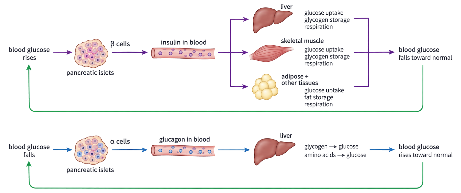
Two feedback pathways show pancreatic beta cells releasing insulin after blood glucose rises and alpha cells releasing glucagon after blood glucose falls.

When Glucose Falls, Alpha Cells Use Glucagon to Mobilize Liver Stores

  1. A fall in blood glucose is detected directly by pancreatic alpha cells.
  2. Alpha cells secrete glucagon into the blood.
  3. In the liver, glucagon promotes glycogen breakdown and glucose production from non-carbohydrate substrates.
  4. Glucose is released to the blood.
  5. As blood glucose rises toward the range, glucagon secretion decreases.
Feature Insulin branch Glucagon branch
source beta cells alpha cells
trigger high blood glucose low blood glucose
main effect uptake and storage liver glucose release
net result lowers blood glucose raises blood glucose

Glucagon is a hormone; glycogen is a glucose-storage polymer. Glucagon activates cellular pathways—it is not the enzyme that digests glycogen.

Type 1 and Type 2 Diabetes Disrupt Different Links in Glucose Control

Feature Type 1 diabetes Type 2 diabetes
primary failure autoimmune destruction of beta cells reduced target-cell response to insulin; beta-cell function may later decline
insulin signal little or none produced present but less effective
common management principle replace insulin and monitor glucose improve response and glucose balance through individualized diet, activity and medication
association genetic susceptibility and environmental triggers age, family history, inactivity and excess body fat are risk factors

A risk factor changes probability; it is not a single sufficient cause. Type 2 diabetes should not be reduced to “eating sugar,” and type 1 is not caused by lifestyle.

Both can produce persistent hyperglycaemia, glucose in urine and long-term damage to blood vessels, kidneys, retina and nerves if control remains poor.

A Glucose Tolerance Curve Tests How Quickly the Control System Recovers

  1. Measure fasting blood glucose.
  2. Give a standard oral glucose dose.
  3. Measure blood glucose at defined times after ingestion.
  4. Compare peak height and recovery toward the fasting range.
Curve feature Effective regulation Impaired regulation
starting value within expected fasting range may already be elevated
post-dose peak rises then turns downward often higher or prolonged
later value approaches the starting range remains elevated longer

The curve shows glucose handling under a standardized challenge. Diagnosis uses clinical thresholds and context; one graph alone does not identify the exact cellular cause.

The Hypothalamus Coordinates Opposing Temperature Responses

skin thermoreceptors monitor environmental influence + hypothalamic thermoreceptors monitor blood temperature → hypothalamus integrates the signals → skin, skeletal muscle, liver and endocrine pathways alter heat loss or production

Control feature Core temperature rises Core temperature falls
corrective direction increase heat loss and reduce heat gain reduce heat loss and increase heat production
shut-off condition response fades as temperature returns toward the range response fades as temperature returns toward the range

Human core temperature is regulated near 37 °C but is not fixed at exactly 37.0 °C; normal values vary with time and physiological state.
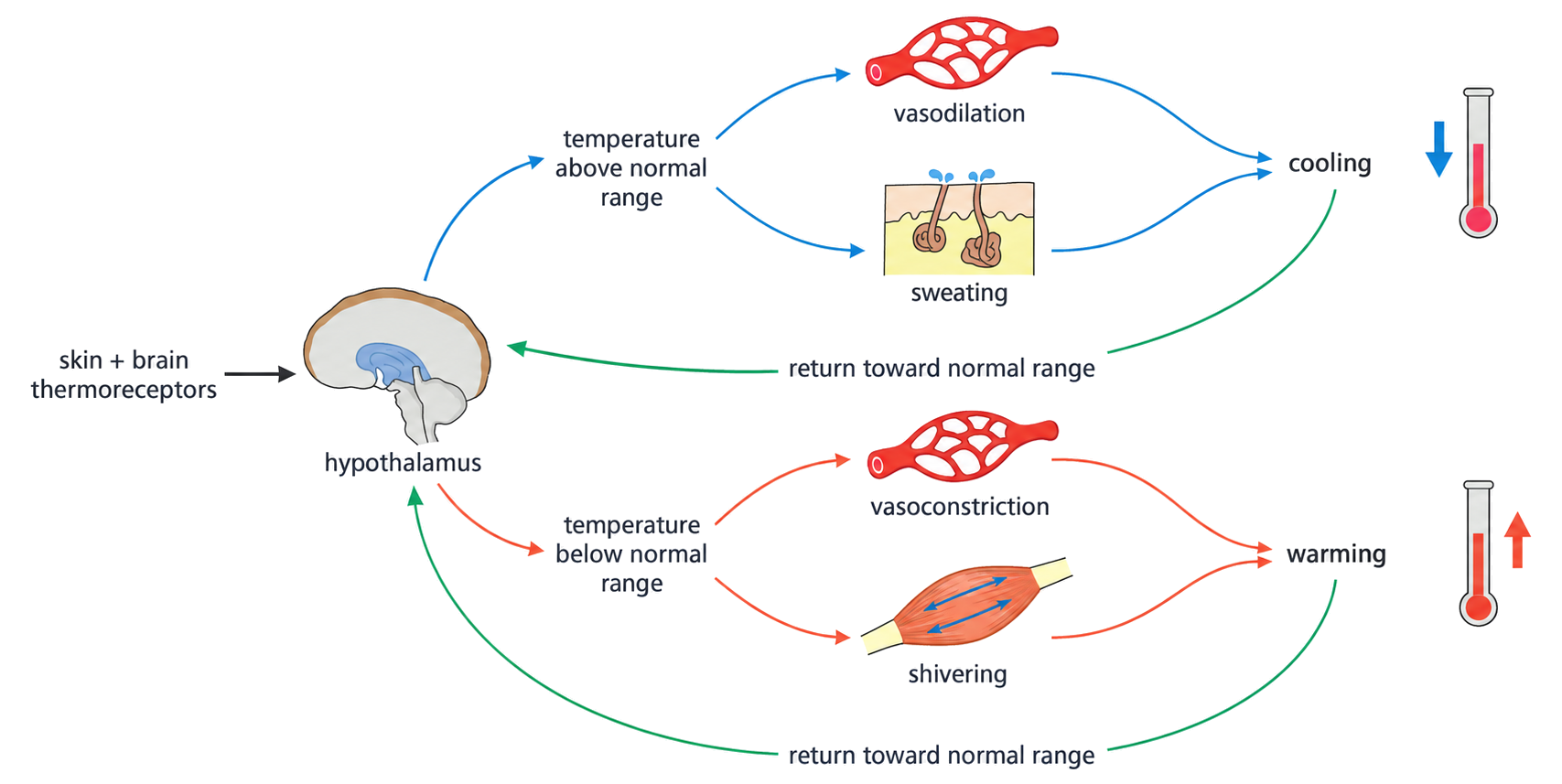
Skin and brain thermoreceptors signal the hypothalamus, which activates cooling above the normal range and warming below it.

A Hot Body Increases Heat Transfer and Evaporative Cooling

Effector response How it cools
skin arterioles dilate more warm blood reaches surface capillaries, increasing heat transfer to surroundings
sweat secretion increases evaporation transfers latent heat from the skin
hair erector muscles relax hairs lie flat, reducing the insulating air layer
behaviour changes shade, reduced activity or exposed skin decreases heat gain and increases loss

Sweating cools only when sweat evaporates. Sweat that drips away without evaporating removes far less heat.

A hot-skin cross-section shows active sweat glands, evaporation, dilated skin blood vessels and relaxed hair muscles.

A Cold Body Conserves Heat and Raises Metabolic Heat Production

Effector response How it warms or conserves heat
skin arterioles constrict less warm blood reaches surface capillaries, reducing heat loss
sweating decreases less evaporative heat is lost
skeletal muscles shiver rapid contractions increase respiration and heat production
liver metabolic activity rises greater respiration releases more heat
brown adipose tissue uncouples respiration fuel energy is released as heat rather than captured mainly in ATP

Brown fat is especially important in newborns, whose large surface-area-to-volume ratio causes rapid heat loss.

Raised hairs have little insulating effect in humans because human body hair is sparse; the mechanism is more effective in furred mammals.

Homeostasis Uses the Same Logic with Different Sensors and Effectors

regulated variable leaves its acceptable range → receptor detects the direction of deviation → coordinator selects an opposing response → effectors alter transfer, storage or metabolism → deviation shrinks → corrective signal declines

System Sensor / coordinator High-side response Low-side response
blood glucose pancreatic islet cells beta cells, insulin, uptake and storage alpha cells, glucagon, liver glucose release
core temperature thermoreceptors and hypothalamus vasodilation and sweating vasoconstriction, shivering and metabolic heat

For any homeostatic explanation, name the variable, direction of deviation, detector, signal, effector action and why that action reverses the deviation.

Kidneys Excrete Metabolic Waste and Regulate Water and Ion Balance

HL only
Kidney job What is controlled Example
excretion removal of metabolic waste from the body urea leaves in urine
osmoregulation balance of water and dissolved solutes urine volume and concentration change with water status

renal artery brings blood → nephrons filter and modify fluid → renal vein carries adjusted blood away; collecting ducts deliver urine → renal pelvis → ureter → bladder → urethra

Excretion is not the same as egestion. Urea is a metabolic waste made inside the body; undigested food leaving the gut was never part of the internal environment.
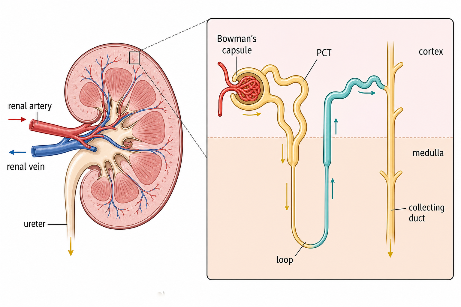
A kidney-to-nephron overview shows renal blood vessels, ureter, cortex, medulla, Bowman’s capsule, proximal tubule, loop of Henle and collecting duct.

Glomerular Pressure Filters Small Solutes but Retains Cells and Most Proteins

HL only
  1. A wide afferent arteriole supplies the glomerulus and a narrower efferent arteriole maintains high hydrostatic pressure.
  2. Water and small solutes cross fenestrated capillary endothelium, the basement membrane and filtration slits between podocyte processes.
  3. Blood cells and most plasma proteins are too large to cross and remain in the blood.
  4. The resulting glomerular filtrate enters Bowman’s capsule and flows into the proximal convoluted tubule.

Ultrafiltration is driven by blood hydrostatic pressure generated by the heart. The filtration step itself is not powered by ATP in podocytes.
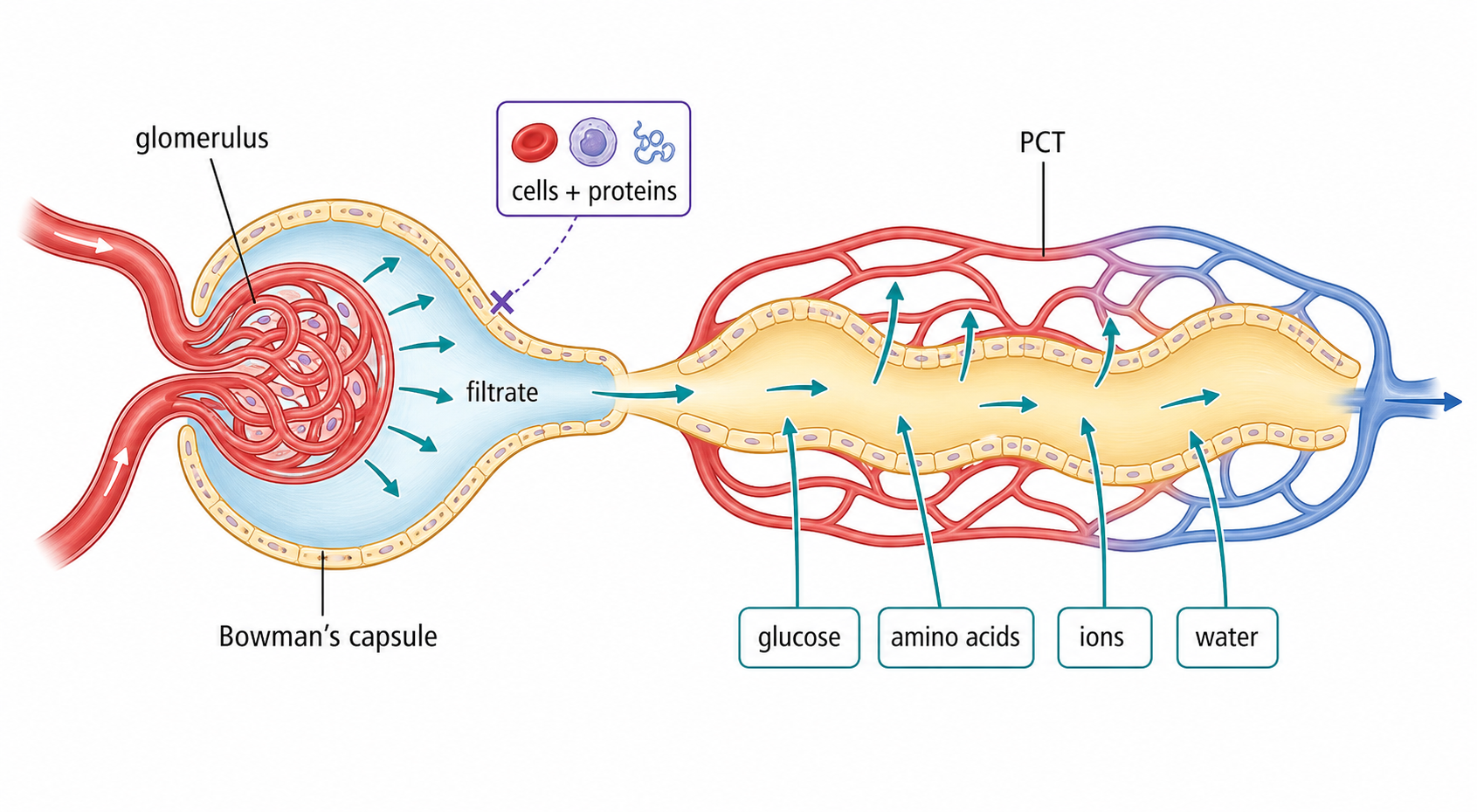
A renal corpuscle diagram shows pressure filtration from the glomerulus into Bowman’s capsule while cells and large proteins remain in blood.

Filtrate Resembles Plasma Until Tubules Select What Returns to Blood

HL only
Substance Blood plasma entering glomerulus Fresh glomerular filtrate Final urine in health
blood cells present absent absent
most plasma proteins present absent or trace absent or trace
glucose and amino acids present present normally reabsorbed, so absent or trace
water and ions present present variable amounts
urea present present concentrated for excretion

Filtration separates mainly by size; selective reabsorption separates by biological value and regulated need. Urine is therefore not simply “filtered blood.”

Glucose in urine can appear when blood glucose is high enough to exceed tubular reabsorptive capacity. Successful diabetes control can restore urine glucose to absent or trace levels.

Proximal-Tubule Cells Are Built for High-Capacity Reabsorption

HL only
Cell feature Contribution to reabsorption
one-cell-thick epithelium short transport distance
apical brush-border microvilli large surface area facing filtrate
many mitochondria ATP supply for active transport
basolateral membrane infoldings large area for pumps and carrier proteins
close capillary network removes reabsorbed substances and maintains gradients

The PCT reabsorbs all normal filtered glucose and amino acids, much sodium and other ions, and much water. Urea is retained more strongly in the tubular fluid.

A proximal convoluted tubule epithelial cell shows apical microvilli, many mitochondria, basolateral membrane folds, sodium–potassium pumps and nearby capillary.

Sodium Pumping Drives Cotransport and Water Reabsorption in the PCT

HL only
  1. Basolateral sodium–potassium pumps use ATP to move Na⁺ from the PCT cell into tissue fluid.
  2. The resulting low intracellular Na⁺ concentration lets Na⁺ enter from the filtrate down its electrochemical gradient.
  3. Na⁺-dependent cotransport carries glucose or amino acids through the apical membrane.
  4. Glucose, amino acids and ions leave across the basolateral side into tissue fluid and capillaries.
  5. Water follows the reabsorbed solutes by osmosis.

Glucose uptake at the apical membrane is secondary active transport: the cotransporter does not use ATP directly, but it depends on the Na⁺ gradient maintained by an ATP-powered pump.

Reabsorption means movement from nephron filtrate back to blood. Secretion is the opposite transfer—from blood or tubule cells into the tubular fluid.

Opposite Permeabilities Let the Loop of Henle Build a Medullary Gradient

HL only
Limb Water permeability NaCl movement Effect on tubular fluid
descending high low solute permeability water leaves by osmosis; fluid becomes more concentrated
thin ascending impermeable NaCl diffuses out fluid becomes less concentrated
thick ascending impermeable Na⁺ and Cl⁻ actively transported out fluid becomes still more dilute; medulla gains solute

The loop’s main function is to establish the hypertonic medulla that later permits water conservation from collecting ducts; it does not respond directly to ADH by inserting aquaporins.
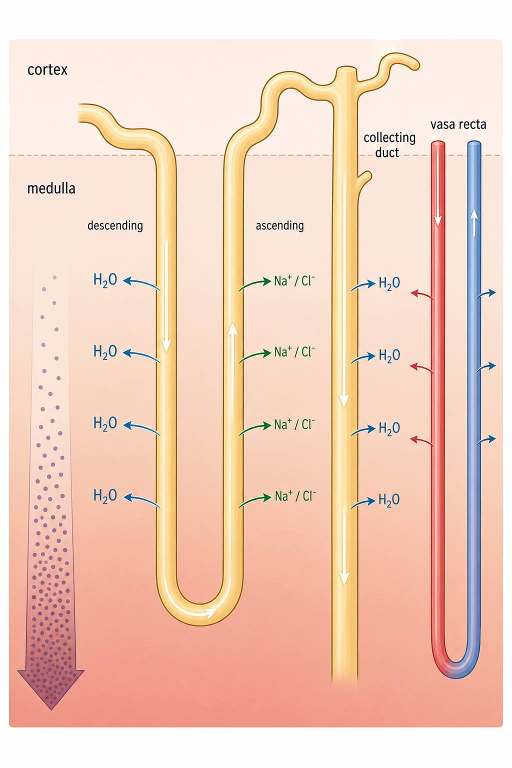
The loop of Henle shows water leaving the descending limb, sodium chloride leaving the water-impermeable ascending limb and increasing medullary concentration toward the hairpin bend.

Countercurrent Flow Multiplies a Small Local Difference Along the Medulla

HL only
  1. Ascending-limb salt transport makes nearby medullary interstitial fluid more concentrated while water stays in that limb.
  2. Water leaves the adjacent descending limb by osmosis until its filtrate approaches the local interstitial concentration.
  3. Continuous flow brings new less-concentrated filtrate into the descending limb and moves concentrated filtrate around the bend.
  4. Repeating the local effect along the loop creates a large cortex-to-inner-medulla gradient.

The vasa recta supplies the loop tissue and carries away reabsorbed water while its countercurrent arrangement limits washout of the gradient.

Longer loops extend farther into the medulla and can support a steeper gradient, increasing the potential to produce concentrated urine.

The Medullary Gradient Creates an Opportunity, Not a Guarantee, for Water Recovery

HL only

The loop of Henle makes interstitial osmotic concentration increase down the medulla. Collecting ducts pass through this gradient, so water has a potential route from tubular fluid into the medulla and then blood.

Condition Collecting-duct permeability Consequence
few aquaporins in apical membrane low most water remains in tubular fluid
many aquaporins in apical membrane high water leaves by osmosis down the medullary gradient

The gradient supplies the driving force; ADH changes the permeability. Neither mechanism alone explains regulated water reabsorption.

High Blood Osmotic Concentration Triggers ADH and Concentrated Urine

HL only
  1. Water loss or high salt intake raises blood osmotic concentration.
  2. Hypothalamic osmoreceptors detect the change; thirst increases and the posterior pituitary releases more ADH.
  3. ADH binds receptors on collecting-duct cells and promotes insertion of aquaporins into the apical membrane.
  4. Collecting ducts become more permeable, so water leaves by osmosis into the hypertonic medulla and returns to blood.
  5. A small volume of concentrated urine is produced; correction reduces the ADH signal.

ADH is synthesized by hypothalamic neurons and stored and released from the posterior pituitary. The posterior pituitary does not synthesize it.
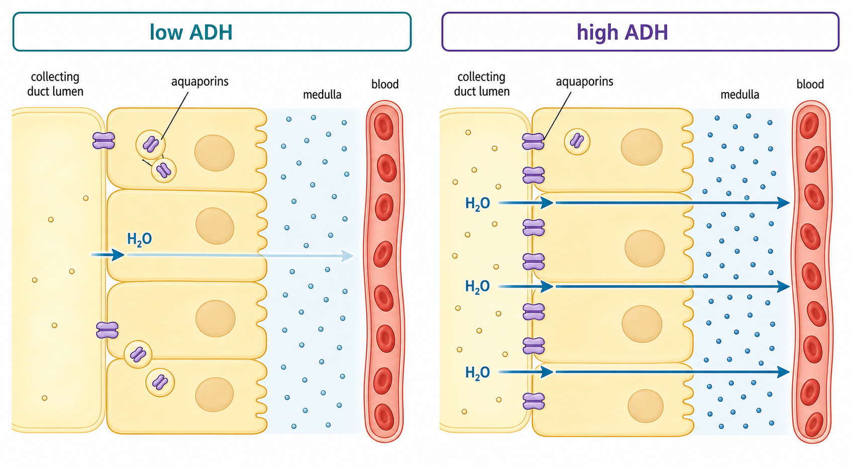
Low- and high-ADH collecting duct cells show aquaporins stored in vesicles or inserted into the apical membrane and the resulting difference in water movement.

Low Blood Osmotic Concentration Reduces ADH and Produces Dilute Urine

HL only
  1. Excess water lowers blood osmotic concentration.
  2. Hypothalamic osmoreceptor stimulation falls, so the posterior pituitary releases less ADH and thirst declines.
  3. Aquaporins are removed from the collecting-duct apical membrane into vesicles.
  4. Collecting ducts become less permeable, so less water leaves the tubular fluid.
  5. A large volume of dilute urine removes excess water; recovery reduces the deviation.
Water status ADH Collecting-duct permeability Urine
dehydrated / salty intake high high small volume, concentrated
excess water low low large volume, dilute

Concentrated urine contains less water per amount of solute; it does not mean the kidney has excreted more total waste in every case.

Arterioles Redistribute a Limited Cardiac Output as Organ Activity Changes

HL only
Physiological state Flow increases toward Flow can decrease toward Reason
vigorous exercise active skeletal muscle, heart and skin when heat loss is needed gut and kidneys oxygen delivery, metabolite removal and thermoregulation take priority
fight or flight skeletal muscle and heart much of the gut epinephrine prepares rapid action
after a meal at rest digestive organs relatively inactive muscle supports secretion, absorption and processing
sleep selected brain regions vary; diaphragm remains supplied most skeletal muscle metabolic demand changes by tissue

Constriction of arterioles raises resistance and reduces flow into a capillary bed; dilation lowers resistance and increases flow. Different vascular beds can respond differently to the same hormone because receptors and local signals differ.

Renal flow may fall during prolonged vigorous exercise, but it cannot remain severely reduced indefinitely because excretion and osmoregulation must continue.

Kidney Homeostasis Separates Filtration, Recovery, Gradient Building and Final Water Control

HL only

renal blood reaches glomerulus → pressure filters water and small solutes → PCT selectively returns useful substances → loop of Henle builds a medullary solute gradient → collecting-duct permeability sets final water recovery → urine leaves while adjusted blood returns to circulation

If asked about… Start with…
cells or proteins absent from filtrate filtration barrier and molecular size
glucose absent from urine selective reabsorption and transport maximum
salty medulla opposite loop-limb permeability and countercurrent multiplication
concentrated urine high ADH plus an existing medullary gradient
organ blood flow during exercise local arteriole resistance and changing metabolic demand

Do not collapse the nephron into one action. Filtration is pressure-driven and non-selective for small solutes; PCT recovery is selective and transport-dependent; the loop builds the gradient; ADH controls how fully collecting ducts use it.

Homeostasis definition

6 marks

Explain how the pH of blood is kept constant during exercise.

Negative feedback loops

4 marks

Discuss the use of positive and negative feedback to control levels of variables.

Blood glucose regulation

8 marks

Explain the control of blood glucose concentrations in humans.

Diabetes exam focus

5 marks

Outline type II diabetes.

Thermoregulation exam focus

8 marks

Explain the control of body temperature in humans.

Thermoregulation mechanisms

1 mark

Outline one change that happens in the human body in response to a rise in body temperature above 36.4∘C36.4^{\circ} \mathrm{C}.

Kidney role

HL only

8 marks

Explain the role of the kidney in osmoregulation.

Glomerulus, Bowman's capsule, PCT

HL only

8 marks

Explain the presence of glucose in the urine of a diabetic person and its absence in the urine of a person with type I diabetes that is being successfully treated.

Loop of Henle

HL only

1 mark

What is the function of the loop of Henle?

Osmoregulation by collecting ducts

HL only

7 marks

Explain the hormonal control of osmoregulation in the kidney by negative feedback.

Blood supply changes

HL only

1 mark

What is a reason for the changes in blood flow during exercise?