DNA replication copies genetic information through template strands, complementary pairing, enzyme action, proofreading, and laboratory amplification or separation techniques used to analyse DNA.
Syllabus
First assessment 2025
Topic
D1.1
Level
HL
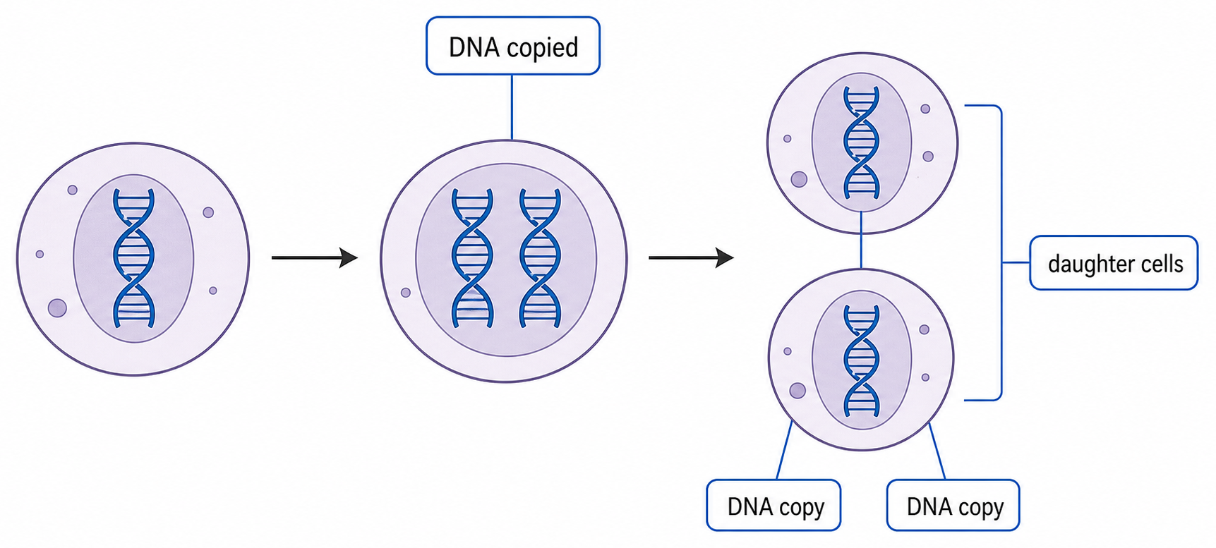
DNA Must Be Copied Before a Cell Divides
DNA replication produces a second DNA molecule with the same base sequence as the original, apart from rare errors.
chromosome DNA → replication during interphase → two DNA copies → cell division → one complete genome enters each daughter cell
Biological context
Why replication is required
reproduction
genetic information passes to new cells or offspring
growth
cell number increases without losing the genome
tissue replacement
new cells inherit the instructions needed for their function
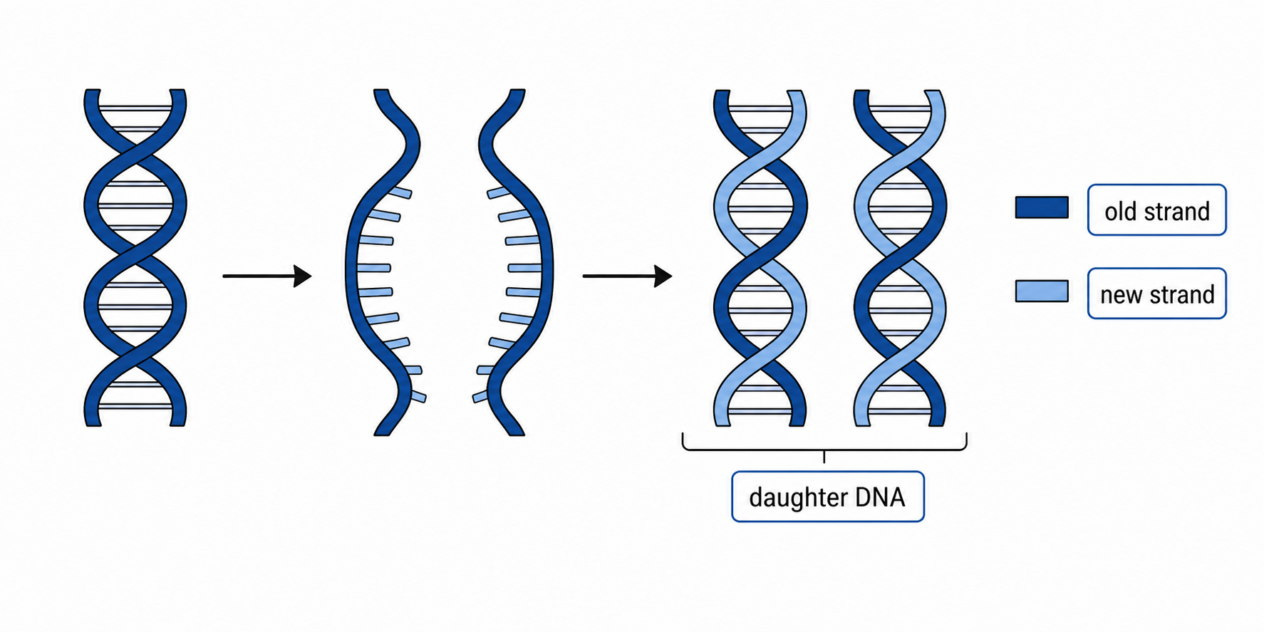
Each Old DNA Strand Templates a New Complement
Exposed template base
New nucleotide added
A
T
T
A
C
G
G
C
The sequence of each original strand determines a complementary new sequence. The two products therefore preserve the original base-pair sequence.
Semi-conservative means that each daughter double helix contains one parental strand and one newly synthesized strand—not that half of every strand is old.
Meselson and Stahl Tested the Strand-Distribution Models
E. coli were first grown with ¹⁵N so their DNA was heavy, then transferred to ¹⁴N medium. DNA from successive generations was separated by density-gradient centrifugation.
Sample
Observed DNA bands
What the result shows
before transfer
one heavy band
all DNA initially contains ¹⁵N
after one replication
one intermediate band
every molecule contains heavy and light material
after two replications
one intermediate + one light band
hybrid molecules persist while fully light molecules appear
The one-generation intermediate band rejects conservative replication, which predicts separate heavy and light bands. The two-generation pattern matches semi-conservative copying.
The isotopes label nitrogen in DNA; they do not make replication radioactive. Both ¹⁵N and ¹⁴N are stable isotopes.
Helicase Exposes Templates; DNA Polymerase Builds Backbones
1
Helicase unwinds the double helix and breaks the hydrogen bonds between complementary bases, exposing two template strands.
2
Free DNA nucleotides align with exposed bases by complementary base pairing.
3
DNA polymerase joins adjacent nucleotides by forming phosphodiester bonds in the sugar–phosphate backbone of each new strand.
Enzyme
Bonds affected
Job
helicase
hydrogen bonds between bases
separates the two strands
DNA polymerase
phosphodiester bonds within a new strand
links nucleotides into a backbone
Helicase does not elongate DNA. It opens the template; DNA polymerase performs strand synthesis.
Reconstruct the Core Replication Argument
need two genomes → helicase separates the original strands → each old strand acts as a template → complementary nucleotides pair → DNA polymerase links the new backbone → two matching double helices form
Feature to check
Correct outcome
sequence
complementary copying preserves the base-pair sequence
strand origin
each product has one old strand and one new strand
biological result
a complete genome can pass to each daughter cell
A model explains how copying could occur; the Meselson–Stahl band pattern provides experimental evidence that the one-old-one-new strand distribution actually occurs.
PCR Selects a DNA Region with Two Primers
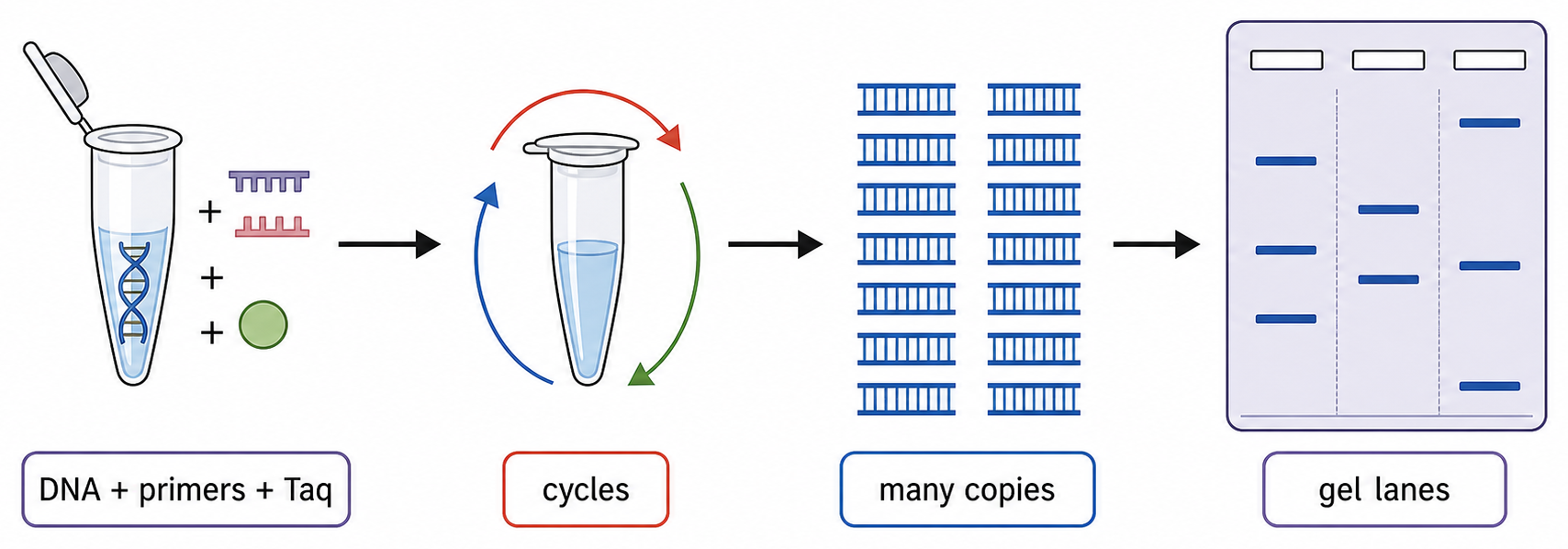
The polymerase chain reaction (PCR) amplifies a selected DNA region in vitro, producing enough copies for detection or analysis.
Component
Job
template DNA
contains the target sequence
two DNA primers
bind on opposite strands at the target's ends and provide free 3′ ends
free DNA nucleotides
become the new strands
Taq polymerase
extends primers and remains active after high-temperature heating
Primer sequences determine which interval is copied. The target's flanking sequences must therefore be known well enough to design complementary primers.
One PCR Cycle Separates, Primes and Extends DNA
1
Denaturation, about 95 °C: hydrogen bonds break and double-stranded DNA separates into templates.
2
Annealing, a lower temperature: primers bind to complementary sequences flanking the target.
3
Extension, about 72 °C: Taq polymerase adds nucleotides to each primer's 3′ end, synthesizing new DNA 5′→3′.
Heating begins the next cycle by separating the newly formed double strands. A thermal cycler repeats the temperature sequence automatically.
Cooling does not make DNA polymerase copy the target by itself. Cooling permits primer binding; polymerase extends only after a primer supplies a paired 3′ end.
Repeated PCR Cycles Amplify—But Not Perfectly
Nn=N0(2n)
In the ideal early cycles, each target molecule produces two copies per cycle. Starting from one target, 30 perfect doublings would give about one billion copies.
Taq polymerase comes from the thermophilic bacterium Thermus aquaticus. Its heat stability prevents the enzyme from being destroyed during repeated denaturation.
Limitation
Consequence
primers bind a similar non-target sequence
the wrong interval can be amplified
contamination supplies foreign DNA
contaminant DNA can also be copied
reagents become limiting and products reanneal
later cycles fall below ideal doubling
Taq lacks proofreading
some newly copied molecules contain errors
An Electric Field Separates DNA Fragments through a Gel
DNA samples are loaded into wells near the negative electrode of a porous agarose gel immersed in conducting buffer.
Phosphate groups give DNA a net negative charge, so fragments migrate through the gel toward the positive electrode when voltage is applied.
Fragment
Movement through gel pores
Final tendency after the same time
shorter
less hindered; moves faster
farther from the wells
longer
more hindered; moves slower
nearer the wells
DNA is colourless. A stain or labelled probe is needed to reveal separated fragments as bands.
For DNA fragments, charge gives a common migration direction; fragment length is the main variable responsible for separation through the gel.
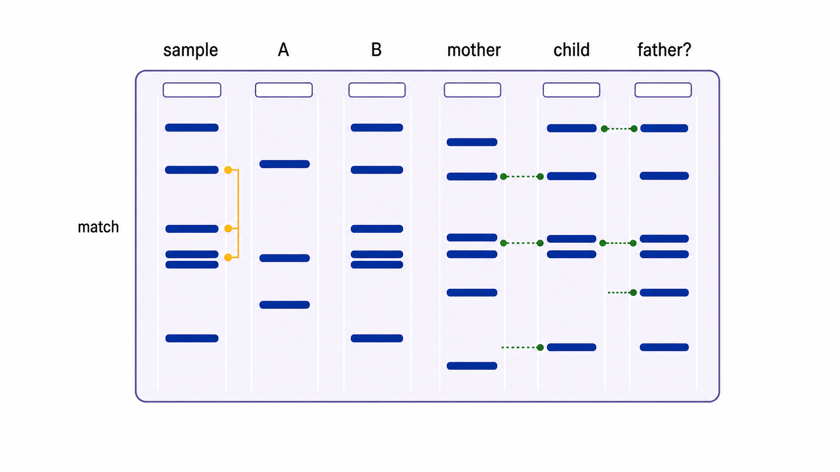
Band Position Reports Fragment Length; Alignment Reports a Match
Observation
Interpretation
band farther from wells
shorter DNA fragments
bands at the same height in two lanes
fragments of the same measured length
more intense band
more stained DNA at that position, not a longer fragment
Compare lanes horizontally, one band position at a time. A profile comparison uses a pattern across many variable markers, not one matching band.
A same-height band supports a shared fragment length; it does not by itself prove that every base in the fragments is identical.
DNA Profiles Compare Several Variable Markers
collect and preserve samples → extract DNA → amplify selected STR or VNTR markers by PCR → separate products by electrophoresis → compare band or peak patterns
Result
Supported conclusion
Not established by the profile alone
questioned sample differs at a reliable marker
that reference individual is excluded
who left the sample
all tested markers match
the samples may share a source
when or how the DNA arrived
Match strength increases when more independent, variable markers agree and when the matching pattern is rare in the relevant population.
Contamination, mixed samples, degraded DNA and laboratory error can weaken the inference. Controls and careful sample handling are part of the evidence.
A Child's Markers Must Be Accounted for by Both Parents
At each tested marker, a child inherits one allele from the biological mother and one from the biological father.
Start with the child's pattern → account for bands or alleles shared with the known parent → compare every remaining child marker with the possible other parent.
Comparison
Inference
a required child marker is absent from the possible parent
exclude that person at that marker, after checking data quality
all non-maternal child markers are present in the possible father
parentage is supported, with strength depending on marker number and frequencies
Close relatives share more DNA than unrelated people. Parentage conclusions use multiple markers and probability, not a visual impression from one or two bands.
Follow DNA from a Tiny Sample to a Qualified Conclusion
Stage
Question answered
Key control
primer design
which region will be copied?
complementary, target-specific flanking sequences
PCR
is there enough target DNA to analyse?
positive and negative controls; contamination prevention
electrophoresis
what fragment lengths are present?
ladder and correct electrode orientation
profile comparison
are patterns consistent with a shared source or inheritance?
enough informative markers and relevant frequencies
PCR changes quantity; electrophoresis changes position; interpretation changes neither. Each step supports a different claim.
A laboratory match is evidence of consistency. The final conclusion must remain limited by sample quality, contamination risk, marker informativeness and the biological question being tested.
DNA Polymerase Extends a Strand Only at Its 3′ End
HL only
The two DNA strands are antiparallel: one runs 5′→3′ while its complement runs 3′→5′.
DNA polymerase forms a phosphodiester bond between the growing strand's free 3′-OH and the incoming nucleotide. New DNA therefore grows only 5′→3′.
Polymerase action
Direction
reads template
3′→5′
synthesizes new strand
5′→3′
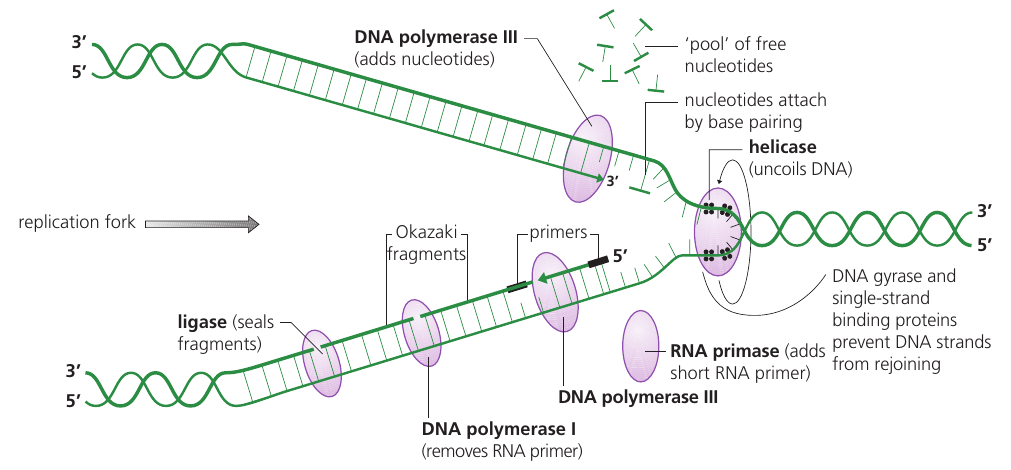
Antiparallel Templates Force Two Replication Patterns
HL only
Both templates are exposed at the same fork, but DNA polymerase can extend each new strand only 5′→3′.
On one template, 5′→3′ synthesis follows the advancing fork, so polymerase can remain behind helicase and synthesize continuously.
On the opposite template, 5′→3′ synthesis points away from the fork. Newly exposed template must be copied in short sections that begin repeatedly near the fork.
The lagging strand is discontinuous because of antiparallel geometry and polymerase directionality—not because its polymerase is slower.
Leading Is Continuous; Lagging Uses Okazaki Fragments
HL only
Feature
Leading strand
Lagging strand
relation to fork movement
synthesis proceeds toward the fork
each fragment is synthesized away from the fork
synthesis pattern
continuous
discontinuous
RNA primers at one fork
one initial primer
repeated primers
DNA product before processing
one continuous new strand
multiple Okazaki fragments
ligase requirement
not required to join Okazaki fragments
joins processed fragments
Both new strands are synthesized simultaneously and both grow 5′→3′. The difference is continuous versus fragment-by-fragment synthesis.
The leading strand also needs an RNA primer to begin. Repeated priming is distinctive of the lagging strand.
Primase Starts; DNA Polymerase III Extends
HL only
1
DNA polymerase III cannot start a strand from unlinked nucleotides; it needs an existing paired chain with a free 3′-OH.
Primase is an RNA polymerase that synthesizes a short RNA primer complementary to the DNA template. It can start without a pre-existing 3′ end.
2
DNA polymerase III binds at the primer's 3′ end and adds complementary DNA nucleotides, extending the new strand 5′→3′.
New strand
Primase action
DNA polymerase III action
leading
makes one initial primer
extends continuously
lagging
makes a new primer for each fragment
extends each Okazaki fragment
DNA Polymerase I Replaces Primers; Ligase Seals Nicks
HL only
1
DNA polymerase I removes each RNA primer and replaces its RNA nucleotides with DNA nucleotides.
After replacement, adjacent DNA sections contain the correct nucleotides but remain separated by a nick: one missing phosphodiester bond in the sugar–phosphate backbone.
2
DNA ligase catalyses formation of the missing phosphodiester bond, joining adjacent DNA fragments into one continuous strand.
Enzyme
Changes nucleotide identity?
Joins a final backbone gap?
DNA polymerase I
yes—RNA is replaced with DNA
leaves a nick between sections
DNA ligase
no
yes—seals the nick
Replication Enzymes Hand the Growing DNA from One Job to the Next
HL only
1
Helicase: separates the parental strands at the replication fork.
2
Primase: makes an RNA primer that supplies a free 3′-OH.
3
DNA polymerase III: extends complementary DNA from the primer.
4
DNA polymerase I: removes the RNA primer and replaces it with DNA.
5
DNA ligase: seals the remaining nick between DNA sections.
Material present
Next enzyme needed
paired template but no starting chain
primase
RNA primer with free 3′ end
DNA polymerase III
RNA segment embedded before DNA
DNA polymerase I
adjacent DNA sections with a nick
DNA ligase
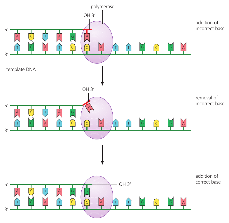
DNA Polymerase III Proofreads the Growing 3′ End
HL only
1
A newly added nucleotide that does not pair correctly distorts the polymerase–DNA structure and stalls extension.
2
DNA polymerase III shifts the 3′ end to its proofreading site, where exonuclease activity removes the mismatched nucleotide.
3
The corrected 3′ end returns to the polymerizing site; the complementary nucleotide is added and 5′→3′ synthesis resumes.
Proofreading greatly reduces replication errors and therefore mutation rate, but it cannot make replication completely error-free.
Solve a Replication Fork from Direction to Fidelity
HL only
Clue at the fork
Consequence
polymerase can add only to a 3′ end
every new segment grows 5′→3′
synthesis can follow helicase
leading strand is continuous
synthesis must restart as template is exposed
lagging strand uses repeated primers and Okazaki fragments
an RNA primer remains
DNA polymerase I replaces it with DNA
two DNA sections meet at a nick
ligase seals the phosphodiester bond
the terminal base is mismatched
DNA polymerase III removes and replaces it
helicase opens → primase starts → DNA polymerase III extends → DNA polymerase I replaces RNA → ligase seals → polymerase III proofreading corrects many terminal mismatches
Directionality creates the leading–lagging difference. Enzyme handoffs solve that geometry, while complementary pairing plus proofreading preserves sequence fidelity.
DNA replication
[ 8 ]
Growth in living organisms includes replication of DNA. Explain DNA replication.
Semi-conservative replication
[ 3 ]
Outline the reason that DNA replication is described as semi-conservative.
Role of helicase and DNA polymerase
[ 1 ]
What is a function of the enzyme helicase?
PCR and gel electrophoresis
[ 4 ]
Describe the polymerase chain reaction (PCR).
Applications exam focus
[ 4 ]
Outline the process of DNA profiling.
DNA polymerase directionality
HL only
[ 1 ]
How does DNA replicate?
Leading vs. lagging strand
HL only
[ 1 ]
What is a difference between the leading and lagging strands in DNA replication?
Functions in replication
HL only
[ 3 ]
Describe the function of three named enzymes involved in DNA replication.
DNA proofreading
HL only
[ 4 ]
Explain how mutation is avoided during DNA replication.