EduNinja
(a)

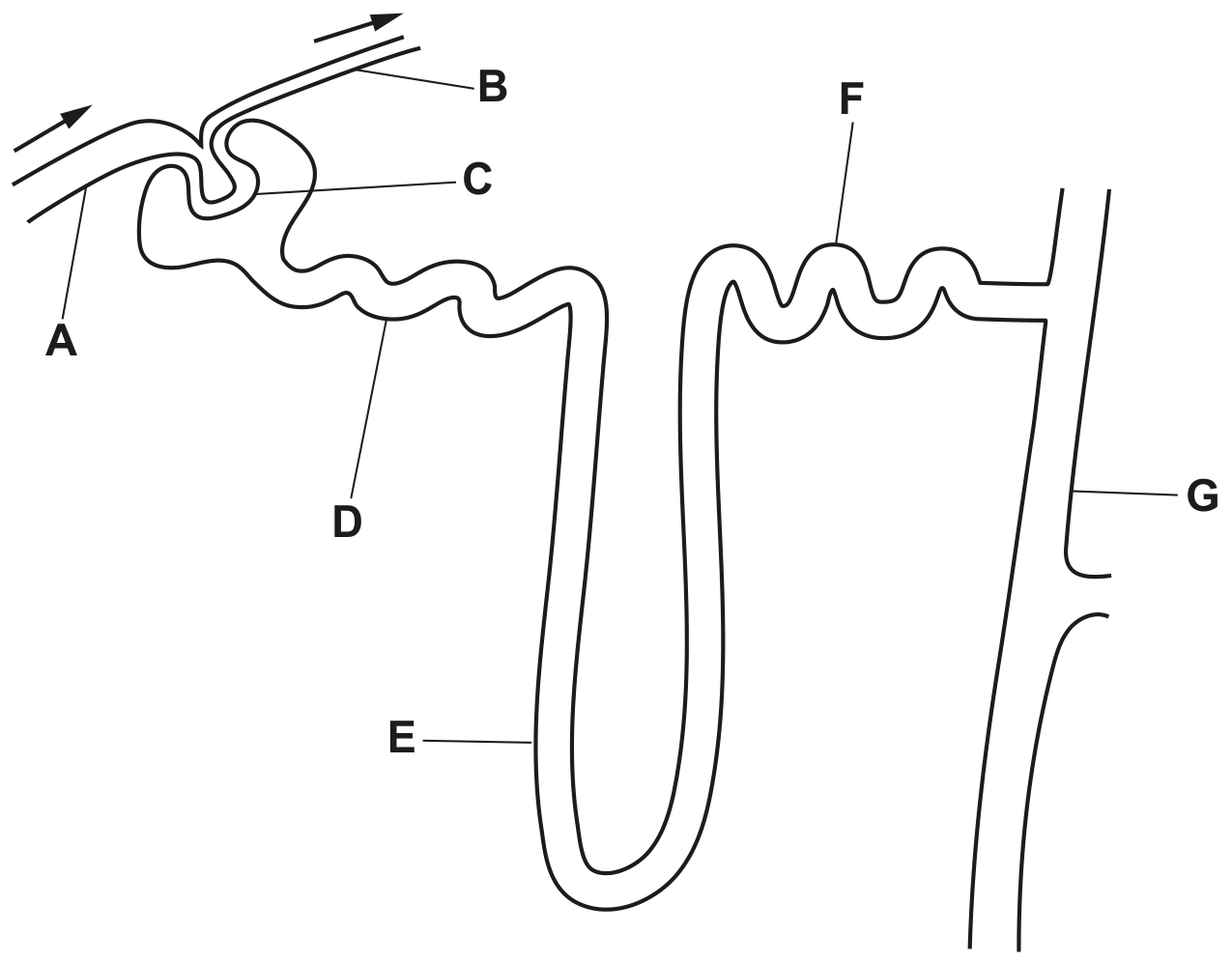
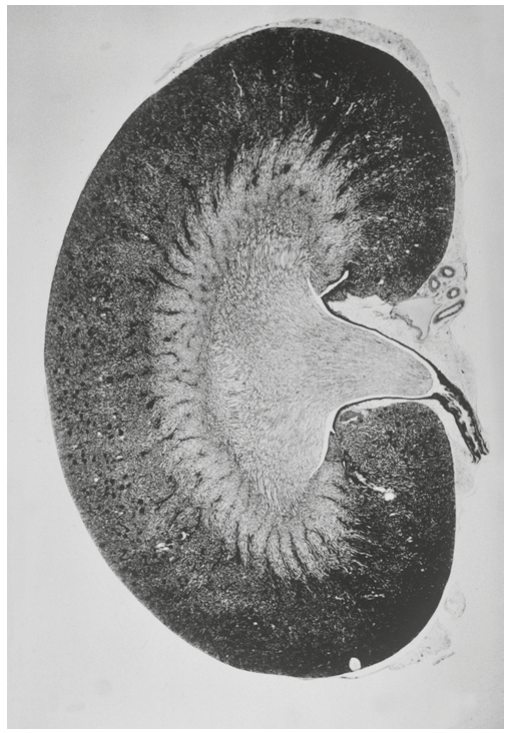
Fig. 1.1 shows a section through a kidney.
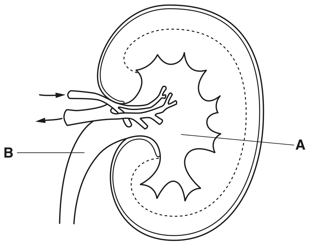
Fig. 1.1

Fig. 1.1

[ 5 ]
(i)

With reference to Fig. 1.1, name structures A and B.

A

B

[ 2 ]
(ii)

On Fig. 1.1, use label lines and letters to label where:

U - ultrafiltration occurs

L - the loop of Henle is found
C-blood urea concentration is low.

[ 3 ]
(a)

The water potential of mammalian blood needs to be maintained within narrow limits so that cells function efficiently. This process is called osmoregulation.

The relative medullary thickness (RMT) indicates the proportion of a kidney that is composed of medullary tissue.

 RMT = thickness of medulla  kidney size ×10\text { RMT }=\frac{\text { thickness of medulla }}{\text { kidney size }} \times 10

Table 1.1 shows the relationship between the RMT and the concentration of urine produced by four mammals from different habitats.

Table 1.1

Table 1.1

[ 2 ]
(i)

Name the parts of the nephron that are located in the medulla.

[ 2 ]
(a)

Fig. 1.1 shows a cell of one of the collecting ducts of the kidney.

Fig. 1.1

Fig. 1.1

Name membrane protein A and cell structure B.
A
B

[ 2 ]
(a)

Fig. 1.1 is a diagram of a kidney nephron and some of its blood vessels.

Fig. 1.1

Fig. 1.1

With reference to Fig. 1.1, complete Table 1.1 using the letters A - G.
Each letter may be used once, more than once or not at all.

Table 1.1

Table 1.1

[ 4 ]
(a)

The Bowman's capsule of a nephron is involved in ultrafiltration.
Fig. 1.1 is a diagram of part of a Bowman's capsule and glomerulus.

Fig. 1.1

Fig. 1.1

With reference to Fig. 1.1, complete Table 1.1 using the letters A - F.
Each letter may be used once, more than once or not at all.

Table 1.1

Table 1.1

[ 4 ]
(a)

Fig. 1.1 is a diagram of a kidney nephron and some of its blood vessels.

Fig. 1.1

Fig. 1.1

With reference to Fig. 1.1, complete Table 1.1 using the letters A - G.
Each letter may be used once, more than once or not at all.

Table 1.1

Table 1.1

[ 4 ]
(a)

Fig. 6.1 shows part of the Bowman's capsule of a kidney nephron.

Fig. 6.1

Fig. 6.1

Name structures A, B and C.
A

B

C

[ 3 ]
(a)

Fig. 5.1 is a photomicrograph of part of the cortex of a kidney.

Fig. 5.1

Fig. 5.1

[ 2 ]
(i)

On Fig. 5.1, use label lines and letters to label:

G - the glomerulus
L - the lumen of the Bowman's (renal) capsule.

[ 2 ]
(a)

Fig. 5.1 is a photograph of a section through a kidney.

Fig. 5.1

Fig. 5.1

On Fig 5.1, use label lines and letters to label the regions of the kidney where the following are located:

L - loop of Henlé

D - distal convoluted tubule

P - podocyte cells.

[ 3 ]
(a)

During contraction, muscles use up ATP very quickly. For a short period of time, ATP can be resynthesised using creatine phosphate, as shown in Fig. 5.1.

 ADP + creatine phosphate  ATP + creatine \text { ADP + creatine phosphate } \longrightarrow \text { ATP + creatine }

The creatine formed as a result of the resynthesis of ATP is converted to creatinine. Creatinine production in the body stays fairly constant. Creatinine becomes part of the glomerular filtrate during ultrafiltration in the kidney nephrons.

[ 1 ]
(i)

Name the main filtration barrier in the nephron that allows creatinine to pass into the Bowman's capsule but stops red blood cells from passing through.

[ 1 ]
0