C2.2 Neural signalling

Neural signalling transmits information through neuron structure, ion gradients, action potentials, synapses and chemical modulation of postsynaptic responses, linking cellular mechanisms to rapid communication in nervous systems.

Syllabus
First assessment 2025
Topic
C2.2
Level
HL

Learning objectives

C2.2.1Neurons• Neurons carry electrical impulses in the nervous system• Motor, sensory, and relay neurons differ in axon, dendrite, and cell body arrangementC2.2.2Resting potential generation• Sodium-potassium pumps use ATP to move 3 Na⁺ out and 2 K⁺ in• Ion gradients make the resting axon polarized at about -70 mVC2.2.3Nerve impulses as action potentials• A nerve impulse is a propagated action potential along a nerve fibre• Stimulus-triggered sodium influx reverses membrane polarityC2.2.4Variation in impulse speed• Larger axon diameter lowers resistance and increases impulse speed• Myelin sheaths and nodes of Ranvier enable faster saltatory conductionC2.2.5Synapses as junctions• Synapses connect neurons to neurons, muscles, or glands• Chemical synapses transmit one way across a narrow synaptic cleftC2.2.6Neurotransmitter release• Action potentials open voltage-gated Ca²⁺ channels in presynaptic terminals• Ca²⁺ causes vesicle fusion and neurotransmitter exocytosis into the cleftC2.2.7Excitatory postsynaptic potential• Neurotransmitters diffuse and bind receptors on the postsynaptic membrane• EPSPs depolarize the membrane and make threshold more likelyC2.2.8(HL)—Depolarization and repolarization• Threshold opens voltage-gated Na⁺ channels, causing rapid depolarization• Voltage-gated K⁺ channels then repolarize or briefly hyperpolarize the axonC2.2.9(HL)—Action potential propagation• Local currents from Na⁺ diffusion depolarize the next axon region• Refractory regions behind the impulse help ensure one-way propagationC2.2.10(HL)—Oscilloscope traces• Oscilloscope traces show resting potential, threshold, and action potential phases• Stimulus intensity is encoded by impulse frequency, not action potential sizeC2.2.11(HL)—Saltatory conduction• Myelin insulates axons and ion exchange occurs mainly at nodes of Ranvier• Saltatory conduction jumps node to node and greatly increases speedC2.2.12(HL)—Exogenous chemicals• Exogenous chemicals can mimic, block, or prolong neurotransmitter effects• Neonicotinoids bind insect acetylcholine receptors; cocaine blocks dopamine reuptakeC2.2.13(HL)—Inhibitory neurotransmitters• Inhibitory neurotransmitters open Cl⁻ entry or K⁺ exit channels• Hyperpolarization makes threshold harder to reachC2.2.14(HL)—Summation• Postsynaptic neurons integrate EPSPs and IPSPs from many synapses• Temporal and spatial summation determine whether threshold is reachedC2.2.15(HL)—Pain perception• Pain uses free nerve endings with TRP ion channels in skin and other tissues• Heat, acid, capsaicin, or tissue damage can trigger impulses perceived as painC2.2.16(HL)—Consciousness• Consciousness emerges from coordinated activity across interacting brain regions• EEG, MRI, and fMRI provide evidence for neural correlates of conscious processing

Neuron Shape Matches the Direction of Information Flow

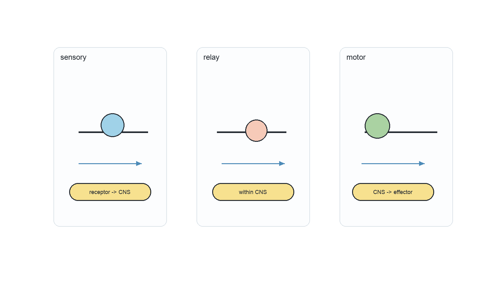
Neuron Information route Decisive structural pattern
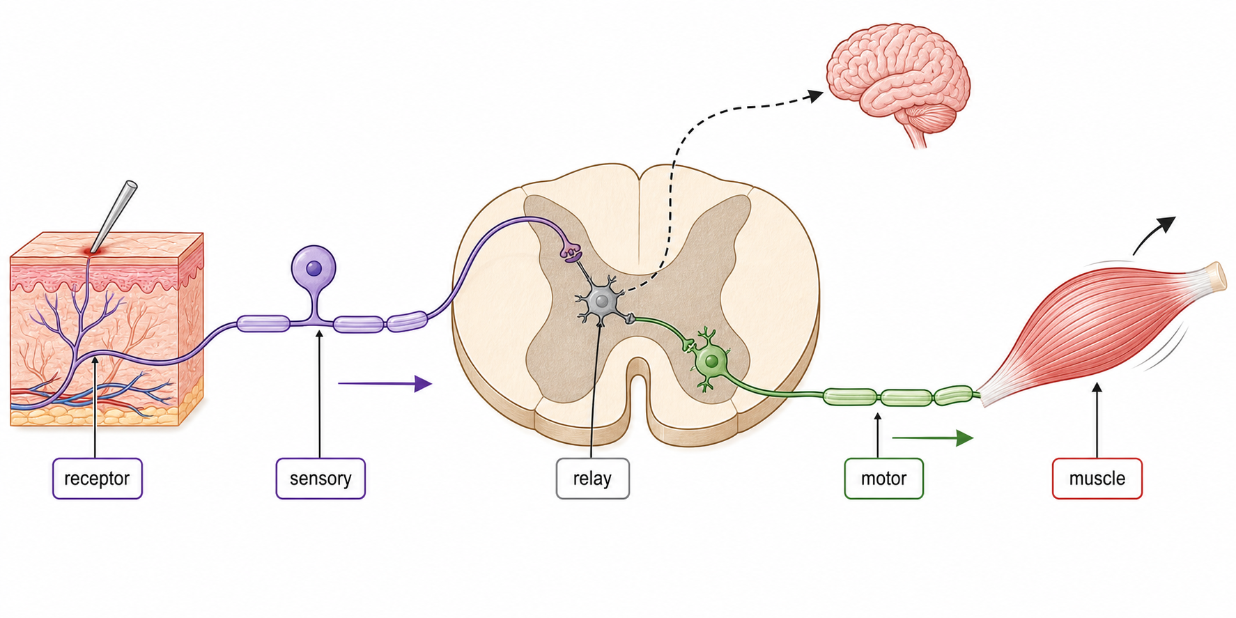
sensory receptor → CNS long dendron toward an off-centre cell body; axon continues into CNS
relay within CNS many short dendrites and a short axon connect neurons locally
motor CNS → muscle or gland many dendrites around the cell body; one long axon reaches an effector
Sensory, relay and motor neurons compared by cell-body position, fibre arrangement and direction of impulse flow.

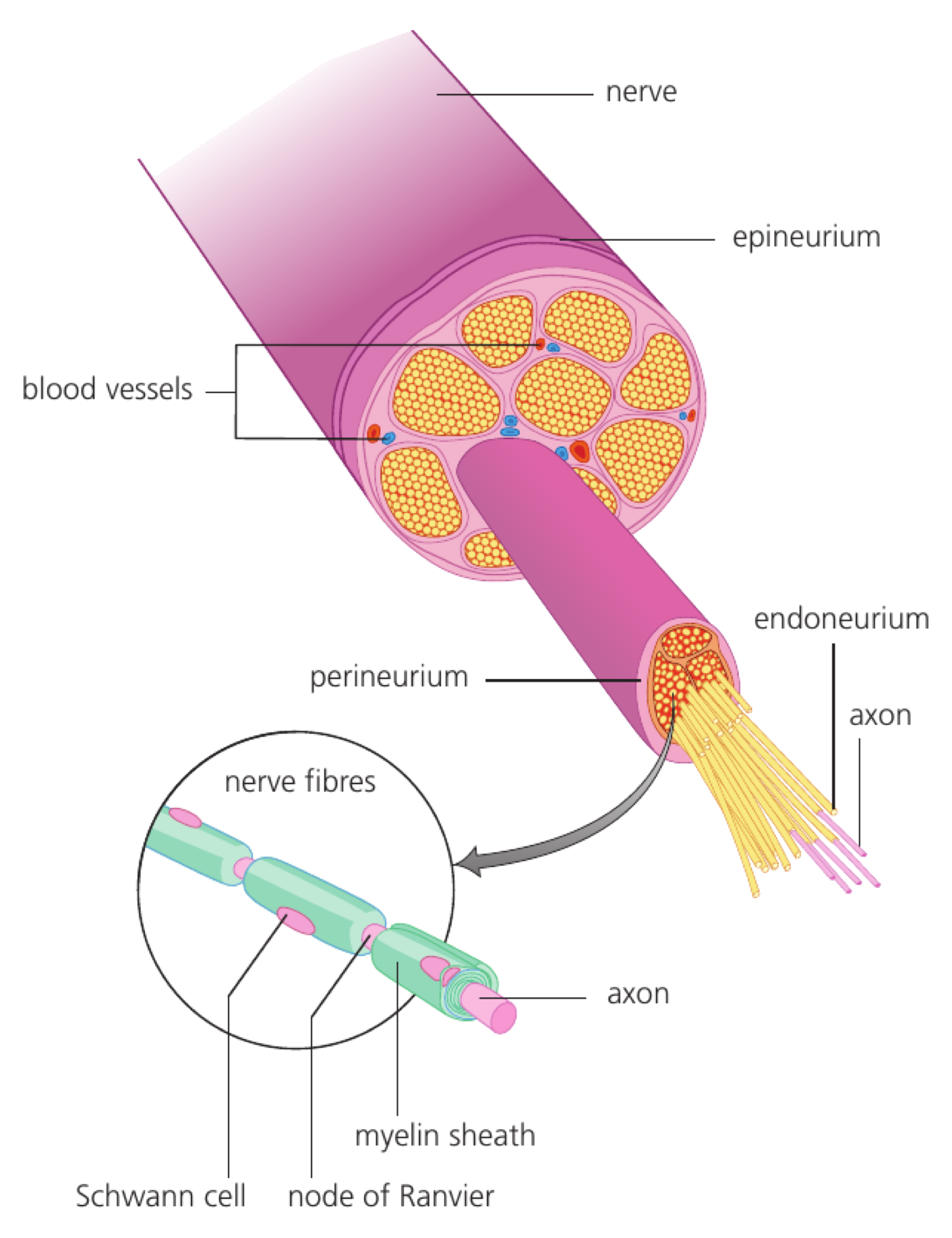
Dendrites and dendrons carry impulses toward the cell body; an axon carries impulses away from it. A nerve is a bundle of many nerve fibres, not one neuron.

Resting Potential Stores Energy in Unequal Ion Distributions

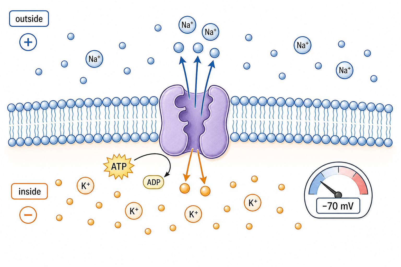
At rest, an axon's inside is about −70 mV relative to the outside. The membrane is polarized because ions and large negative solutes are distributed unequally across it.

The sodium–potassium pump hydrolyses ATP to move 3 Na⁺ out for every 2 K⁺ in, against both concentration gradients. It establishes and maintains high Na⁺ outside and high K⁺ inside.

At rest the membrane is much more permeable to K⁺ than to Na⁺. K⁺ leakage leaves behind impermeant negative charge, so the inside becomes negative. The pump maintains the gradients that this leakage continually uses.

A resting axon is shown with a negative interior relative to the positively charged exterior.

A Nerve Impulse Is a Regenerated Action Potential

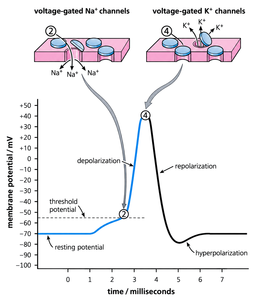
An action potential is a brief reversal of membrane potential, typically from about −70 mV to about +40 mV. A nerve impulse is this action potential repeatedly regenerated along a nerve fibre.

  • Below threshold, the membrane returns to rest without firing.
  • At or above threshold, a full action potential occurs.
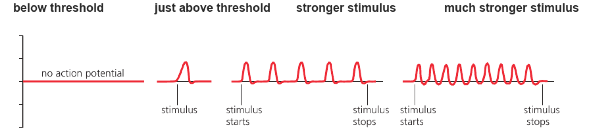
  • Stronger stimuli do not make larger action potentials; they produce a higher frequency of standard-sized impulses.
Traces show no action potential below threshold, one standard-sized action potential just above threshold, and higher impulse frequency with stronger sustained stimuli.

Ion flow through open channels is passive, down electrochemical gradients. ATP is used by pumps to maintain and restore those gradients, not to push each ion through an open channel.

Axon Design Sets the Speed of an Impulse

Structural change Immediate physical effect Consequence for speed
larger axon diameter lower resistance to local current inside the axon adjacent membrane reaches threshold sooner
myelin sheath raises membrane resistance and limits ion exchange to nodes of Ranvier local current travels farther before another action potential is generated

Within both myelinated and unmyelinated groups, conduction velocity increases with diameter. At the same diameter, a myelinated axon is much faster; giant unmyelinated axons partly compensate through size.

Diameter and myelination are independent adaptations. Neither changes the amplitude of each action potential; they change how quickly the next region reaches threshold.

A Chemical Synapse Has a Sender and a Receiver

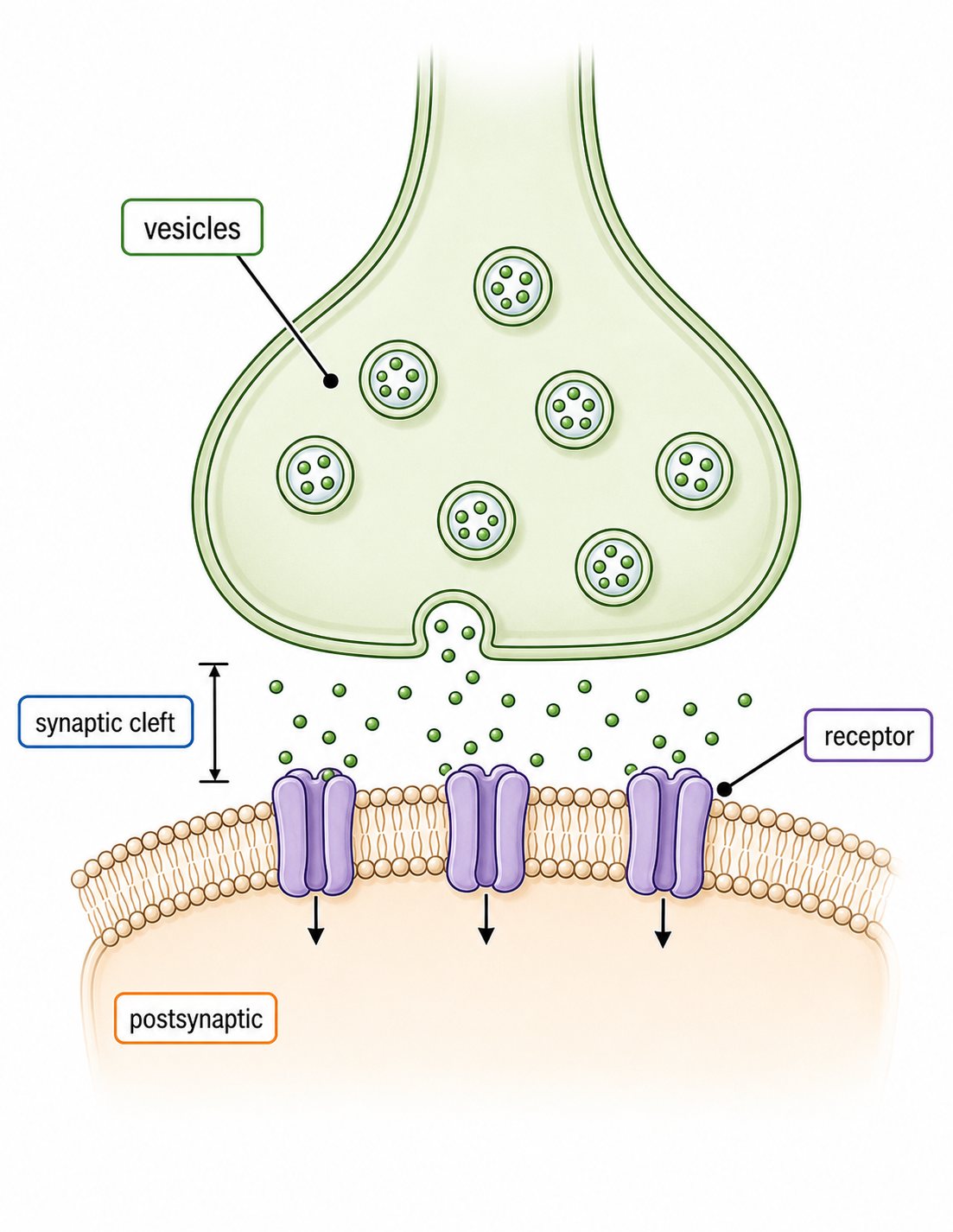
  • The presynaptic terminal stores neurotransmitter in vesicles and releases it.
  • The synaptic cleft is a narrow extracellular gap, typically about 20 nm.
  • The postsynaptic membrane carries receptors that convert the chemical signal into a membrane response.

Transmission is normally one-way because release machinery is presynaptic while matching receptors are postsynaptic. Synapses can connect neuron to neuron, neuron to muscle, or neuron to a gland cell.

A presynaptic terminal contains neurotransmitter vesicles above a narrow synaptic cleft and a postsynaptic membrane bearing receptor proteins.

Calcium Converts an Arriving Impulse into Secretion

  1. An action potential depolarizes the presynaptic terminal.
  2. Voltage-gated Ca²⁺ channels open.
  3. Ca²⁺ diffuses into the terminal down its electrochemical gradient.
  4. Ca²⁺ triggers neurotransmitter vesicles to dock and fuse with the presynaptic membrane.
  5. Exocytosis releases neurotransmitter, creating a high concentration beside the cleft.

This is an electrical → chemical conversion. Ca²⁺ acts as an intracellular signal that couples membrane depolarization to vesicle fusion.

Presynaptic structure Contribution
mitochondria supply ATP for vesicle cycling and ion-gradient maintenance
Golgi apparatus and vesicles process, package and store transmitter components
presynaptic membrane provides voltage-gated Ca²⁺ channels and the fusion surface

An EPSP Moves the Postsynaptic Membrane toward Threshold

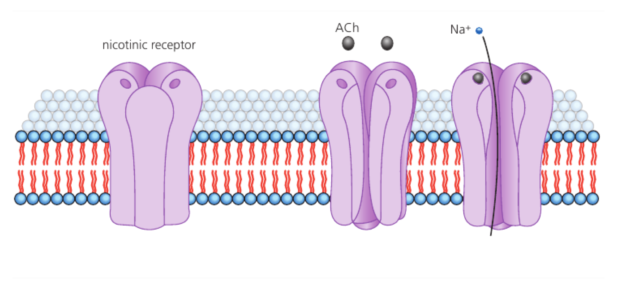
neurotransmitter diffuses across the cleft → binds a complementary postsynaptic receptor → ligand-gated cation channels open → Na⁺ entry depolarizes the membrane → an excitatory postsynaptic potential (EPSP) forms

An EPSP is graded: more transmitter can produce a larger local depolarization. It makes an action potential more likely, but only a summed depolarization reaching threshold triggers the all-or-none spike.

Acetylcholine binding opens a postsynaptic nicotinic receptor channel and allows sodium ions to enter.

The signal must be removed. At cholinergic synapses, acetylcholinesterase hydrolyses acetylcholine; receptors close, and choline can be taken up for reuse. Other transmitters may be removed by reuptake or diffusion.

SL Summary: Trace the Signal into the Next Cell

Within a neuron: ion gradients create a resting potential; threshold opens a regenerative membrane event; axon diameter and myelin change how quickly it travels.
Across a synapse: arriving depolarization opens Ca²⁺ channels → Ca²⁺ triggers exocytosis → transmitter diffuses → postsynaptic receptors open channels → an EPSP moves the next cell toward threshold.

If transmission fails here… Inspect…
resting voltage cannot be maintained ATP supply, Na⁺/K⁺ pump and membrane permeability
presynaptic impulse arrives but no transmitter appears voltage-gated Ca²⁺ channels and vesicle fusion
transmitter reaches the cleft but no EPSP forms receptor match, postsynaptic channel and transmitter removal

Channel Timing Draws the Action-Potential Trace

HL only
Trace phase Main channel event Ion movement and voltage change
threshold and rising phase voltage-gated Na⁺ channels open rapidly Na⁺ enters; positive feedback causes steep depolarization
peak Na⁺ channels inactivate; delayed K⁺ channels are open inward current falls and outward K⁺ current dominates
falling phase voltage-gated K⁺ channels remain open K⁺ leaves; membrane repolarizes
undershoot K⁺ channels close slowly continued K⁺ exit briefly hyperpolarizes the membrane
An action-potential trace links sodium-channel opening to depolarization and potassium-channel opening to repolarization and hyperpolarization.

The Na⁺/K⁺ pump maintains the long-term gradients, but it does not cause the rapid falling phase. Repolarization is mainly produced by K⁺ leaving through voltage-gated channels.
Local Current Regenerates the Impulse Ahead

HL only

Na⁺ entry makes one axon region positive inside. Positive charge spreads through the axoplasm toward the adjacent negative region, while return current flows outside the axon. This local current depolarizes the next membrane region.

If the adjacent region reaches threshold, its voltage-gated Na⁺ channels open and a new full-sized action potential forms. The signal is regenerated rather than a single electrical pulse fading with distance.

Sodium entering a depolarized axon region creates local current toward the adjacent polarized membrane.

The Refractory Region Prevents Backward Regeneration

HL only

Immediately after an action potential, voltage-gated Na⁺ channels are inactivated and cannot reopen until the membrane has repolarized. This interval is the absolute refractory period.

Local current spreads both ways, but only the membrane ahead is excitable. The region behind has just fired and is refractory, so the action potential normally regenerates in one direction.

During the later relative refractory period, the membrane is hyperpolarized and a stronger stimulus is needed. Refractory time therefore also places an upper limit on action-potential frequency.

Refractoriness is primarily a channel-state phenomenon. Restoring bulk Na⁺ and K⁺ concentration gradients is slower and is maintained by the Na⁺/K⁺ pump over many impulses.

Read an Oscilloscope Trace as Membrane Events

HL only

An oscilloscope trace plots membrane potential (mV) on the y-axis against time (ms) on the x-axis. A flat line near −70 mV is resting potential; each spike is one action potential recorded at the electrode.

Trace evidence Inference
gradual rise toward threshold graded depolarization is summating
steep rise after threshold regenerative Na⁺ influx
fall from the peak Na⁺ channels inactivate and K⁺ exits
brief dip below rest K⁺ channels close slowly; hyperpolarization
more equal-height spikes per second a stronger or more persistent stimulus is encoded by frequency

Compare spike number and spacing, not spike height, when judging stimulus intensity. Once threshold is reached, action-potential amplitude is all-or-none.

Myelin Makes Propagation Saltatory

HL only
  • Myelin electrically insulates internodes and reduces current leakage across their membranes.
  • Voltage-gated channels are concentrated at exposed nodes of Ranvier.
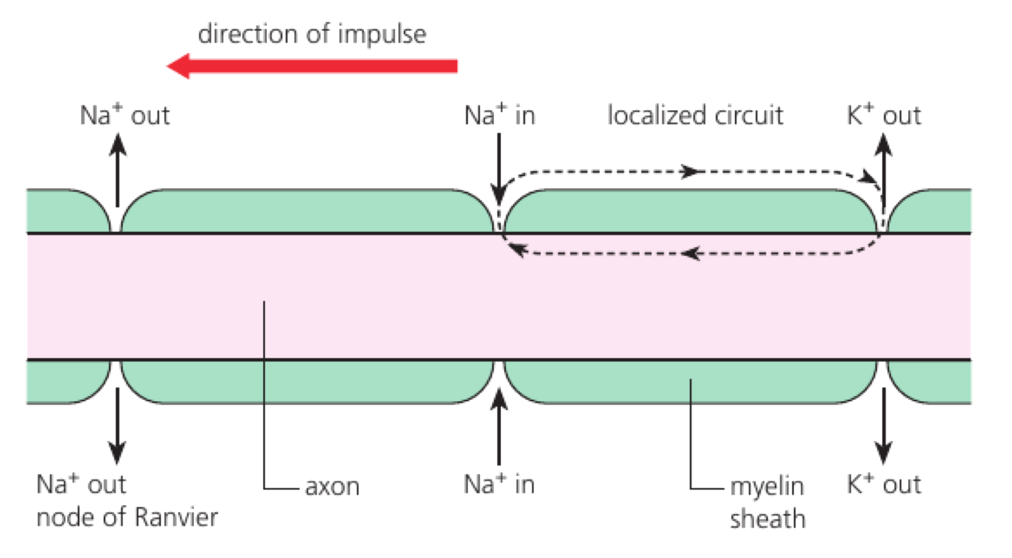
  • Local current travels rapidly beneath the myelin and depolarizes the next node.
  • A new action potential forms at that node, so conduction appears to jump node to node.

Saltatory conduction is faster and more energy-efficient than regenerating an action potential at every adjacent patch of membrane, because far less membrane exchanges ions.

Local current travels beneath myelin from one node of Ranvier to the next, where sodium entry regenerates the action potential.

Exogenous Chemicals Disrupt Different Synaptic Steps

HL only

An exogenous chemical originates outside the body. At a synapse it can mimic a transmitter, block a receptor, change release, prevent breakdown, or block reuptake.

Chemical Molecular target Immediate synaptic effect
neonicotinoid insect nicotinic acetylcholine receptor binds persistently and disrupts cholinergic transmission; acetylcholinesterase cannot remove it
cocaine dopamine reuptake transporter dopamine remains in the cleft longer, so postsynaptic signalling is prolonged and amplified
Cocaine blocks dopamine reuptake transporters, leaving more dopamine in the synaptic cleft to bind postsynaptic receptors.

The same behavioural label—‘stimulant’—does not identify a molecular mechanism. Predict the effect by locating the chemical's target in the release–receptor–removal cycle.

An IPSP Moves the Membrane Away from Threshold

HL only

At an inhibitory synapse, transmitter binding opens channels that make the postsynaptic interior more negative. The resulting inhibitory postsynaptic potential (IPSP) reduces the probability of reaching threshold.

Open channel Ion movement Effect on membrane potential
Cl⁻ channel Cl⁻ enters the neuron adds negative charge inside
K⁺ channel K⁺ leaves the neuron removes positive charge from inside

EPSPs depolarize toward threshold; IPSPs hyperpolarize or oppose depolarization. Both are graded local potentials, not smaller action potentials.
Summation Turns Many Graded Inputs into One Firing Decision

HL only
Summation Inputs being combined
temporal repeated postsynaptic potentials arriving close together from the same synapse
spatial postsynaptic potentials arriving at about the same time from different synapses

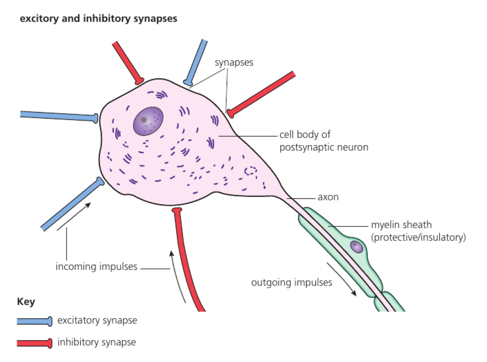
The axon initial segment integrates EPSPs and IPSPs across space and time. If net depolarization reaches threshold, a full action potential begins; if inhibition or decay keeps it below threshold, no action potential occurs.
Summation combines graded potentials. Action potentials themselves do not add into a larger spike.
Multiple excitatory and inhibitory synapses converge on one postsynaptic neuron before an outgoing impulse is produced.

Nociceptors Convert Threatening Conditions into Pain Signals

HL only

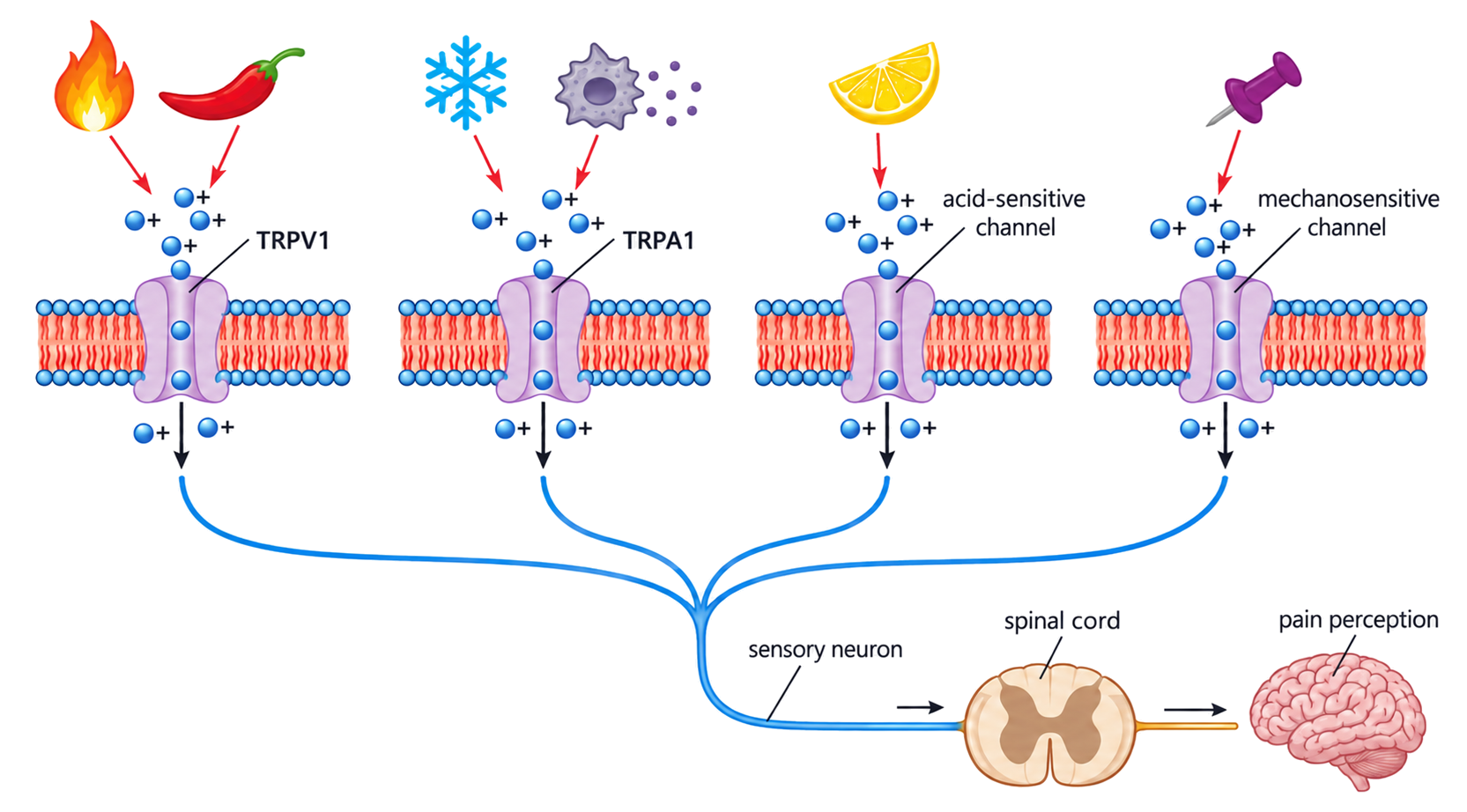
noxious heat, cold, acid, chemicals or tissue damage → matching TRP or other sensory ion channel opens in a free nerve ending → positive ions enter → receptor potential reaches threshold → action potentials travel through sensory pathways → brain activity produces pain perception

TRPV1 responds to damaging heat and capsaicin; TRPA1 responds to several intense cold, reactive and damage-associated signals. Inflammation can lower activation thresholds, making normally tolerable warmth painful.

Nociception is neural detection and signalling of potentially damaging stimuli. Pain is the conscious perception that emerges after central processing; they are related but not identical.

Heat, capsaicin, cold, damage-associated chemicals, acid and mechanical force open stimulus-sensitive channels in free sensory nerve endings, initiating signals toward spinal cord and brain.

Consciousness Is Inferred from Coordinated Brain Activity

HL only

Consciousness is the subjective awareness associated with perception, thought and reflection. It is an emergent property: no single neuron contains it; it depends on coordinated interactions across changing brain networks.

Method What it measures Strongest contribution Limitation
EEG summed electrical activity detected at the scalp very fine timing of changing neural states poor spatial localization
structural MRI signals from hydrogen nuclei in a magnetic field detailed brain anatomy does not directly measure neural activity
fMRI blood-oxygen-level-dependent change linked to local activity maps active regions and coordinated networks indirect and slower than neuronal firing

More complex, changing and coordinated activity patterns are associated with conscious states compared with deep anaesthesia or minimally conscious states. These are neural correlates: evidence of association, not proof that one recorded region alone creates subjective experience.

HL Summary: From Channel State to Neural Decision

HL only

Along an axon: Na⁺-channel opening regenerates depolarization ahead; K⁺-channel opening repolarizes; refractory Na⁺ channels prevent backward firing; myelin carries local current rapidly between nodes.

At the next neuron: EPSPs and IPSPs are graded. Their spatial and temporal sum determines whether threshold is reached. Drugs alter the decision by acting at specific release, receptor, breakdown or reuptake steps.

Across scales: stimulus-sensitive channels can begin a pain pathway; coordinated activity across many regions correlates with conscious processing. A molecular event becomes perception only through successive cellular and network interactions.

Observation Most direct interpretation
spike amplitude unchanged but spacing decreases stimulus intensity is encoded by frequency
conduction slows after myelin loss local current leaks and more membrane must depolarize continuously
transmitter remains in the cleft breakdown or reuptake has been inhibited
many EPSPs arrive but no spike occurs IPSPs or decay keep net potential below threshold

Neurons exam focus

5 marks

Draw a labelled diagram of a motor neuron.

Resting potential generation

4 marks

Outline how neurons generate a resting potential.

Nerve impulses as action potentials

3 marks

Outline how nerve impulses are transmitted along a nerve fibre.

Variation in impulse speed

1 mark

The diagrams represent sections through different axons. Which axon has the slowest speed of impulse?

Synapses as junctions

2 marks

Distinguish between the presynaptic and postsynaptic membranes by giving two differences.

Neurotransmitter release

3 marks

Describe how neurotransmitters are released from a presynaptic neuron membrane.

Excitatory postsynaptic potential

3 marks

Describe how an excitatory postsynaptic potential is generated.

Up to one additional mark is available for the construction of your answer.

Depolarization and repolarization

HL only

4 marks

Explain the role of membranes in the depolarization and repolarization of axons.

Action potential propagation

HL only

4 marks

Describe what occurs in a neuron when an action potential is propagated along the axon.

Oscilloscope traces

HL only

1 mark

State the apparatus used to show the resting and action potentials.

Saltatory conduction

HL only

3 marks

Explain saltatory conduction.

Exogenous chemicals

HL only

6 marks

Discuss how drugs can affect the release and reception of chemical messengers at synapses.

Summation exam focus

HL only

3 marks

Explain the effect of the interaction between the activities of excitatory and inhibitory presynaptic neurons at synapses on the central nervous system.