C2.2.10 (HL)—Oscilloscope traces

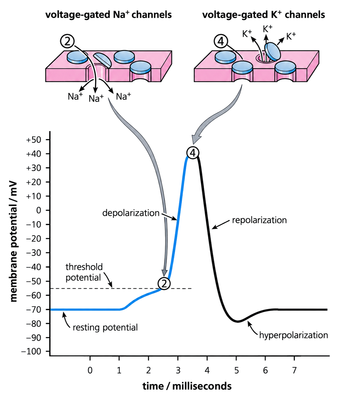
Oscilloscope traces display resting and action potentials as voltage changes over time, allowing depolarization and repolarization to be identified, linking cellular mechanisms to rapid communication in nervous systems.

Syllabus
First assessment 2025
Objective
C2.2.10
Level
HL

Exam analysis

Chance of appearing1%of analysed past papers
Latest appearanceNovember 2025
Most common paperPaper1B
Typical marks1

Common command terms

  • State
  • Annotate

Recent exam appearances

November 2025Paper1B ["HL"] · TZ13(a)[ 1 ]C2.2.10 (HL)—Oscilloscope traces
Practice this objective

Coverage 2025–2025 · Updated 15 Jul 2026

Oscilloscope Traces Show Action-Potential Timing

HL only

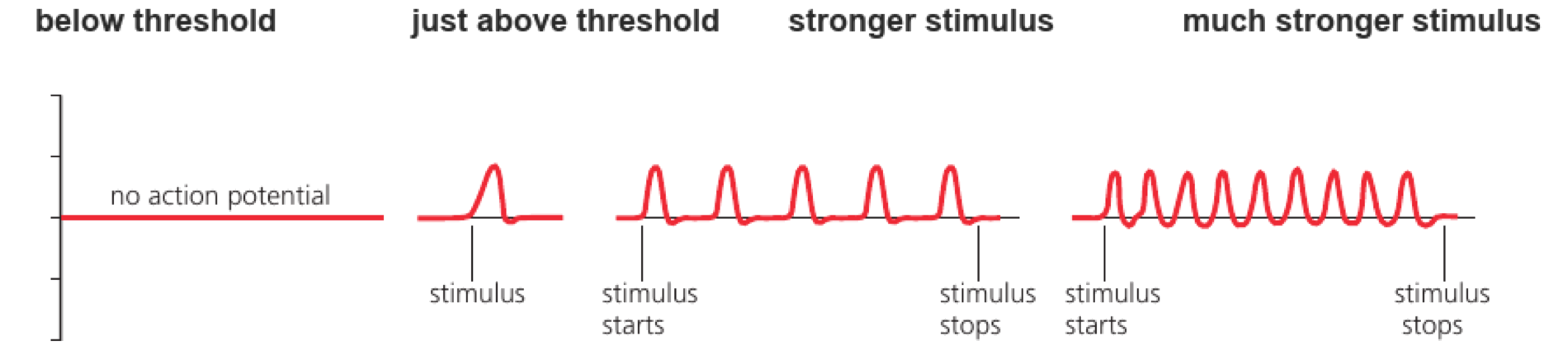
An oscilloscope trace displays voltage against time, allowing action-potential threshold, amplitude, duration and frequency to be compared.

The vertical scale reports membrane potential and the horizontal scale reports time. Repeated spikes indicate frequency; baseline shifts or noise must be distinguished from true action potentials. Reading the trace means linking each voltage change to a stage of channel opening or closing.
Read a trace in order:

  • calibrate axes
  • locate threshold and peak
  • measure duration or interval
  • calculate frequency from repetition

If five spikes occur in 0.1 seconds, the firing frequency is 50 Hz, not an amplitude of five units.
A taller trace is not automatically a stronger stimulus; check what the axis represents.
Oscilloscope traces

HL only

Assessment in practice

1 marks
How it is assessed

This objective is assessed through structured response, commonly using State / Annotate.

Command terms

State / Annotate

What earns marks

Build the answer around this relationship: An oscilloscope can show resting and action potentials.

Representative question

Question 1

[Maximum number: 1]

State the apparatus used to show the resting and action potentials.

Neural Control And Evidence

HL only
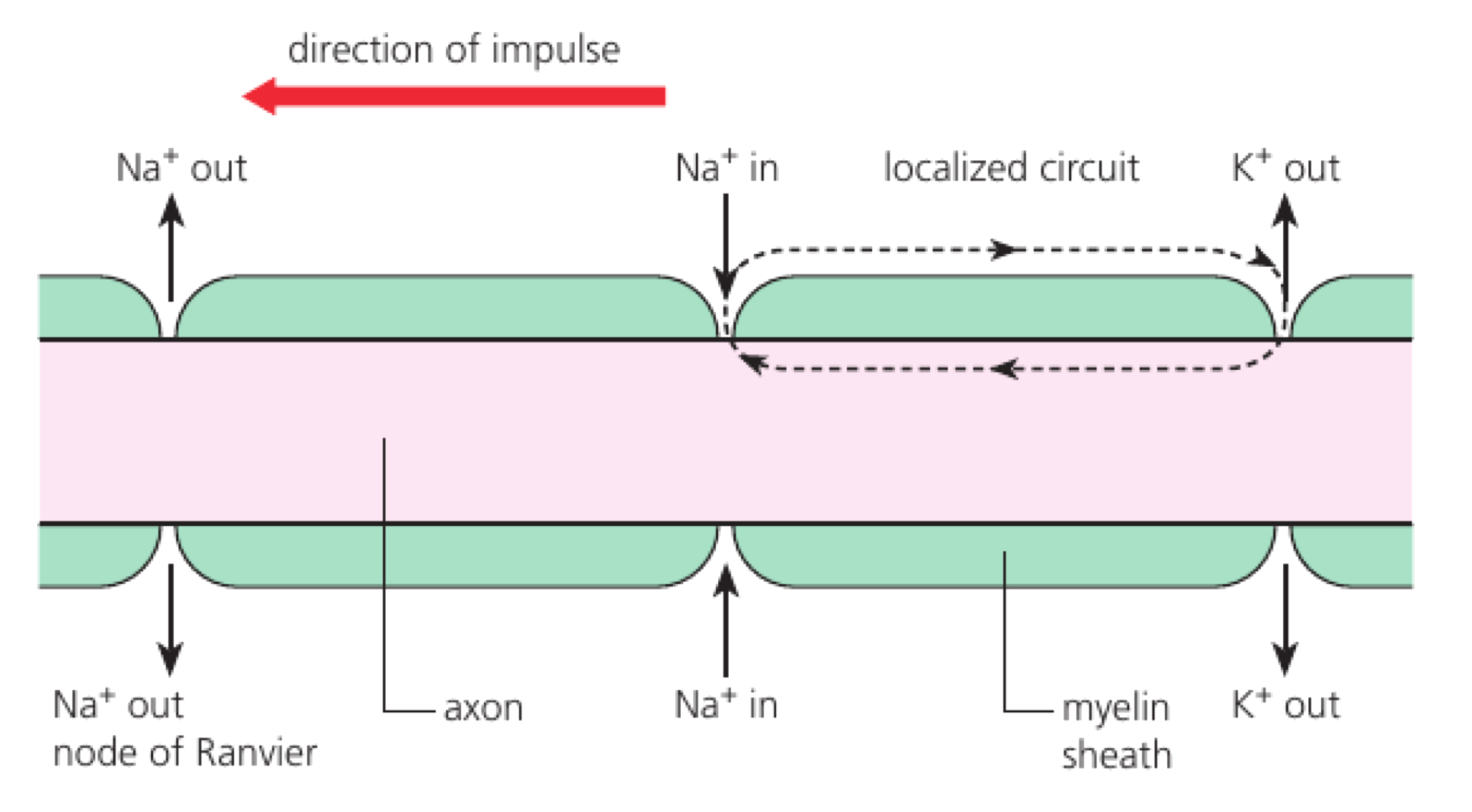
  • At threshold, voltage-gated Na+ channels depolarize the axon; K+ channels then repolarize it. Local currents propagate the impulse and the refractory period enforces direction.
  • Action potentials are all-or-nothing; stimulus intensity is encoded by frequency. Myelin enables fast saltatory conduction between nodes of Ranvier.
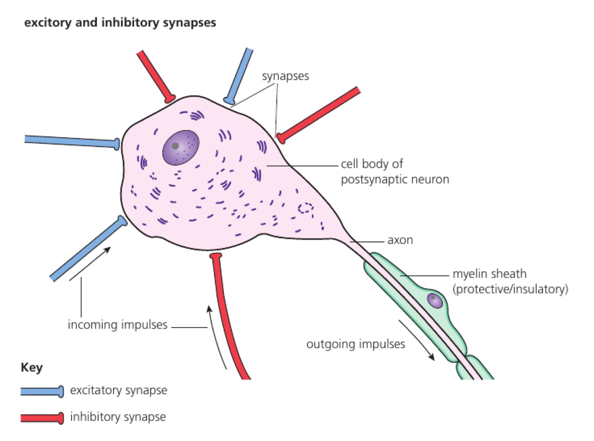
  • Synapses integrate excitatory and inhibitory inputs through spatial and temporal summation. Chemicals may mimic, block or prolong neurotransmitter action.
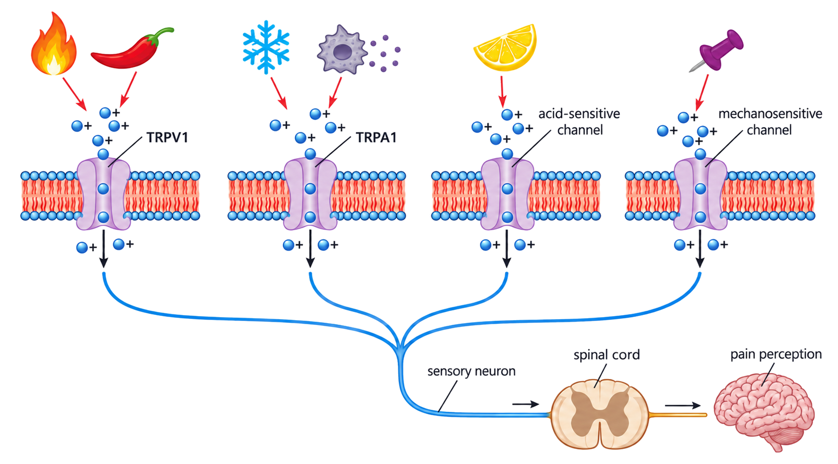
  • Pain receptors use TRP channels that respond to heat, acid, capsaicin or tissue damage.
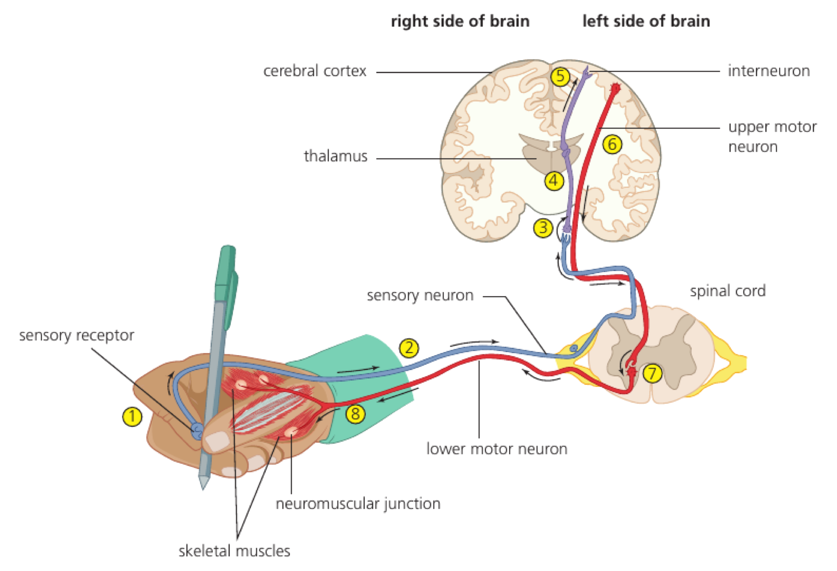
  • EEG, MRI and fMRI link conscious processing to coordinated activity across brain regions, providing neural correlates rather than a single consciousness centre.

Concept essentials

  • An oscilloscope can show resting and action potentials.
  • The trace records membrane potential as voltage over time.
  • Depolarization appears as the rising phase of an action-potential trace.
  • Trace interpretation links graph shape to ion movement across the axon membrane.