Q BankQuestion BankDocsDocuments

C2.2 Neural signalling

Neural signalling transmits information through neuron structure, ion gradients, action potentials, synapses and chemical modulation of postsynaptic responses, linking cellular mechanisms to rapid communication in nervous systems.

Syllabus
First assessment 2025
Topic
C2.2
Level
HL

Neurons Carry Information through Electrical and Chemical Signals

Neurons are specialized cells that receive, integrate and transmit information using membrane potentials and neurotransmitters.

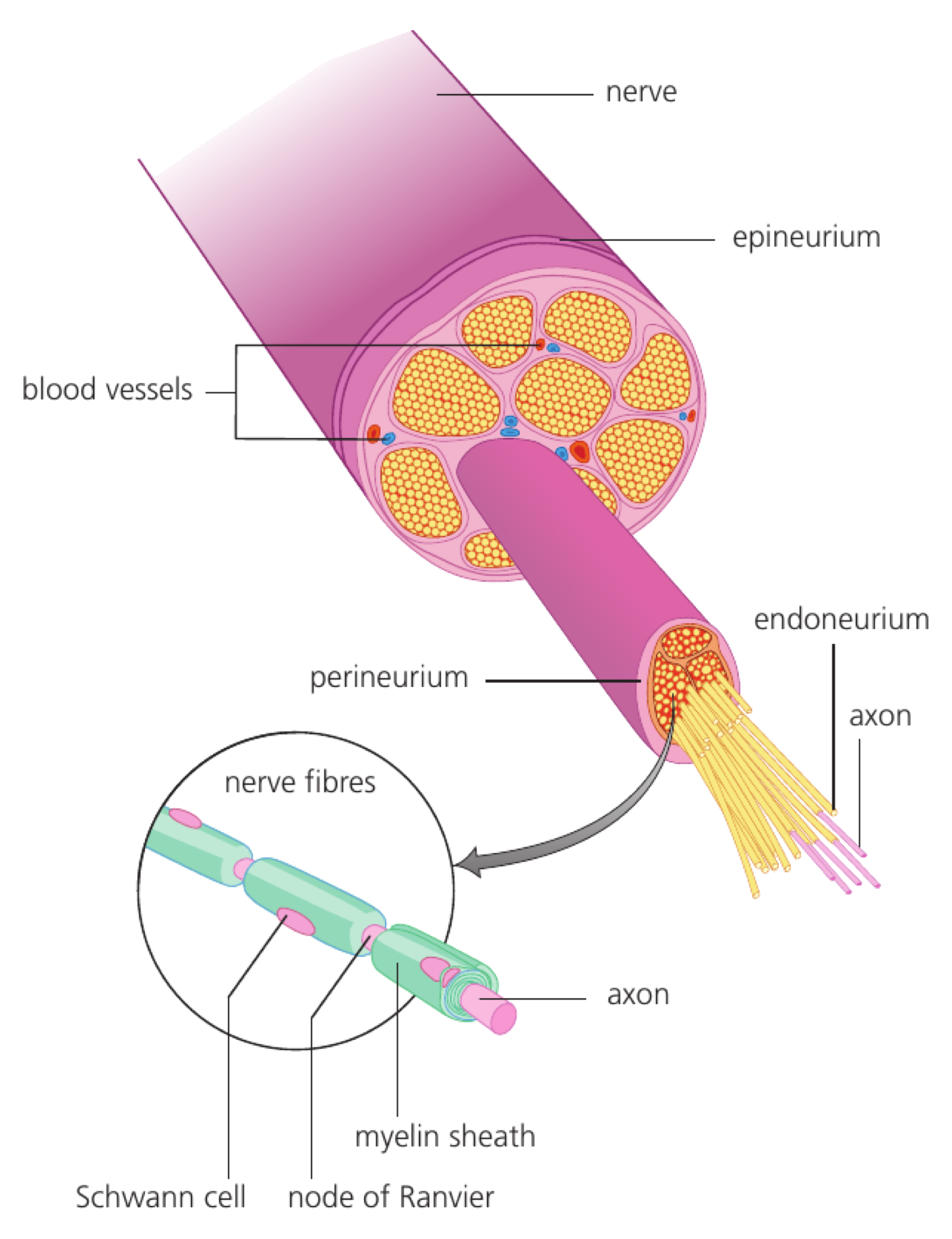
Dendrites receive inputs, the cell body integrates them, and an axon carries action potentials to terminals. Ion channels and synapses connect electrical changes inside one neuron with chemical communication between neurons. This division of labour lets the cell preserve a signal while passing it to the next stage.

Identify the functional regions:

  • dendrites and cell body: input
  • axon hillock: decision to fire
  • axon: impulse propagation
  • terminals: transmitter release

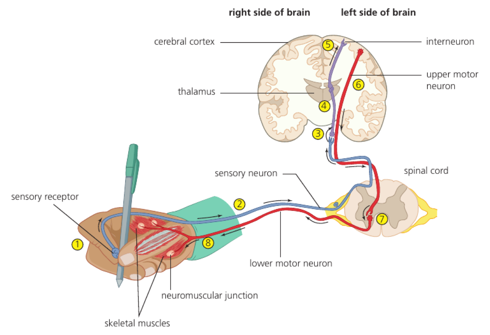
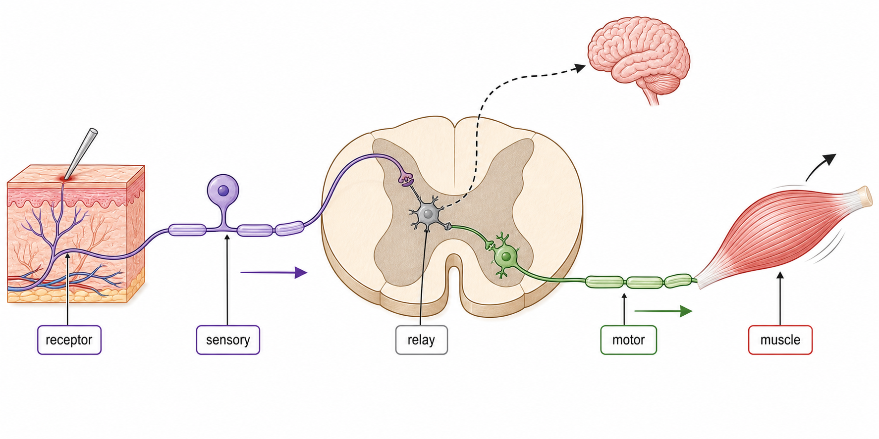
A sensory neuron converts pressure at the skin into receptor-potential changes and action potentials that reach the central nervous system.

Neuron shape varies with function; not every neuron has the same dendrite or axon arrangement.

Neurons exam focus

Assessment in practice

1–2 marks
How it is assessed

This objective is assessed through experimental design, commonly using Label / Draw / State.

Command terms

Label / Draw / State / Distinguish / Identify

What earns marks

Build the answer around this relationship: Dendrites receive signals and conduct them toward the cell body.

Representative question

Question 1

[Maximum number: 5]

Draw a labelled diagram of a motor neuron.

Resting Potential Stores an Ion Gradient

A resting neuron has a stable negative membrane potential created by ion gradients and selective membrane permeability.

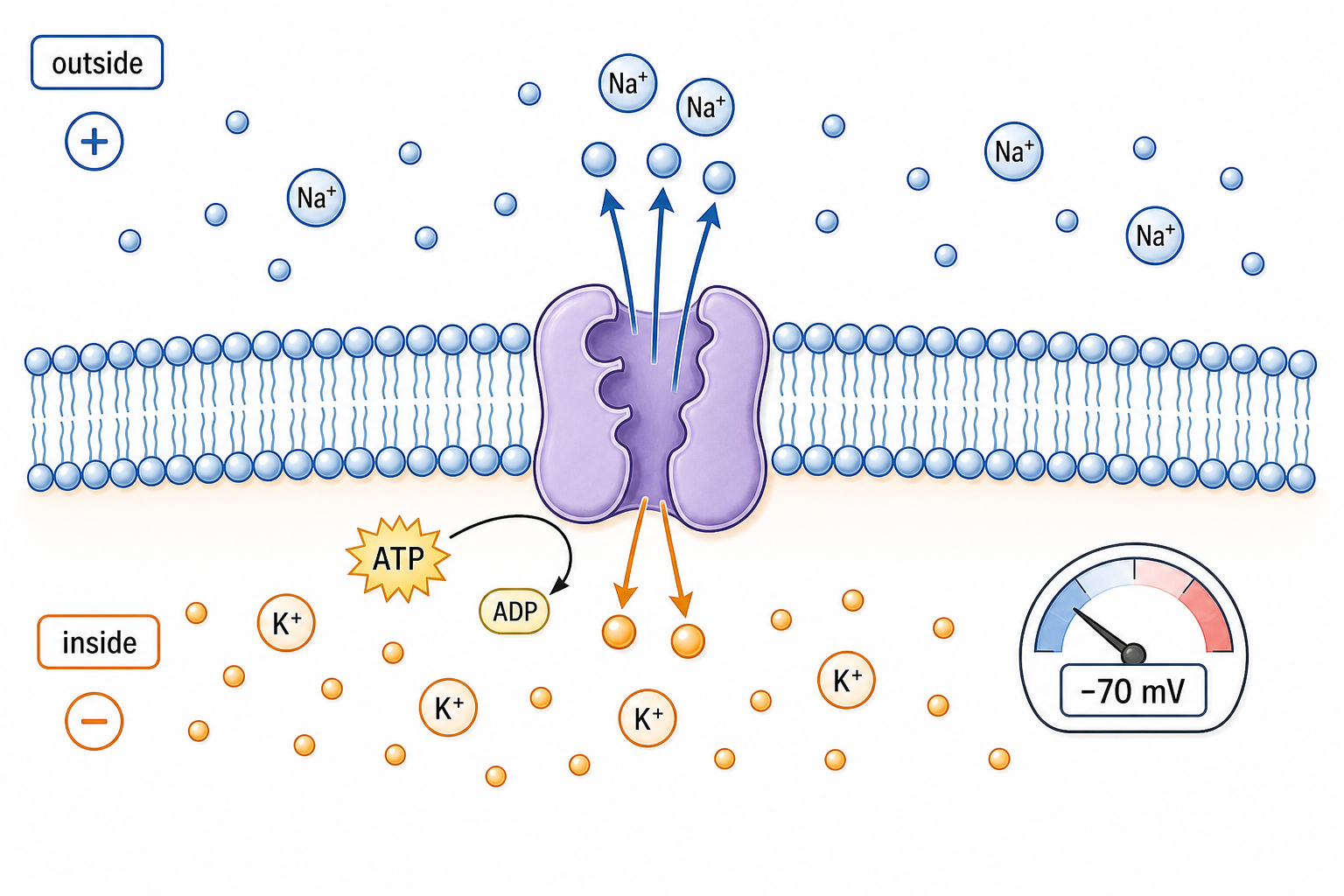
The sodium–potassium pump maintains high K⁺ inside and Na⁺ outside. Leak channels let more K⁺ leave than Na⁺ enter, leaving the inside relatively negative until electrical and chemical forces balance. At rest, selective ion permeability and active transport keep the membrane ready to respond to a new stimulus.

Explain resting potential with:

  • ion concentration gradients
  • selective leak permeability
  • sodium–potassium pump
  • electrical attraction opposing diffusion

If K⁺ leak channels close, the membrane potential shifts because the main outward current has changed.

The pump maintains gradients over time; it does not produce each action potential directly.

Resting potential generation

Assessment in practice

1–2 marks
How it is assessed

This objective is assessed through structured response, commonly using Define / Outline / Explain.

Command terms

Define / Outline / Explain / State / Label

What earns marks

Build the answer around this relationship: The resting axon is negative inside relative to outside.

Representative question

Question 1

[Maximum number: 4]

Outline how neurons generate a resting potential.

An Action Potential Is an All-or-none Nerve Impulse

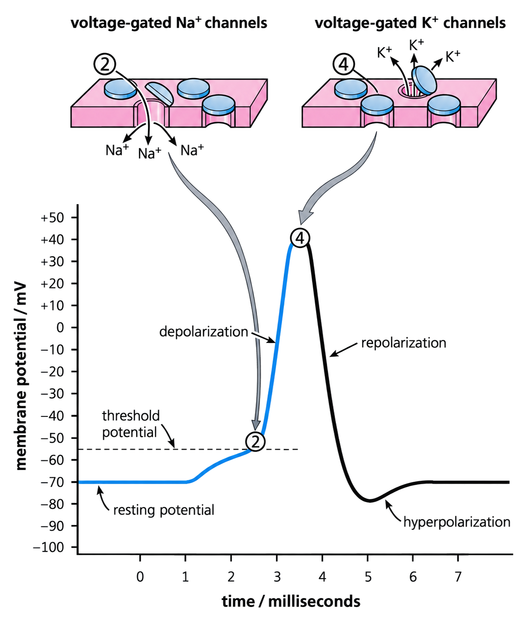
An action potential is a brief, threshold-triggered reversal of membrane potential that propagates along an axon.

Voltage-gated sodium channels open rapidly during depolarization, then inactivate while potassium channels open to repolarize the membrane. Refractory periods prevent immediate re-firing in the same segment. The threshold is a condition, not a fixed promise: if it is not reached, the full action potential does not start.

Trace the phases:

  • threshold
  • Na⁺ influx and depolarization
  • K⁺ efflux and repolarization
  • hyperpolarization and recovery

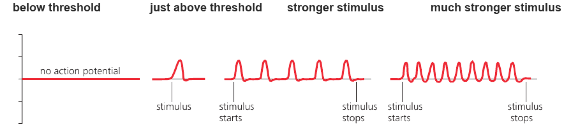
A stimulus below threshold produces no full impulse, while a threshold stimulus produces an impulse with a similar amplitude.

Stronger stimuli are coded mainly by impulse frequency, not by a larger action-potential amplitude.

Nerve impulses as action potentials

Assessment in practice

1–8 marks
How it is assessed

This objective is assessed through structured response, commonly using Explain / Outline.

Command terms

Explain / Outline

What earns marks

Build the answer around this relationship: Action potentials are regenerated along neuron membranes.

Representative question

Question 1

[Maximum number: 3]

Outline how nerve impulses are transmitted along a nerve fibre.

Impulse Speed Depends on Axon Design

Nerve impulse speed varies with axon diameter, myelination and membrane properties.

A larger diameter lowers internal resistance, while myelin reduces ion leakage and concentrates voltage-gated channels at nodes. Both changes let depolarization influence a distant membrane more quickly. A faster impulse therefore reduces delay, but it does not change the information carried by the action-potential sequence.

Compare speed factors:

  • diameter and internal resistance
  • myelin insulation
  • node spacing
  • temperature and channel kinetics

A myelinated motor axon can conduct faster than a thin unmyelinated axon even when both carry the same action-potential amplitude.

A faster impulse does not mean a stronger impulse; speed and amplitude are different properties.

Variation in impulse speed

Assessment in practice

1 marks
How it is assessed

This objective is assessed through multiple choice, commonly using Describe / Determine / Calculate.

Command terms

Describe / Determine / Calculate / Suggest / Outline

What earns marks

Build the answer around this relationship: Larger axon diameter is associated with faster impulse transmission.

Representative question

Question 1

[Maximum number: 1]

The diagrams represent sections through different axons. Which axon has the slowest speed of impulse?

A
B

A Synapse Connects One Cell to Another

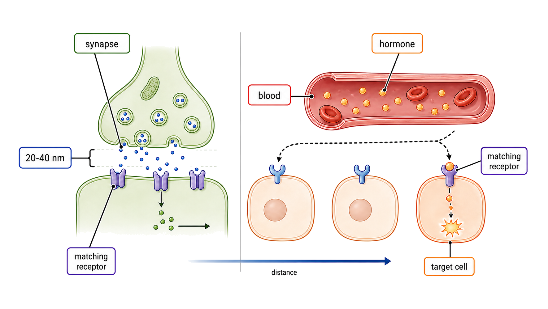
A synapse is a junction where a presynaptic cell communicates with a postsynaptic cell, usually through neurotransmitter release.

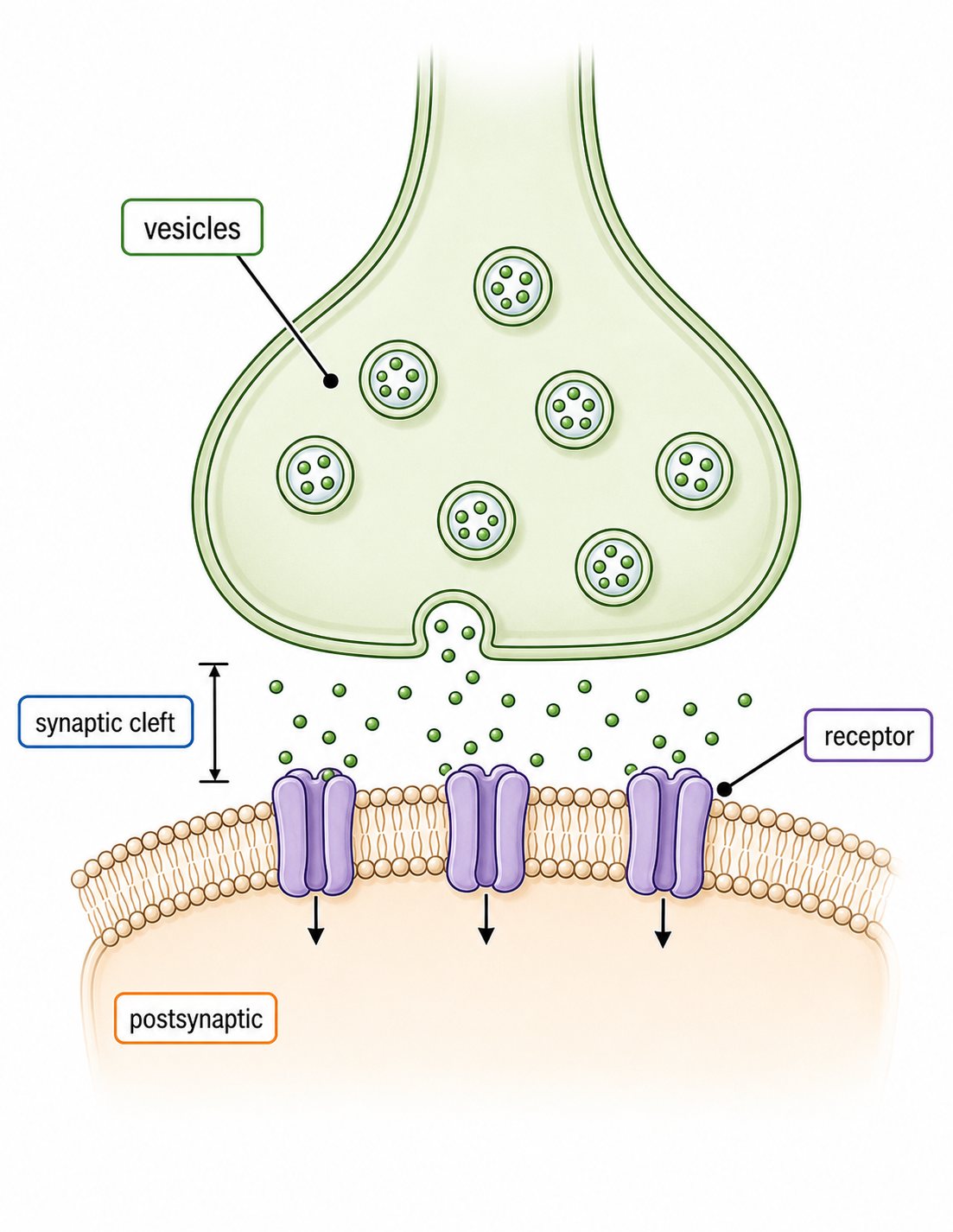
The synaptic cleft separates membranes, creating a one-way delay in chemical synapses. Receptors on the postsynaptic membrane convert transmitter binding into ion-flow or intracellular effects. The synapse is a junction, so the signal must be converted before it can cross to the next cell.

Describe a chemical synapse:

  • presynaptic terminal
  • vesicles and cleft
  • postsynaptic receptors
  • signal termination

An action potential at a motor neuron terminal releases transmitter that binds receptors on a muscle fibre.

A synapse is not simply a physical gap; receptor type and transmitter removal determine the response.

Synapses as junctions

Assessment in practice

1–6 marks
How it is assessed

This objective is assessed through structured response, commonly using Explain / Describe / State.

Command terms

Explain / Describe / State / Identify / Distinguish / Suggest

What earns marks

Build the answer around this relationship: Synapses transmit signals chemically between neurons.

Representative question

Question 1

[Maximum number: 2]

Distinguish between the presynaptic and postsynaptic membranes by giving two differences.

Calcium Triggers Neurotransmitter Release

Arrival of an action potential at a presynaptic terminal opens voltage-gated calcium channels and triggers vesicle fusion.

Ca²⁺ entry binds release machinery, causing exocytosis of neurotransmitter into the synaptic cleft. Enzymatic breakdown, reuptake or diffusion then terminates the signal. This one-way sequence explains why changing calcium entry can alter how strongly a synapse communicates.

Trace release:

  • action potential arrives
  • Ca²⁺ channels open
  • vesicles fuse
  • transmitter binds and is cleared

Blocking presynaptic calcium entry reduces transmitter release even if the action potential still reaches the terminal.

The electrical impulse triggers release; it does not cross the cleft as the same electrical current.

Neurotransmitter release

Assessment in practice

1–3 marks
How it is assessed

This objective is assessed through structured response, commonly using Describe / Explain / Outline.

Command terms

Describe / Explain / Outline / Deduce / Label / State

What earns marks

Build the answer around this relationship: An arriving action potential depolarizes the presynaptic terminal.

Representative question

Question 1

[Maximum number: 3]

Describe how neurotransmitters are released from a presynaptic neuron membrane.

Excitatory Postsynaptic Potentials Move a Neuron toward Threshold

An excitatory postsynaptic potential (EPSP) is a local depolarization that increases the probability of an action potential.

Excitatory transmitter opens channels that allow net positive charge in or reduce outward current. EPSPs decay with distance and time, so several inputs may be needed at the axon hillock. The summed postsynaptic effect determines whether the receiving neuron reaches threshold.

Interpret an EPSP by checking:

  • receptor channel
  • ion movement
  • membrane-potential change
  • effect on threshold

Opening ligand-gated sodium channels at a dendrite produces a small depolarization that can combine with EPSPs from other synapses.

An EPSP is graded and local; it is not itself an all-or-none action potential.

Excitatory postsynaptic potential

Assessment in practice

1–2 marks
How it is assessed

This objective is assessed through structured response, commonly using Explain / Describe.

Command terms

Explain / Describe

What earns marks

Build the answer around this relationship: Excitatory neurotransmitters bind receptors on the postsynaptic membrane.

Representative question

Question 1

[Maximum number: 3]

Describe how an excitatory postsynaptic potential is generated.

Up to one additional mark is available for the construction of your answer.

SL Transfer: Explain Core Neural Signalling

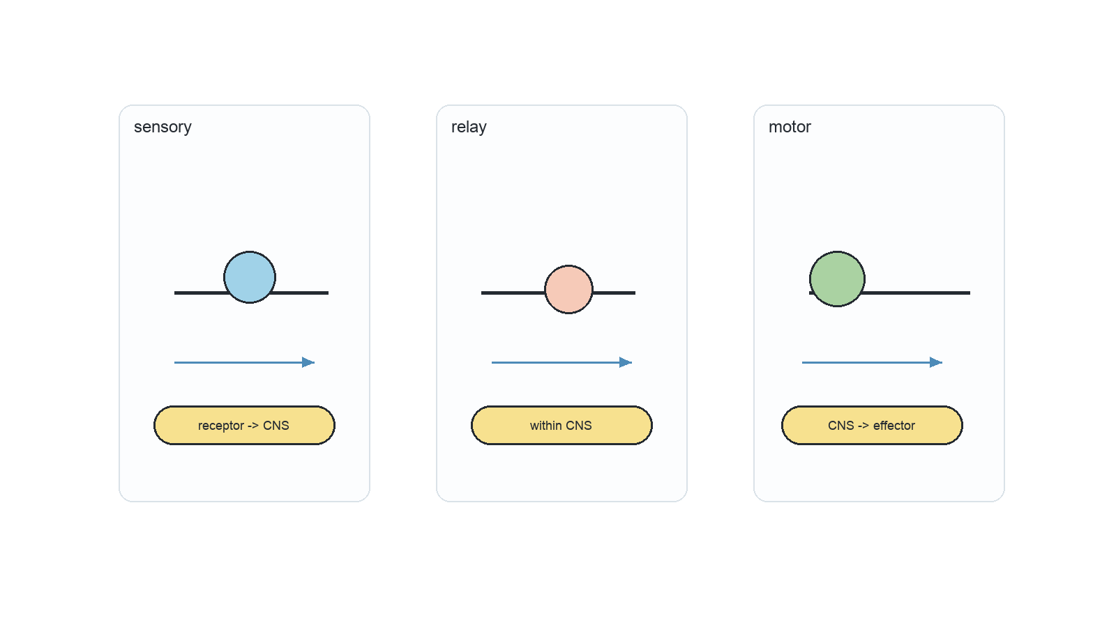
Neurons carry electrical impulses in the nervous system; motor, sensory, and relay neurons differ in axon, dendrite, and cell body arrangement. Sodium-potassium pumps use ATP to move 3 Na+ out and 2 K+ in; ion gradients make the resting axon polarized at about -70 mV. A nerve impulse is a propagated action potential along a nerve fibre; stimulus-triggered sodium influx reverses membrane polarity. Larger axon diameter lowers resistance and increases impulse speed; myelin sheaths and nodes of Ranvier enable faster saltatory conduction. Synapses connect neurons to neurons, muscles, or glands; chemical synapses transmit one way across a narrow synaptic cleft. Action potentials open voltage-gated Ca2+ channels in presynaptic terminals; Ca2+ causes vesicle fusion and neurotransmitter exocytosis into the cleft. Neurotransmitters diffuse and bind receptors on the postsynaptic membrane; EPSPs depolarize the membrane and make threshold more likely.

Depolarization and Repolarization Follow Channel Timing

HL only

Depolarization is the rising phase of an action potential as sodium conductance increases; repolarization returns the membrane toward negative values as potassium conductance rises.

Voltage-gated channels have distinct activation and inactivation timing. The overlap creates a refractory period and ensures the impulse moves forward rather than immediately backward. The changing voltage is evidence of ion movement across the membrane, not a flow of the whole neuron along its axon.

Read the voltage trace with channel states:

  • Na⁺ activation: rising phase
  • Na⁺ inactivation: peak
  • K⁺ activation: falling phase
  • delayed K⁺ closing: undershoot

A sodium-channel blocker removes the rapid rising phase, while a potassium-channel blocker prolongs repolarization.

The membrane does not become permanently positive; ion gradients and channel timing restore the resting state.

Depolarization and repolarization

HL only

Assessment in practice

1 marks
How it is assessed

This objective is assessed through structured response, commonly using Explain / Outline / Describe.

Command terms

Explain / Outline / Describe

What earns marks

Build the answer around this relationship: Sodium ion entry causes depolarization.

Representative question

Question 1

[Maximum number: 4]

Explain the role of membranes in the depolarization and repolarization of axons.

An Action Potential Propagates without Losing Amplitude

HL only

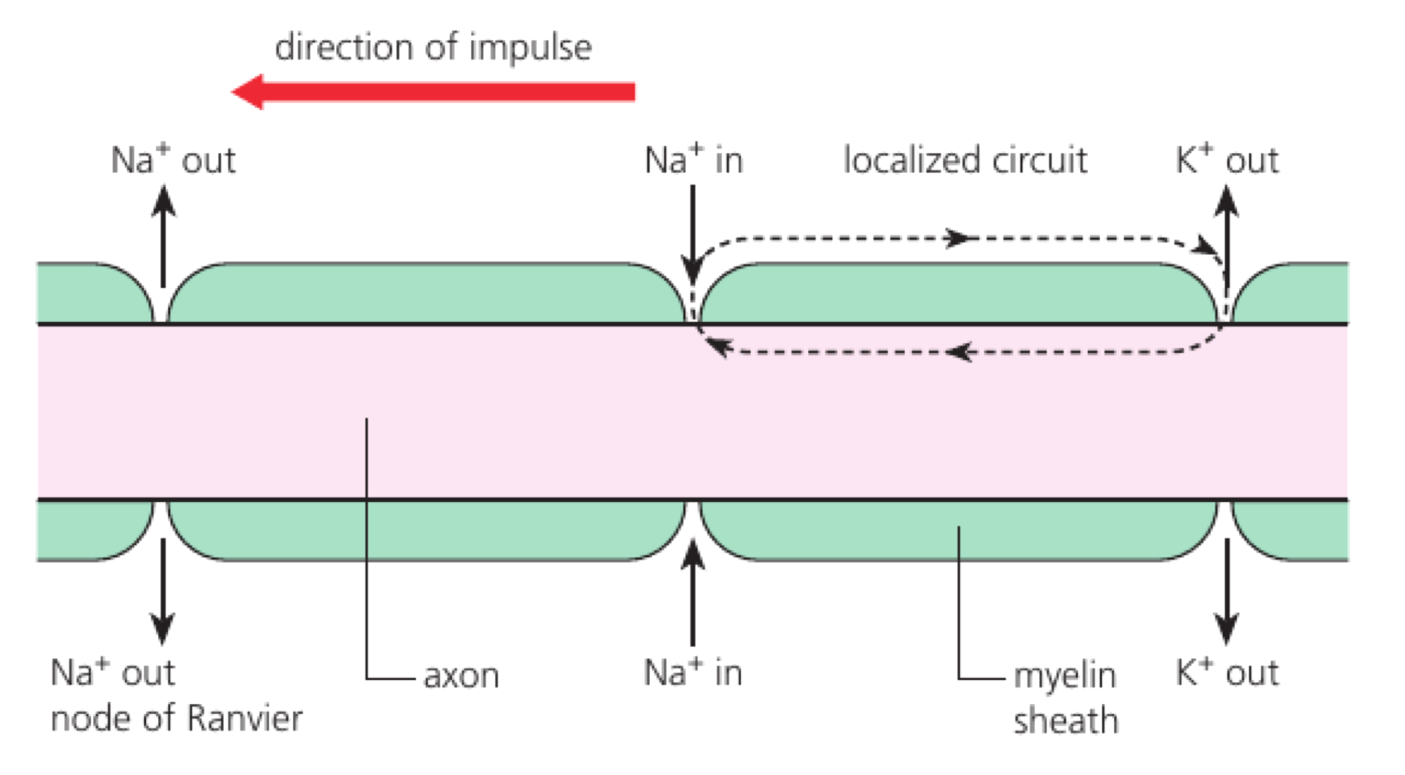
An action potential propagates because local current from one depolarized membrane segment brings the next segment to threshold.

Voltage-gated channels open sequentially along the axon. The refractory region behind the wave prevents immediate reactivation, so the impulse travels forward while being regenerated at each segment. Because each active segment triggers the next, the impulse is regenerated rather than used up as it travels.
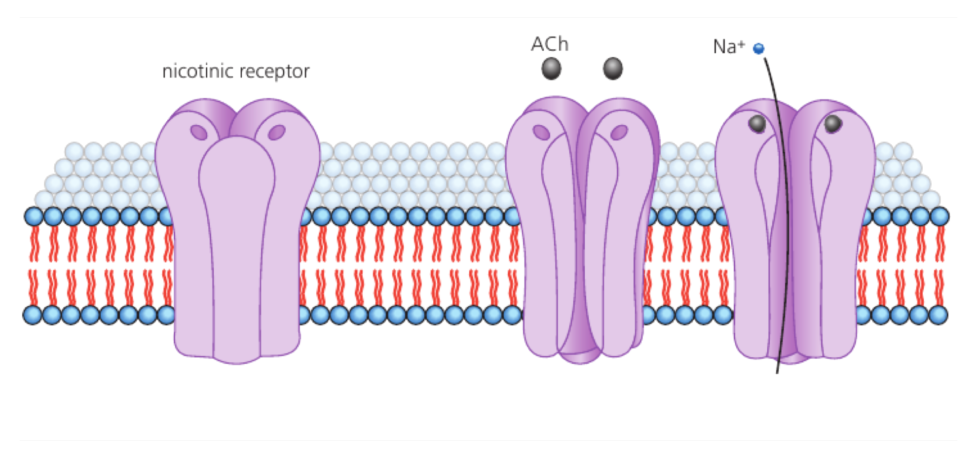
Explain propagation:

  • local current spreads
  • next segment reaches threshold
  • channels regenerate the spike
  • refractory region enforces direction

A cut axon cannot pass the impulse beyond the damaged membrane because the next excitable segment is no longer connected.

Propagation speed and direction come from membrane organization, not from a particle travelling unchanged down the axon.

Action potential propagation

HL only

Assessment in practice

4–7 marks
How it is assessed

This objective is assessed through structured response, commonly using Explain / Describe.

Command terms

Explain / Describe

What earns marks

Build the answer around this relationship: Local current from one depolarized region triggers adjacent membrane.

Representative question

Question 1

[Maximum number: 4]

Describe what occurs in a neuron when an action potential is propagated along the axon.

Oscilloscope Traces Show Action-Potential Timing

HL only

An oscilloscope trace displays voltage against time, allowing action-potential threshold, amplitude, duration and frequency to be compared.

The vertical scale reports membrane potential and the horizontal scale reports time. Repeated spikes indicate frequency; baseline shifts or noise must be distinguished from true action potentials. Reading the trace means linking each voltage change to a stage of channel opening or closing.

Read a trace in order:

  • calibrate axes
  • locate threshold and peak
  • measure duration or interval
  • calculate frequency from repetition

If five spikes occur in 0.1 seconds, the firing frequency is 50 Hz, not an amplitude of five units.

A taller trace is not automatically a stronger stimulus; check what the axis represents.

Oscilloscope traces

HL only

Assessment in practice

1 marks
How it is assessed

This objective is assessed through structured response, commonly using State / Annotate.

Command terms

State / Annotate

What earns marks

Build the answer around this relationship: An oscilloscope can show resting and action potentials.

Representative question

Question 1

[Maximum number: 1]

State the apparatus used to show the resting and action potentials.

Myelin Enables Saltatory Conduction

HL only

Saltatory conduction makes an action potential appear to jump between nodes of Ranvier along a myelinated axon.

Myelin insulates internodes and reduces capacitance, while voltage-gated channels are concentrated at nodes. Local current travels rapidly beneath myelin and brings the next node to threshold. The insulating layer increases effective conduction speed by reducing how much membrane must exchange charge directly.

Compare myelinated conduction:

  • internodes insulated
  • nodes regenerate impulses
  • fewer membrane regions depolarize
  • speed and efficiency increase

Demyelination exposes membrane and increases current leakage, slowing or blocking conduction even if the neuron’s channels remain present.

The impulse is regenerated at nodes; it does not literally jump through empty space.

Saltatory conduction

HL only

Assessment in practice

1–2 marks
How it is assessed

This objective is assessed through structured response, commonly using Outline / Explain.

Command terms

Outline / Explain

What earns marks

Build the answer around this relationship: Myelin electrically insulates sections of the axon.

Representative question

Question 1

[Maximum number: 3]

Explain saltatory conduction.

Exogenous Chemicals Can Alter Neural Signalling

HL only

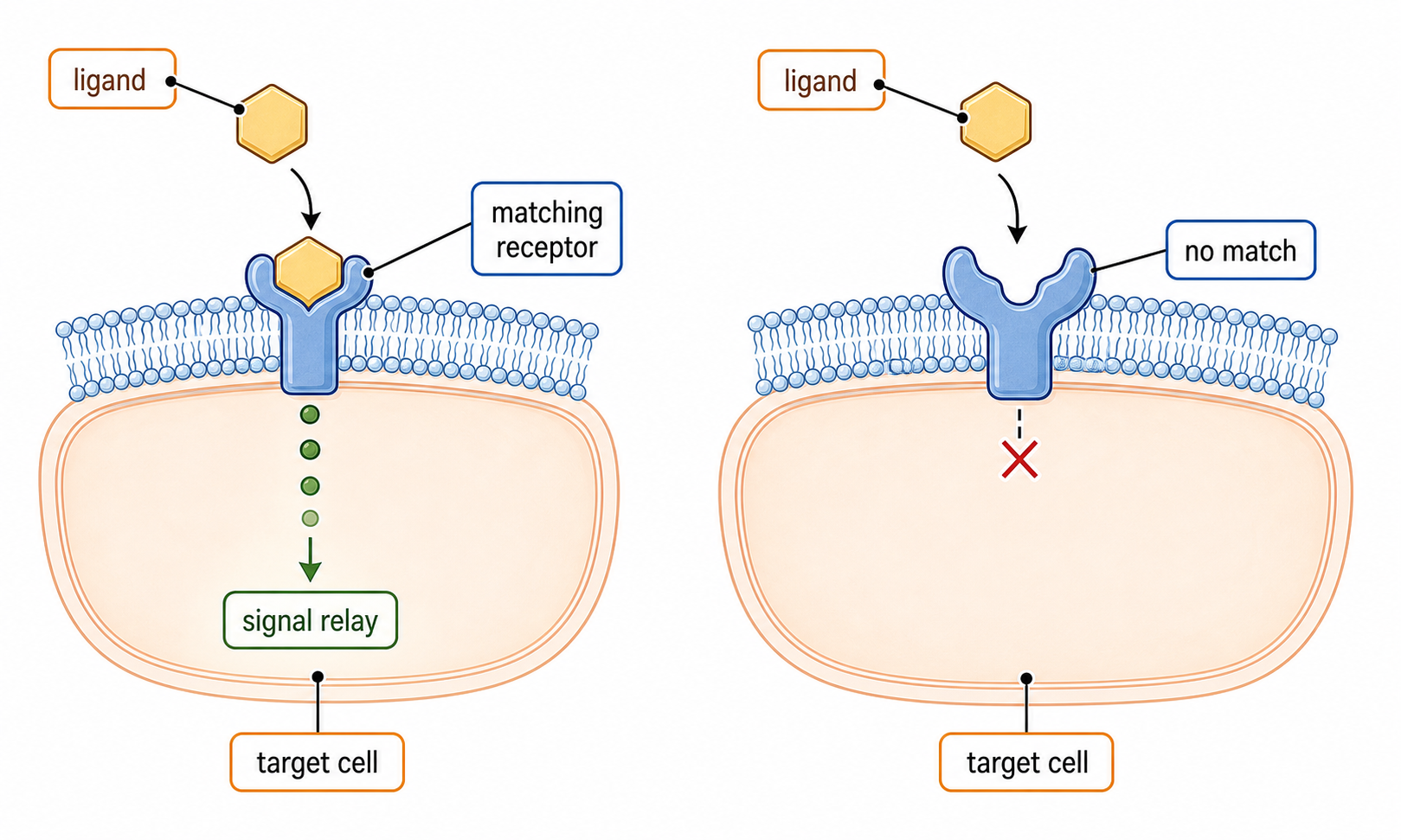
Exogenous chemicals can change neural signalling by mimicking transmitters, blocking receptors, altering reuptake or changing ion channels.
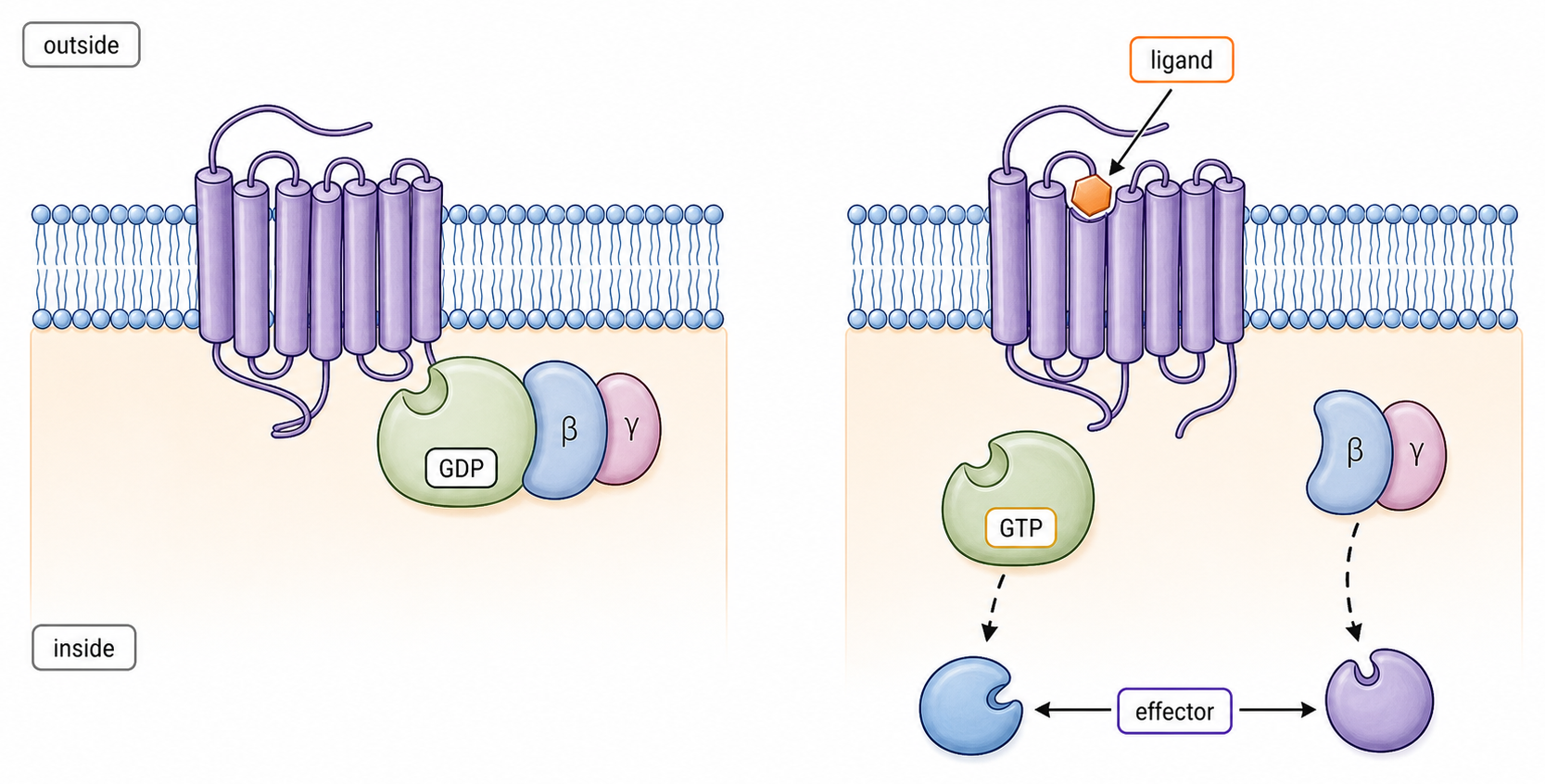
The effect depends on dose, receptor affinity, tissue distribution and clearance. A chemical can increase or decrease signalling without changing the neuron’s DNA. A chemical can change signalling by altering receptors or channels, even when the neuron's wiring is unchanged.

Classify a chemical action:

  • agonist mimics a signal
  • antagonist blocks a receptor
  • reuptake inhibitor prolongs a signal
  • channel modifier changes excitability

A reuptake inhibitor can leave neurotransmitter in the synaptic cleft longer, increasing postsynaptic stimulation.

A chemical’s presence does not prove a single behavioural effect; receptor distribution and dose determine the outcome.

Exogenous chemicals

HL only

Assessment in practice

1–3 marks
How it is assessed

This objective is assessed through essay response, commonly using Explain / State / Suggest.

Command terms

Explain / State / Suggest / Outline / Describe / Discuss / Label

What earns marks

Build the answer around this relationship: Cocaine blocks dopamine reuptake and prolongs postsynaptic stimulation.

Representative question

Question 1

[Maximum number: 6]

Discuss how drugs can affect the release and reception of chemical messengers at synapses.
Inhibitory Neurotransmitters Reduce Firing Probability

HL only

Inhibitory neurotransmitters make a postsynaptic neuron less likely to reach action-potential threshold.
They may open chloride channels, increase potassium efflux or reduce excitatory channel activity. The resulting hyperpolarization or shunting inhibition opposes excitatory inputs. Inhibition is relational: the same transmitter can have different effects if receptor types differ, and the receiving circuit determines the outcome.

Identify inhibition by checking:

  • receptor and ion channel
  • current direction or conductance
  • membrane-potential effect
  • threshold probability

Opening chloride channels can stabilize the membrane near its resting potential and reduce the effect of a simultaneous EPSP.

Inhibition is not always a large negative voltage; increased conductance can also shunt excitatory current.

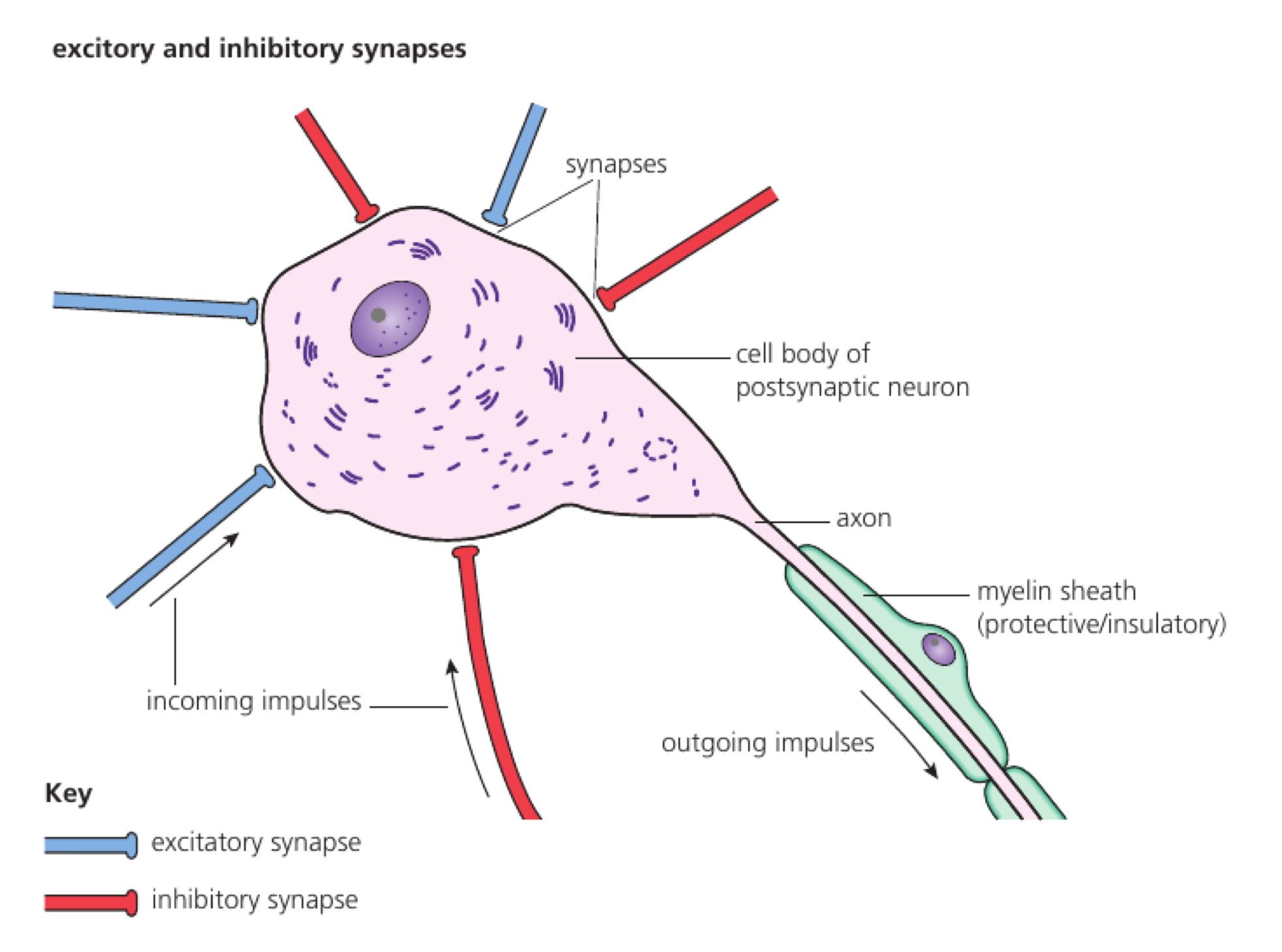
Summation Combines Synaptic Inputs

HL only

Summation combines graded postsynaptic potentials across space and time at the axon hillock.

Spatial summation adds inputs from different synapses; temporal summation adds repeated inputs from one synapse. The net potential reaches threshold only if excitation outweighs inhibition. The result depends on timing as well as size, because inputs that overlap can combine before the threshold decision.

Separate the two forms:

  • spatial: different locations
  • temporal: different times
  • EPSPs add
  • IPSPs subtract or shunt

Two small EPSPs arriving together at the axon hillock can reach threshold even though neither would trigger an action potential alone.

The neuron integrates potentials, not neurotransmitter names; receptor effects determine the sign and size.

Summation exam focus

HL only

Assessment in practice

2–3 marks
How it is assessed

This objective is assessed through structured response, commonly using Explain.

Command terms

Explain

What earns marks

Build the answer around this relationship: Summation combines postsynaptic effects from multiple presynaptic neurons.

Representative question

Question 1

[Maximum number: 3]

Explain the effect of the interaction between the activities of excitatory and inhibitory presynaptic neurons at synapses on the central nervous system.

Pain Perception Depends on Nociceptor Signalling

HL only

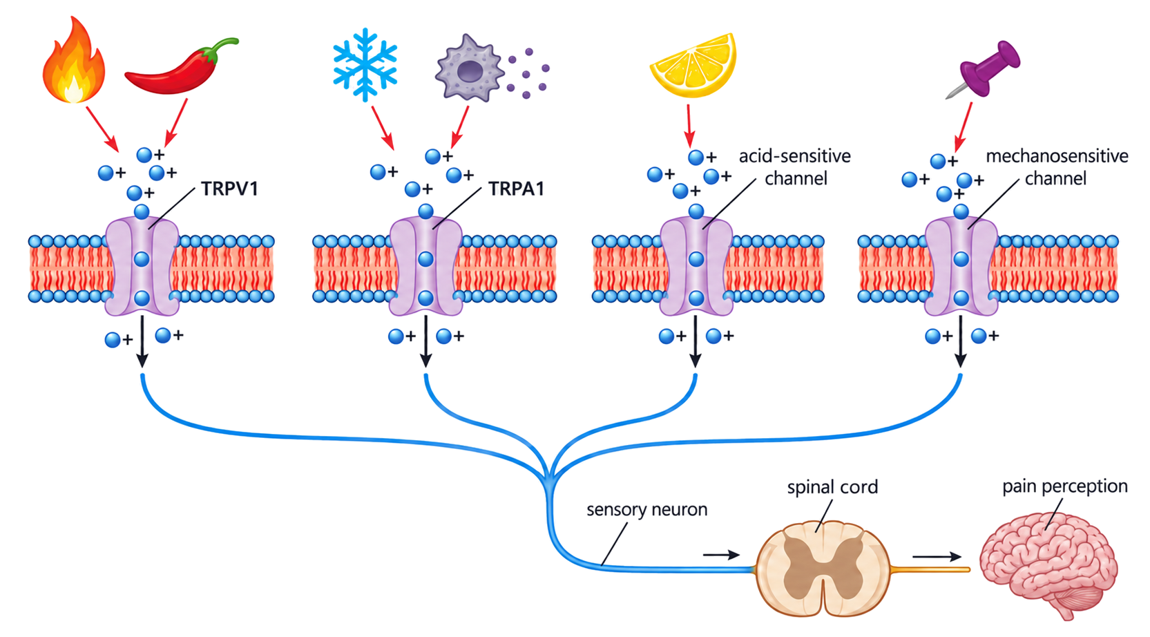
Pain perception begins when nociceptors detect potentially damaging stimuli and transmit signals through sensory pathways to the brain.

Tissue damage releases chemicals that activate or sensitize nociceptors. Spinal circuits and descending brain pathways can amplify or reduce the signal before conscious perception. The pathway shows how a physical stimulus becomes a coded neural signal before the brain interprets it as pain.

Trace pain information:

  • damaging stimulus
  • nociceptor transduction
  • sensory neuron impulses
  • spinal and brain processing

Inflammatory mediators can lower a nociceptor’s threshold, so a normally harmless touch becomes painful in injured tissue.

Nociception and conscious pain are related but not identical; the brain’s interpretation matters.

Consciousness Depends on Integrated Brain Activity

HL only

Consciousness is associated with coordinated activity across interacting brain networks rather than one isolated neuron or single region.

Arousal systems regulate wakefulness while cortical and subcortical networks integrate sensory information, memory and self-related processing. Evidence from sleep, anaesthesia and brain injury links changes in network connectivity to conscious state. Conscious awareness is therefore an emergent interpretation of coordinated neural activity, not a single switch in one neuron.

Separate the components:

  • arousal and wakefulness
  • information integration
  • communication among brain regions
  • reportable experience

During deep anaesthesia, widespread connectivity and coordinated responses fall, while local sensory circuits may still respond to stimulation.

A neural response alone does not prove a conscious experience; integration and reportability require additional evidence.

Neural Control And Evidence

HL only
  • At threshold, voltage-gated Na+ channels depolarize the axon; K+ channels then repolarize it. Local currents propagate the impulse and the refractory period enforces direction.
  • Action potentials are all-or-nothing; stimulus intensity is encoded by frequency. Myelin enables fast saltatory conduction between nodes of Ranvier.
  • Synapses integrate excitatory and inhibitory inputs through spatial and temporal summation. Chemicals may mimic, block or prolong neurotransmitter action.
  • Pain receptors use TRP channels that respond to heat, acid, capsaicin or tissue damage.
  • EEG, MRI and fMRI link conscious processing to coordinated activity across brain regions, providing neural correlates rather than a single consciousness centre.
ConceptIB Biology HL