C3.1.9—Pain reflex arcs
Pain reflex arcs links named stimuli, coordinating structures and effectors so body systems or plant tissues produce an integrated biological response.
- Syllabus
- First assessment 2025
- Objective
- C3.1.9
- Level
- SL
Pain reflex arcs links named stimuli, coordinating structures and effectors so body systems or plant tissues produce an integrated biological response.

Coverage 2010–2025 · Updated 16 Jul 2026
A pain withdrawal reflex is an involuntary response integrated in the spinal cord, with skeletal muscle as the effector.
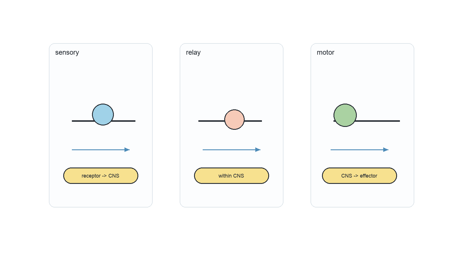
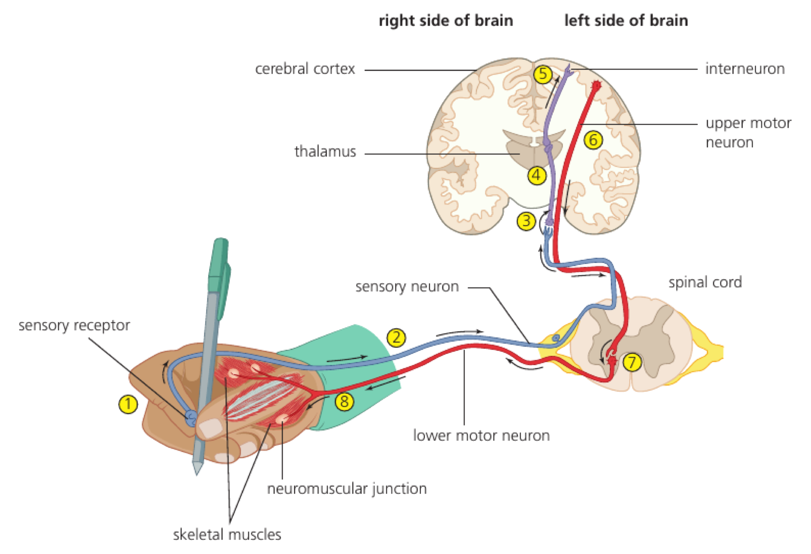
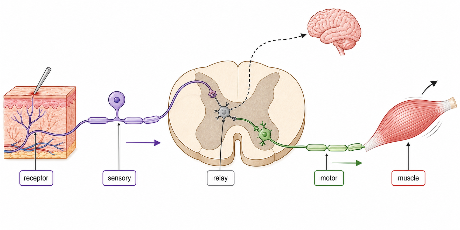
A free sensory nerve ending in the hand detects a damaging stimulus. The sensory neuron enters the spinal cord and synapses with one interneuron in grey matter, which synapses with a motor neuron; the motor neuron stimulates a skeletal muscle to withdraw the hand.
Free nerve ending → sensory neuron → single interneuron in spinal grey matter → motor neuron → skeletal-muscle contraction. The short spinal route starts withdrawal before conscious pain processing is complete.
Touching a hot object activates the pain receptor; flexor muscle contraction withdraws the hand while impulses also travel to the brain and pain becomes conscious.
The reflex does not bypass the CNS—the spinal cord is part of the CNS. The brain can be informed in parallel but is not required to initiate this immediate response.
This objective is assessed through experimental design, commonly using Define / Label / Explain.
Define / Label / Explain / Draw / Identify / Outline / Describe / Annotate
Mark schemes reward named structures, correct direction of information flow and explanations that keep the sequence causal.
Putting the brain before the spinal relay in a rapid pain reflex.
Representative question
Draw a labelled diagram of a reflex arc for a pain withdrawal reflex.
Spinal cord showing white and grey matter
Spinal nerves showing dorsal and ventral roots
Sensory neuron / receptor
Motor neuron / effector
Relay neuron
Arrows showing path from stimulus/receptor to response/effector