C3.1 Integration of body systems

Integrated body systems coordinate neural, hormonal and plant responses through receptors, control centres and effectors, maintaining function across organisms and changing environmental conditions.
Syllabus
First assessment 2025
Topic
C3.1
Level
SL

Learning objectives

C3.1.1System integration• Integration lets interacting parts coordinate an overall biological function• Systems interact across molecular, cellular, organ, organism, and ecosystem levelsC3.1.2Hierarchy in multicellular organisms• Cells form tissues, organs, organ systems, and whole organisms• Emergent properties arise when subsystems interact, such as gut peristalsis and absorptionC3.1.3Integration of organs• Organs are integrated by nervous signals, hormones, and blood transport• Transport links nutrients, gases, wastes, hormones, and energy substrates between organsC3.1.4Brain as information integration organ• The brain integrates sensory input and coordinates complex responses• Cerebral hemispheres, cerebellum, hypothalamus, and medulla have distinct rolesC3.1.5Spinal cord• The spinal cord links brain and peripheral nervous system• It integrates unconscious processes such as reflex arcsC3.1.6Input through sensory neurons• Sensory neurons carry impulses from receptors to spinal cord and brain• Inputs may reach cerebral hemispheres for conscious perceptionC3.1.7Output through motor neurons• Motor neurons carry impulses from CNS to skeletal muscle effectors• Voluntary muscle contraction is coordinated through cerebral hemispheresC3.1.8Nerves as bundles• Nerves are bundles of sensory and motor nerve fibres in connective tissue• Mixed nerves carry impulses both to and from the CNSC3.1.9Pain reflex arcs• Pain reflexes are rapid involuntary withdrawal responses• Reflex arcs use receptors, sensory neurons, relay neurons, motor neurons, and skeletal muscleC3.1.10Cerebellum role• The cerebellum coordinates timing and force of skeletal muscle contractions• It maintains balance, posture, and smooth learned movementsC3.1.11Melatonin and sleep• The pineal gland secretes melatonin according to light-dark cycles• Melatonin modulates sleep timing as part of circadian rhythmsC3.1.12Epinephrine (adrenaline)• Adrenal glands secrete epinephrine during stress or danger• It increases heart rate, ventilation, and respiratory substrate availabilityC3.1.13Hypothalamus and pituitary control• The hypothalamus links nervous inputs to endocrine control• Pituitary hormones regulate glands including thyroid, gonads, adrenals, and mammary glandsC3.1.14Heart rate feedback control• Baroreceptors monitor blood pressure; chemoreceptors monitor CO₂, pH, and O₂• The medulla adjusts sinoatrial node activity by sympathetic and parasympathetic nervesC3.1.15Ventilation rate feedback control• Chemoreceptors detect CO₂-driven pH changes in blood and cerebrospinal fluid• The medulla alters diaphragm and intercostal muscle activity to change ventilation rateC3.1.16Peristalsis control• The CNS controls voluntary swallowing and egestion• The enteric nervous system coordinates involuntary peristalsis in gut smooth muscle

Integration Creates a Function No Part Performs Alone

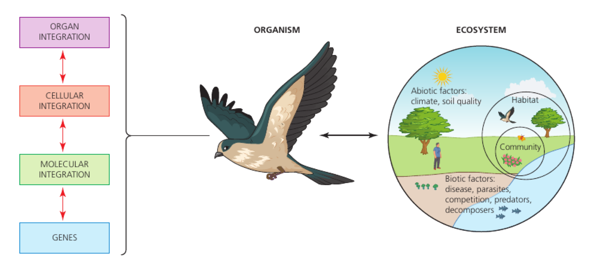
Integration is the coordination of interacting parts so the whole biological system performs an overall function. An emergent property appears through those interactions and is not a property of any isolated part.
Genes and molecular, cellular and organ integration contribute to an organism that also interacts with its ecosystem.
If the claim describes… Classify it as…
what one component can do by itself a component property
what coordinated components achieve together an emergent property
information or material passing between components evidence of integration

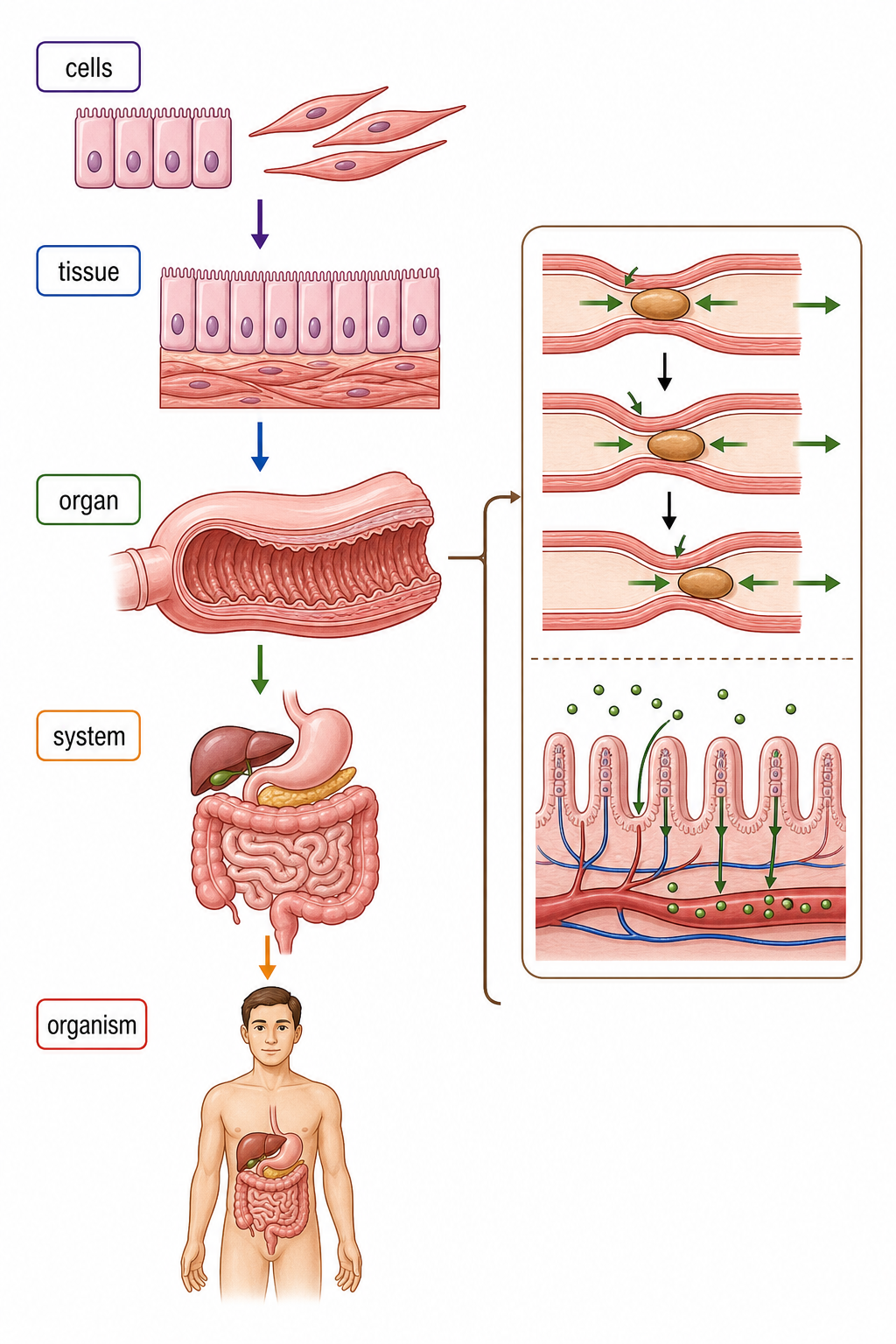
A smooth-muscle cell can contract, but one cell cannot propel a meal through an intestine. Peristalsis emerges when many cells, tissues and neural signals coordinate waves of contraction.

The Body Hierarchy Builds New Capabilities

cells → tissues → organs → organ systems → organism

Each level contains interacting subsystems from the level below; moving upward adds organisation, not merely more material.

Epithelial and smooth-muscle cells form tissues, the tissues form the small intestine, organs form the digestive system and organ systems form an organism.
Level Contribution to the intestine
epithelial tissue forms a selective surface for secretion and absorption
smooth-muscle tissue contracts and relaxes repeatedly
nervous and connective tissues coordinate movement and hold working tissues together
whole organ combines movement, digestion, absorption and transport

The organ's function depends on different tissue types interacting. Calling an organ ‘a group of similar cells’ misses the transition from tissue to organ.

Organs Coordinate through Signals and Shared Transport

Integrating route What travels Characteristic job
nervous signalling action potentials along neurons; neurotransmitter across synapses rapid, precise control of particular effectors
hormonal signalling hormones in blood coordinated effects in target cells with matching receptors
blood transport gases, nutrients, substrates, hormones and wastes physically links organ inputs and outputs

During exercise, motor neurons stimulate skeletal muscle, epinephrine changes several target organs, and blood delivers O₂ and glucose while carrying CO₂ away. No route replaces the others.

Blood reaches many tissues, but a hormone changes only cells with the appropriate receptor. Nervous output is targeted mainly by the physical wiring of the pathway.

Blood Links the Outputs of One Organ to the Needs of Another

Material Important route Why the route matters
glucose and amino acids small intestine → hepatic portal vein → liver → body tissues liver processes and buffers nutrient supply before wider distribution
O₂ lungs → red blood cells → respiring tissues supports aerobic ATP production
CO₂ respiring tissues → blood → lungs removes a respiratory waste and helps restore pH
urea liver → blood → kidneys transfers nitrogenous waste to the organ that excretes it

Long-chain lipids are rebuilt into triglycerides and packaged with protein before entering lymph, then the blood near the heart. Their hydrophobicity changes the transport route.

Transport integrates organs because one organ's output becomes another organ's substrate, signal or waste load. The circulation is the connecting route; it does not itself perform digestion, respiration or excretion.

Different Brain Regions Integrate Different Kinds of Information

Region Main integration role
cerebral hemispheres conscious perception, memory, planning and voluntary motor commands
cerebellum compares intended movement with sensory feedback to refine timing, force, posture and balance
hypothalamus links nervous information and blood conditions to autonomic and endocrine control
medulla adjusts involuntary functions including heart rate and ventilation
External and sectional brain views identify the cerebral hemispheres, cerebral cortex, hypothalamus, pituitary gland and cerebellum.

The brain is an integration organ because it receives, compares, stores and redistributes information. A region's role is not an isolated behaviour; it contributes to coordinated networks.

The Spinal Cord Is Both a Relay and a Local Integrating Centre

Spinal-cord role Information route Outcome
relay sensory input ascends to the brain; motor commands descend to peripheral neurons links brain and body
local integration sensory neuron synapses through relay neuron(s) to a motor neuron rapid unconscious response can begin without waiting for the brain

A spinal reflex is integrated in the spinal cord, but sensory impulses can still travel to the brain. Rapid withdrawal and later conscious pain are therefore compatible, not alternative explanations.

Spinal circuits also contribute to involuntary processes such as urination, defecation and some sweating. ‘Unconscious’ means not under direct voluntary control; it does not mean unregulated.

A Voluntary Action Closes the Loop from Receptor to Muscle

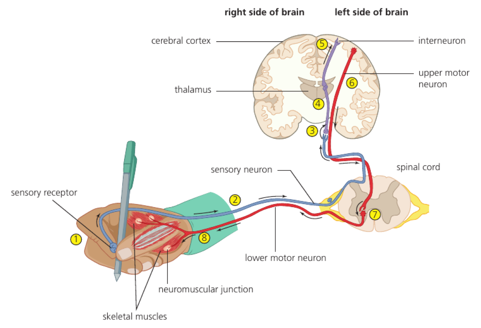
receptor transduces a stimulus → sensory neuron carries impulses into the CNS → relay neurons in brain networks support conscious perception → an upper motor neuron carries a command downward → a lower motor neuron reaches skeletal muscle → neurotransmitter at the neuromuscular junction triggers muscle action potentials and contraction

Sensory input from a receptor travels to the spinal cord and cerebral cortex, while upper and lower motor neurons carry output to a skeletal muscle.
Fibre Direction relative to CNS Function
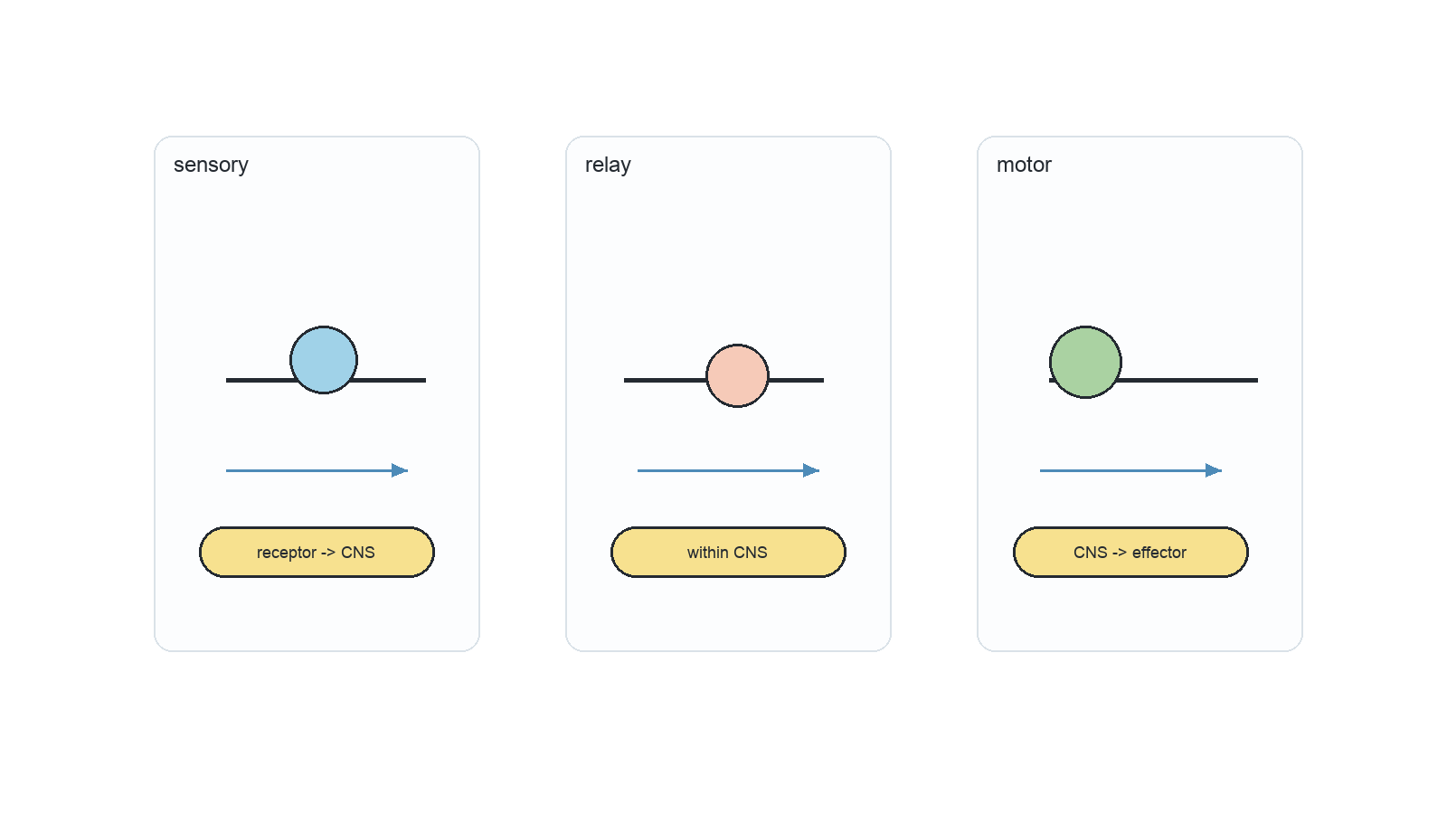
sensory (afferent) receptor → CNS delivers information for integration
motor (efferent) CNS → skeletal muscle delivers the coordinated response

The cerebral hemispheres coordinate voluntary action, but they do not contract the muscle directly. The final output is carried by motor neurons to the neuromuscular junction.

A Mixed Nerve Bundles Many Fibres in Protective Layers

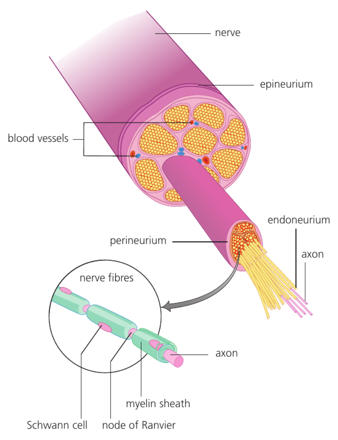
Structure What it encloses
endoneurium one nerve fibre
perineurium one fascicle, or bundle of fibres
epineurium several fascicles plus blood vessels, forming the whole nerve
A peripheral nerve cutaway shows many axons grouped into fascicles, surrounded by endoneurium, perineurium and epineurium.

A neuron is one cell. A nerve fibre is a neuron's long conducting process, usually an axon. A nerve is a connective-tissue package containing many fibres.

A mixed nerve can carry sensory impulses toward the CNS and motor impulses away from it at the same time because those signals travel in different fibres.

Withdrawal Begins before Conscious Pain Is Interpreted

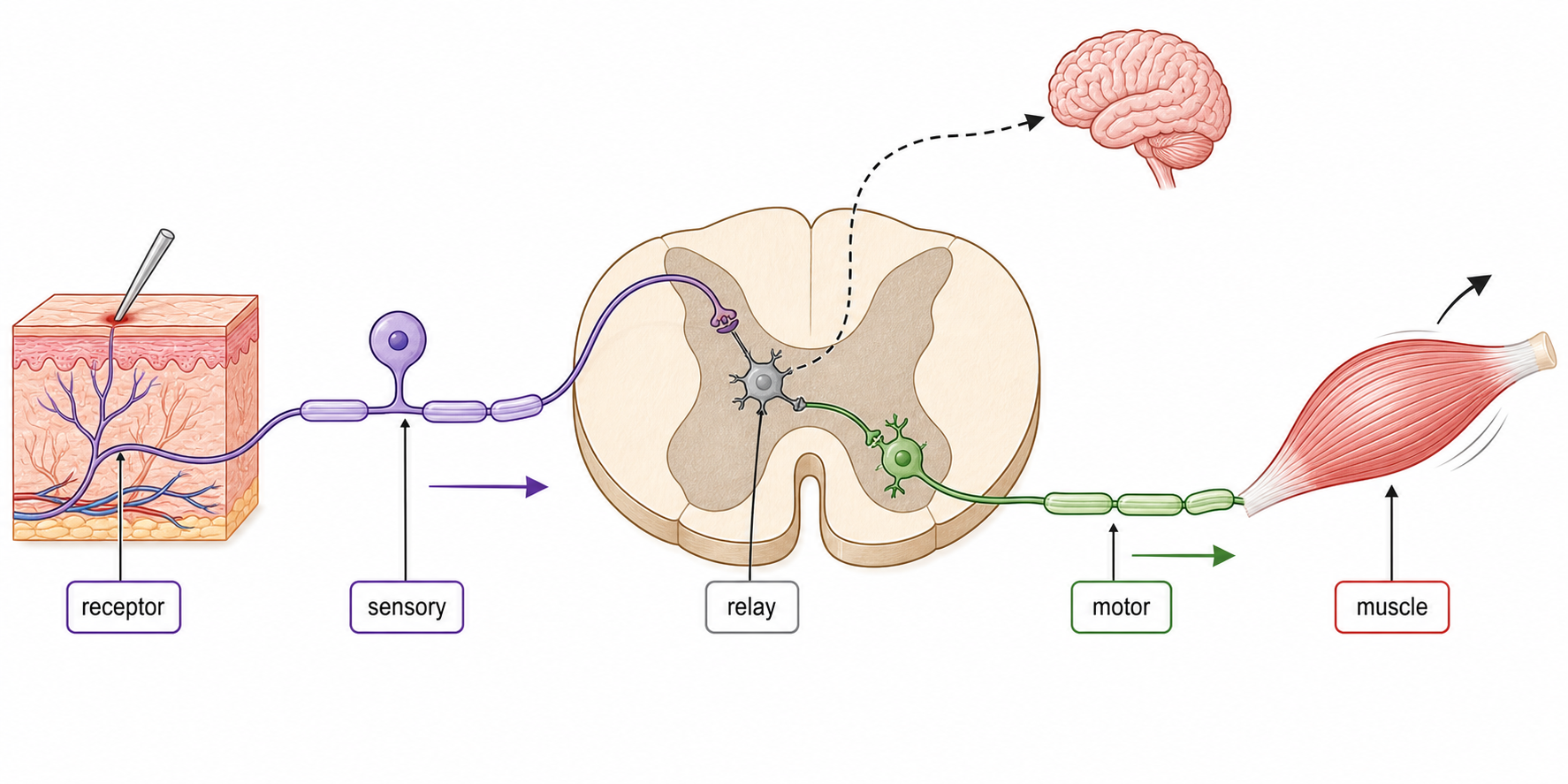
  1. A damaging stimulus activates a pain receptor in the skin.
  2. A sensory neuron carries impulses into the spinal cord.
  3. A relay neuron transfers the signal to a motor neuron.
  4. The motor neuron stimulates a flexor skeletal muscle.
  5. The muscle contracts and withdraws the limb.
A pain receptor in skin sends an impulse through a sensory neuron to a spinal relay neuron and motor neuron, contracting a flexor muscle to withdraw the hand while information also travels to the brain.
Route Earliest outcome
spinal reflex arc rapid involuntary withdrawal
ascending pathways to brain conscious pain, awareness and memory
descending input from brain may modify the response after integration

The reflex trades deliberation for speed: a predictable protective response starts through a short spinal pathway while the brain receives and interprets the same event.

The Cerebellum Fine-Tunes Movement; It Does Not Initiate It

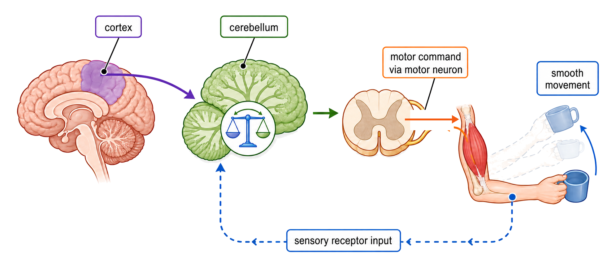
intended movement from cerebral cortex + sensory feedback about position and balance → cerebellum compares intention with performance → timing and force of motor output are adjusted → movement becomes smooth and posture remains stable

The cerebral cortex supplies an intended motor command, sensory receptors return information about the movement, and the cerebellum adjusts output so the movement becomes smooth.
Cerebellar contribution Example
timing activates muscles in the correct sequence
force prevents contraction from overshooting or undershooting
balance and posture uses receptor input to correct body position
learned precision refines writing, speech and practiced hand movements

The cerebellum coordinates and corrects skeletal-muscle contraction. Voluntary movement is initiated elsewhere, particularly in the cerebral cortex.

Melatonin Aligns Sleep Timing with the Light–Dark Cycle

A circadian rhythm is an approximately 24-hour pattern in physiology or behaviour. The pineal gland secretes melatonin as a hormonal timing signal within this clock system.

Environmental state Melatonin secretion Likely effect
darkness, including longer nights rises supports sleepiness and night-time timing
light falls supports preparation for wakefulness
abruptly shifted light–dark cycle internal rhythm and local time are misaligned jet lag persists while the clock resets

Melatonin modulates the timing of sleep; it is not a sedative switch that alone causes every transition into sleep. Light resets a continuing biological rhythm rather than producing an immediate, complete change.

Epinephrine Coordinates Organs for Immediate High Demand

During danger or stress, the adrenal medulla releases epinephrine into the blood. One hormone then coordinates target organs for vigorous activity.

Target response Immediate change Contribution to intense muscle contraction
heart heart rate and cardiac output increase delivers O₂ and glucose more rapidly
ventilation system breathing rate and depth increase increases gas exchange
liver glycogen is broken down and glucose enters blood raises available respiratory substrate
skeletal muscle and other responsive tissues metabolic pathways are shifted toward rapid ATP demand supports forceful activity

The important outcome is matched supply and demand: circulation, ventilation and fuel mobilisation rise together, allowing a higher rate of aerobic respiration and ATP production.

Epinephrine is an amino-acid-derived catecholamine, not a peptide hormone. It acts only in cells with appropriate adrenergic receptors.

The Hypothalamus Uses Two Routes to Control Pituitary Output

Pituitary part Hypothalamic route What the pituitary does
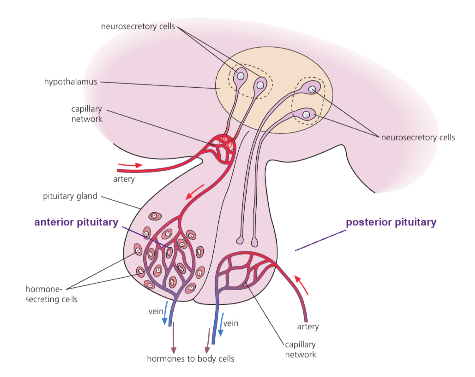
anterior releasing or inhibiting hormones travel through portal blood vessels endocrine cells synthesize and release pituitary hormones
posterior axons of hypothalamic neurosecretory cells end in the gland stores and releases ADH and oxytocin made in the hypothalamus
Hypothalamic neurosecretory cells regulate the anterior pituitary through a portal capillary system and send axons directly to the posterior pituitary.

Anterior-pituitary hormones regulate processes including growth, reproduction and homeostasis and can control endocrine glands such as the thyroid, gonads and adrenal cortex.

Calling the pituitary the ‘master gland’ is incomplete: the hypothalamus largely controls its activity, linking neural and endocrine information.

Heart-Rate Control Starts by Identifying the Disturbance

Receptor Variable detected Important locations What a change implies
baroreceptor stretch caused by blood pressure carotid sinus, aortic arch, right atrium pressure/venous return has changed
chemoreceptor mainly CO₂-linked pH; also O₂ carotid bodies, aortic bodies and brain/medulla gas exchange or metabolic demand has changed
  • High arterial pressure increases baroreceptor signalling and calls for a slower heart rate.
  • Low pressure calls for a faster, stronger response.
  • High CO₂ or low pH calls for increased blood flow so CO₂ reaches the lungs faster.

Sensory impulses converge on the cardiovascular centre in the medulla. The medulla integrates the evidence; receptors detect change but do not directly set the SA node.

Antagonistic Nerves Correct SA-Node Firing

Integrated condition Medulla increases output through… Transmitter at SA node SA-node effect
low pressure, high CO₂ or low pH sympathetic cardiac nerves norepinephrine firing rate rises; heart rate increases
high pressure or low CO₂ demand parasympathetic vagus nerve acetylcholine firing rate falls; heart rate decreases

disturbance → receptor firing changes → medulla integrates input → sympathetic or parasympathetic output changes → SA-node rate changes → blood pressure and gas transport move toward the required range

The SA node initiates each heartbeat within cardiac muscle. Autonomic nerves adjust its firing rate; they do not supply every impulse that causes the heart to contract.

Circulating epinephrine can also increase SA-node rate, so nervous and endocrine signals may reinforce one another during stress.

CO₂-Driven pH Feedback Adjusts Ventilation

  1. Cellular respiration raises blood CO₂.
  2. CO₂ forms carbonic acid, increasing H⁺ and lowering pH.
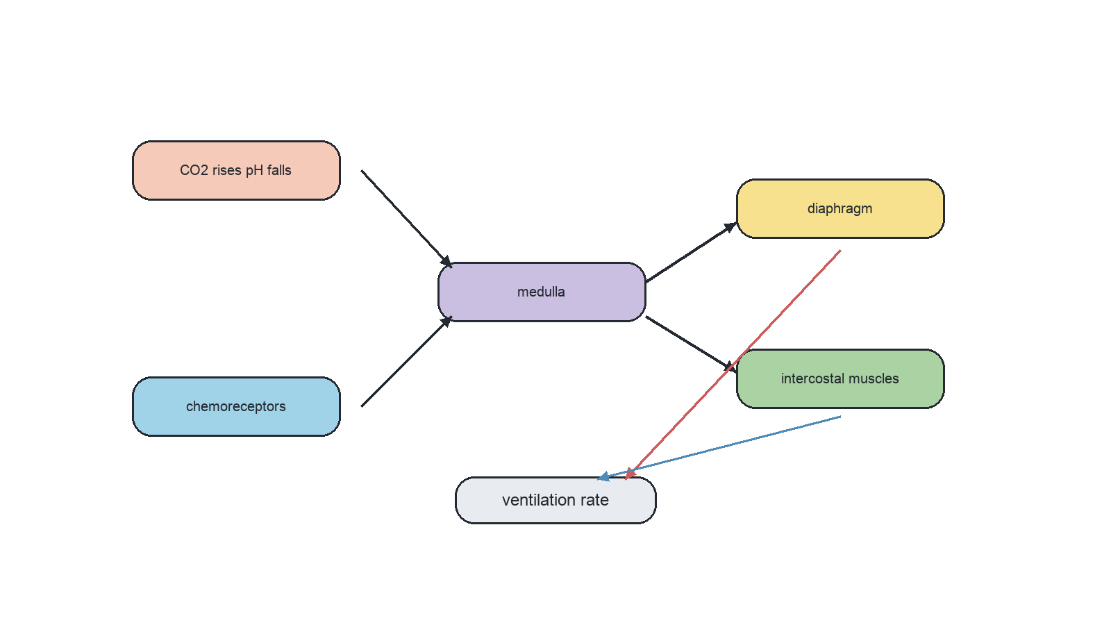
  3. Central and peripheral chemoreceptors increase signalling to respiratory centres in the medulla.
  4. Motor output to the diaphragm and external intercostal muscles increases rate and depth of contraction.
  5. Ventilation removes CO₂ faster, so H⁺ falls and pH moves back toward its set range.
Chemoreceptors in the medulla, aortic bodies and carotid bodies signal the respiratory centre, which controls intercostal muscles and the diaphragm.
Observation Best interpretation
CO₂ rises and pH falls ventilation should increase
exercise stops and CO₂ falls ventilation can decrease gradually
O₂ falls strongly peripheral chemoreceptors add a weaker but important drive

The principal routine stimulus is CO₂-linked H⁺, not a direct measurement of how hard the muscles are working. Voluntary control can temporarily override the automatic rhythm but does not replace it.

The Gut Combines Voluntary Boundaries with Enteric Control

Part of the route Main control Status
start of swallowing CNS and somatic motor pathways voluntary initiation
most movement through gut enteric nervous system, modulated by autonomic/CNS input involuntary
defecation boundary spinal/CNS reflexes plus voluntary control of external sphincter mixed control

Peristalsis is a travelling wave of contraction and relaxation in circular and longitudinal smooth muscle that propels gut contents forward.

The enteric nervous system contains sensory neurons, relay neurons and motor neurons in the gut wall. Local stretch and chemical information can therefore coordinate adjacent regions without a separate conscious command for every contraction.

Enteric is not a synonym for all autonomic control. The enteric nervous system specifically serves the gastrointestinal tract and is one specialised part of involuntary regulation.

SL Summary: Build a Coordinated Response from Routes and Feedback

System architecture: specialised cells form tissues and organs; nervous signals, hormones and circulating materials cross the boundaries between them; coordinated interaction creates emergent functions.

Control route: disturbance or stimulus → receptor evidence → integration centre → targeted motor or endocrine output → effector response → changed internal condition.

Observation Most useful place to inspect
touch is detected but no voluntary movement follows CNS integration, descending motor route or neuromuscular junction
pressure rises but heart rate does not fall baroreceptors, medulla, vagus nerve or SA node
CO₂ rises but breathing remains unchanged chemoreceptors, respiratory centre, phrenic/intercostal output
gut smooth muscle is intact but waves are uncoordinated enteric sensory, relay and motor circuits
Stabilising loop Coordinated response without a fixed set point
heart-rate and ventilation feedback oppose a disturbance epinephrine temporarily raises several outputs together for anticipated demand

System integration

3 marks

Using the diagram, explain the concept of emergent properties of biofilms.

Hierarchy in multicellular organisms

1 mark

Why do multicellular organisms have emergent properties?

Integration of organs

3 marks

Pancreatic secretions contain sodium hydrogen carbonate, making them basic.
Deduce the significance of the response by the pancreas to secretin.

Brain as information integration organ

6 marks

Explain two methods that scientists have used to determine the different functions of the brain.

Spinal cord

1 mark

The spinal cord is involved in the coordination of involuntary actions such as the pain reflex.

Where are synapses between sensory neurons and interneurons found?

Input through sensory neurons

1 mark

Between which structures do sensory neurons carry nerve impulses?

Output through motor neurons

1 mark

What is the main role of nerves in human movement?

Nerves as bundles

1 mark

State the role of the vagus nerve.

Pain reflex arcs

4 marks

Draw a labelled diagram of a reflex arc for a pain withdrawal reflex.

Cerebellum role

2 marks

Describe the use of fMRI to identify the role of the cerebellum.

Melatonin and sleep

2 marks

Outline the role of melatonin in humans.

Epinephrine (adrenaline)

2 marks

Describe two ways in which epinephrine in the body facilitates intense muscle contraction. [2]
1.
2.

Hypothalamus and pituitary control

7 marks

Explain the roles of named hormones in the development and function of the sexual reproductive systems in males and females.

Heart rate feedback control

7 marks

Explain the control mechanism of the heart rate.

Ventilation rate feedback control

8 marks

Explain the causes of a decreased blood pH and its effects on the ventilation rate in humans.

Peristalsis control

3 marks

Outline how food is moved from the stomach to the large intestine.