C2.2 Neural signalling

Neural signalling transmits information through neuron structure, ion gradients, action potentials, synapses and chemical modulation of postsynaptic responses, linking cellular mechanisms to rapid communication in nervous systems.

Syllabus
First assessment 2025
Topic
C2.2
Level
SL

Neurons Carry Information through Electrical and Chemical Signals

A neuron is a specialized cell of the nervous system that conducts electrical impulses along elongated nerve fibres.

The cell body contains cytoplasm and the nucleus. Multiple shorter dendrites usually conduct impulses toward the cell body, while one long axon conducts impulses away toward other neurons or effector cells.
Identify the nucleus and cytoplasm in the cell body, then distinguish the long single axon from the multiple shorter dendrites. Fibre length and branching vary with neuron function.
A motor neuron's long axon can carry an impulse from the central nervous system to a distant muscle, while its dendrites receive inputs from other neurons.

An axon and dendrites are cellular projections, not separate cells. Neuron shape varies, so use fibre number, length and direction of impulse rather than assuming every drawing has the same layout.

Neurons exam focus

Assessment in practice

1–2 marks
How it is assessed

This objective is assessed through experimental design, commonly using Label / Draw / State.

Command terms

Label / Draw / State / Distinguish / Identify

What earns marks

Build the answer around this relationship: Dendrites receive signals and conduct them toward the cell body.

Representative question

Question 1

[Maximum number: 5]

Draw a labelled diagram of a motor neuron.

Resting Potential Stores an Ion Gradient

The resting potential is the negative voltage across a resting neuron's polarized plasma membrane, maintained by sodium and potassium ion gradients.

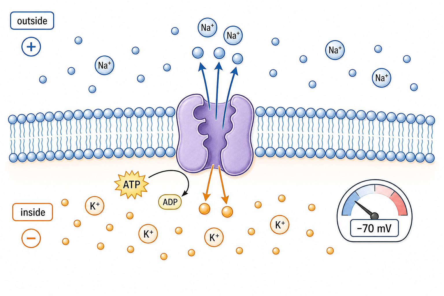
ATP supplies energy to the sodium–potassium pump, which moves Na⁺ out and K⁺ in opposite directions against their gradients. The membrane is more permeable to K⁺ at rest, so more positive charge diffuses out than enters and the inside remains negative relative to outside.

ATP-driven pumping establishes and maintains high Na⁺ outside and high K⁺ inside; selective leak channels create unequal ion movement; separated charge produces membrane polarization and a measurable membrane potential.

If ATP production stops, the pump can no longer maintain the gradients. Existing gradients may persist briefly, but continued leakage gradually erodes the negative resting potential.

The pump maintains the gradients over time; it does not directly create each action potential. A concentration gradient and a voltage gradient both influence ion movement.

Resting potential generation

Assessment in practice

1–2 marks
How it is assessed

This objective is assessed through structured response, commonly using Define / Outline / Explain.

Command terms

Define / Outline / Explain / State / Label

What earns marks

Build the answer around this relationship: The resting axon is negative inside relative to outside.

Representative question

Question 1

[Maximum number: 4]

Outline how neurons generate a resting potential.

An Action Potential Is an All-or-none Nerve Impulse

An action potential is a brief, threshold-triggered reversal of membrane potential that propagates along an axon.

Voltage-gated sodium channels open rapidly during depolarization, then inactivate while potassium channels open to repolarize the membrane. Refractory periods prevent immediate re-firing in the same segment. The threshold is a condition, not a fixed promise: if it is not reached, the full action potential does not start.

Trace the phases:

  • threshold
  • Na⁺ influx and depolarization
  • K⁺ efflux and repolarization
  • hyperpolarization and recovery

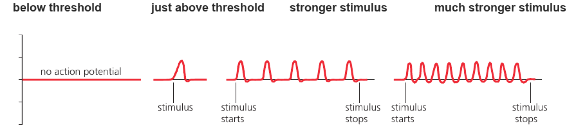
A stimulus below threshold produces no full impulse, while a threshold stimulus produces an impulse with a similar amplitude.

Stronger stimuli are coded mainly by impulse frequency, not by a larger action-potential amplitude.

Nerve impulses as action potentials

Assessment in practice

1–8 marks
How it is assessed

This objective is assessed through structured response, commonly using Explain / Outline.

Command terms

Explain / Outline

What earns marks

Build the answer around this relationship: Action potentials are regenerated along neuron membranes.

Representative question

Question 1

[Maximum number: 3]

Outline how nerve impulses are transmitted along a nerve fibre.

Impulse Speed Depends on Axon Design

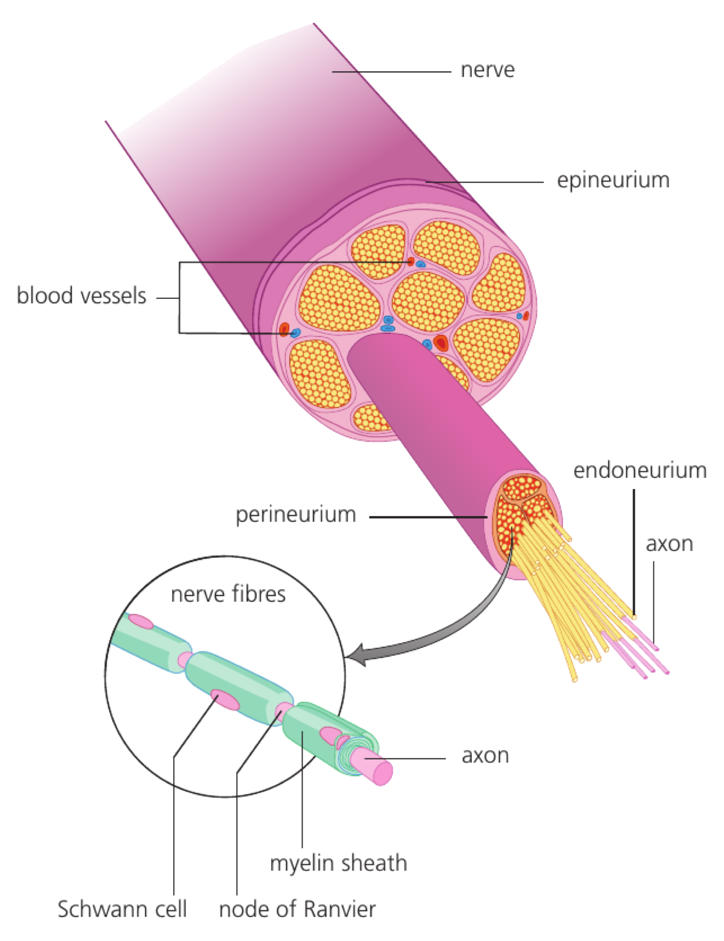
Impulse conduction speed increases with axon diameter and myelination because both allow local currents to influence the next excitable membrane region more rapidly.

Comparison Faster case Mechanistic reason
Giant squid axon vs smaller unmyelinated fibre Giant squid axon Larger diameter lowers internal resistance
Myelinated vs unmyelinated fibre Myelinated fibre Insulation limits leakage and impulses regenerate mainly at nodes

Describe a positive correlation when conduction speed rises as axon diameter rises, and a negative correlation when one variable falls as the other rises. The correlation coefficient r gives direction and strength; R² estimates the proportion of variation in conduction speed explained by the fitted relationship.

If r = 0.90 for diameter and speed, the association is strong and positive; R² = 0.81 means about 81% of the observed variation in speed is explained by the fitted relationship, not that diameter is proven to be the sole cause.

Correlation does not by itself prove causation. Faster impulses do not have larger action-potential amplitudes; speed and signal strength are different properties.

Variation in impulse speed

Assessment in practice

1 marks
How it is assessed

This objective is assessed through multiple choice, commonly using Describe / Determine / Calculate.

Command terms

Describe / Determine / Calculate / Suggest / Outline

What earns marks

Build the answer around this relationship: Larger axon diameter is associated with faster impulse transmission.

Representative question

Question 1

[Maximum number: 1]

The diagrams represent sections through different axons. Which axon has the slowest speed of impulse?

A
B

A Synapse Connects One Cell to Another

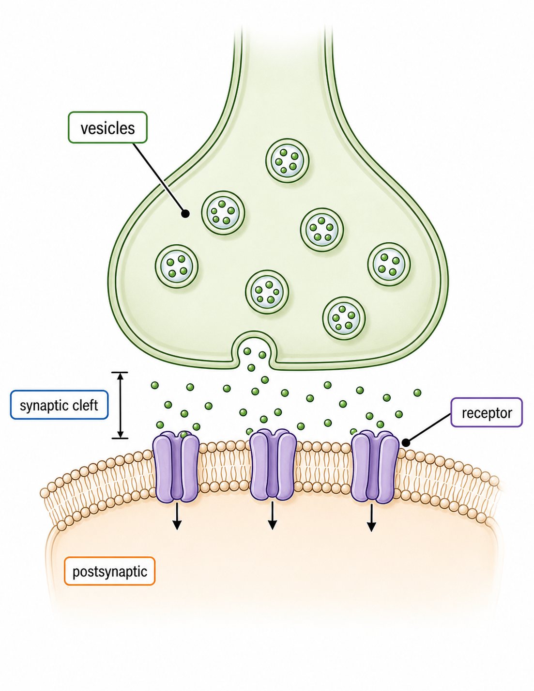
A synapse is a junction where a presynaptic cell communicates with a postsynaptic cell, usually through neurotransmitter release.

The synaptic cleft separates membranes, creating a one-way delay in chemical synapses. Receptors on the postsynaptic membrane convert transmitter binding into ion-flow or intracellular effects. The synapse is a junction, so the signal must be converted before it can cross to the next cell.

Describe a chemical synapse:

  • presynaptic terminal
  • vesicles and cleft
  • postsynaptic receptors
  • signal termination

An action potential at a motor neuron terminal releases transmitter that binds receptors on a muscle fibre.

A synapse is not simply a physical gap; receptor type and transmitter removal determine the response.

Synapses as junctions

Assessment in practice

1–6 marks
How it is assessed

This objective is assessed through structured response, commonly using Explain / Describe / State.

Command terms

Explain / Describe / State / Identify / Distinguish / Suggest

What earns marks

Build the answer around this relationship: Synapses transmit signals chemically between neurons.

Representative question

Question 1

[Maximum number: 2]

Distinguish between the presynaptic and postsynaptic membranes by giving two differences.

Calcium Triggers Neurotransmitter Release

Arrival of an action potential at a presynaptic terminal opens voltage-gated calcium channels and triggers vesicle fusion.

Ca²⁺ entry binds release machinery, causing exocytosis of neurotransmitter into the synaptic cleft. Enzymatic breakdown, reuptake or diffusion then terminates the signal. This one-way sequence explains why changing calcium entry can alter how strongly a synapse communicates.

Trace release:

  • action potential arrives
  • Ca²⁺ channels open
  • vesicles fuse
  • transmitter binds and is cleared

Blocking presynaptic calcium entry reduces transmitter release even if the action potential still reaches the terminal.

The electrical impulse triggers release; it does not cross the cleft as the same electrical current.

Neurotransmitter release

Assessment in practice

1–3 marks
How it is assessed

This objective is assessed through structured response, commonly using Describe / Explain / Outline.

Command terms

Describe / Explain / Outline / Deduce / Label / State

What earns marks

Build the answer around this relationship: An arriving action potential depolarizes the presynaptic terminal.

Representative question

Question 1

[Maximum number: 3]

Describe how neurotransmitters are released from a presynaptic neuron membrane.

Excitatory Postsynaptic Potentials Move a Neuron toward Threshold

An excitatory postsynaptic potential (EPSP) is a graded depolarization that makes the postsynaptic neuron more likely to reach action-potential threshold.

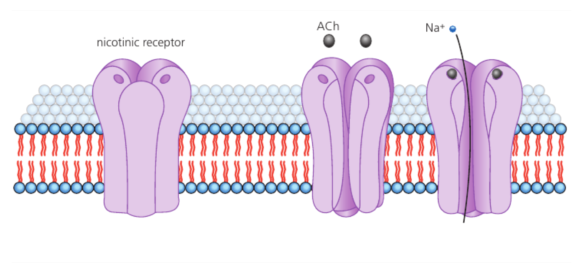
Neurotransmitter released from the presynaptic terminal diffuses across the synaptic cleft and binds a transmembrane receptor. Acetylcholine can open ligand-gated channels, allowing net positive charge to enter and depolarize the postsynaptic membrane.

Sequence: transmitter release → diffusion across cleft → receptor binding → channel opening → local depolarization. Acetylcholine acts at many synapses, including neuromuscular junctions.

One acetylcholine input may produce a small EPSP below threshold; overlapping excitatory inputs can sum at the axon hillock until an action potential is triggered.

An EPSP is local and graded, not an all-or-none action potential. The neurotransmitter does not carry the electrical impulse across the cleft; receptor-controlled ion movement changes voltage.

Excitatory postsynaptic potential

Assessment in practice

1–2 marks
How it is assessed

This objective is assessed through structured response, commonly using Explain / Describe.

Command terms

Explain / Describe

What earns marks

Build the answer around this relationship: Excitatory neurotransmitters bind receptors on the postsynaptic membrane.

Representative question

Question 1

[Maximum number: 3]

Describe how an excitatory postsynaptic potential is generated.

Up to one additional mark is available for the construction of your answer.

SL Transfer: Explain Core Neural Signalling

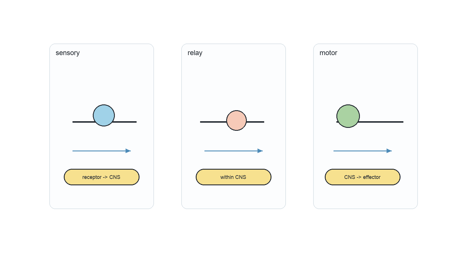
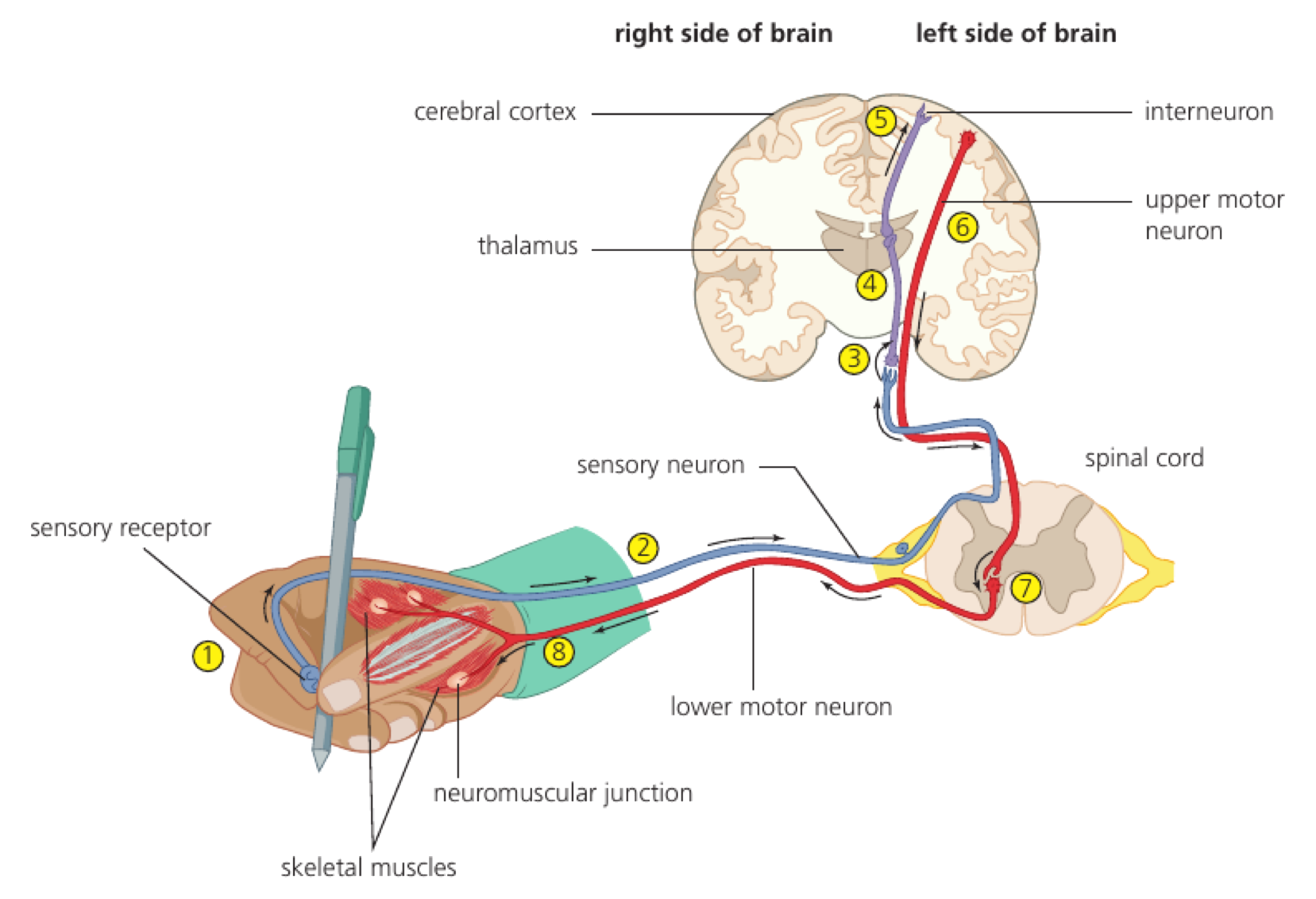
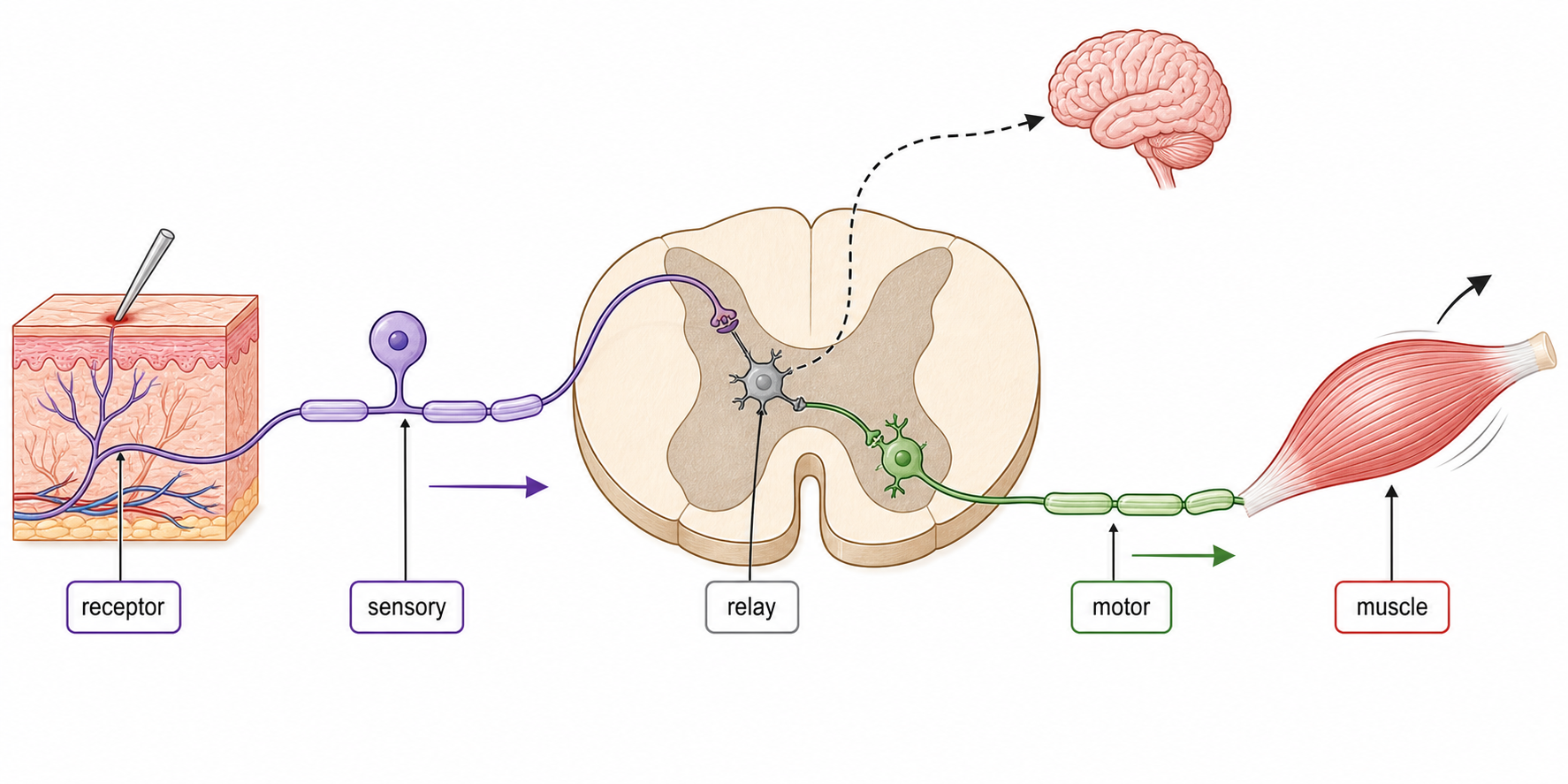
Neurons carry electrical impulses in the nervous system; motor, sensory, and relay neurons differ in axon, dendrite, and cell body arrangement. Sodium-potassium pumps use ATP to move 3 Na+ out and 2 K+ in; ion gradients make the resting axon polarized at about -70 mV. A nerve impulse is a propagated action potential along a nerve fibre; stimulus-triggered sodium influx reverses membrane polarity. Larger axon diameter lowers resistance and increases impulse speed; myelin sheaths and nodes of Ranvier enable faster saltatory conduction. Synapses connect neurons to neurons, muscles, or glands; chemical synapses transmit one way across a narrow synaptic cleft. Action potentials open voltage-gated Ca2+ channels in presynaptic terminals; Ca2+ causes vesicle fusion and neurotransmitter exocytosis into the cleft. Neurotransmitters diffuse and bind receptors on the postsynaptic membrane; EPSPs depolarize the membrane and make threshold more likely.

Objective notes

7 learning objectives
C2.2.1Neurons• Neurons carry electrical impulses in the nervous system• Motor, sensory, and relay neurons differ in axon, dendrite, and cell body arrangement7% of analysed papers 10 papers · 11 questionsViewC2.2.2Resting potential generation• Sodium-potassium pumps use ATP to move 3 Na⁺ out and 2 K⁺ in• Ion gradients make the resting axon polarized at about -70 mV4% of analysed papers 5 papers · 6 questionsViewC2.2.3Nerve impulses as action potentials• A nerve impulse is a propagated action potential along a nerve fibre• Stimulus-triggered sodium influx reverses membrane polarity4% of analysed papers 6 papers · 6 questionsViewC2.2.4Variation in impulse speed• Larger axon diameter lowers resistance and increases impulse speed• Myelin sheaths and nodes of Ranvier enable faster saltatory conduction4% of analysed papers 6 papers · 6 questionsViewC2.2.5Synapses as junctions• Synapses connect neurons to neurons, muscles, or glands• Chemical synapses transmit one way across a narrow synaptic cleft6% of analysed papers 9 papers · 10 questionsViewC2.2.6Neurotransmitter release• Action potentials open voltage-gated Ca²⁺ channels in presynaptic terminals• Ca²⁺ causes vesicle fusion and neurotransmitter exocytosis into the cleft4% of analysed papers 5 papers · 5 questionsViewC2.2.7Excitatory postsynaptic potential• Neurotransmitters diffuse and bind receptors on the postsynaptic membrane• EPSPs depolarize the membrane and make threshold more likely2% of analysed papers 3 papers · 3 questionsView