C3.1 Integration of body systems

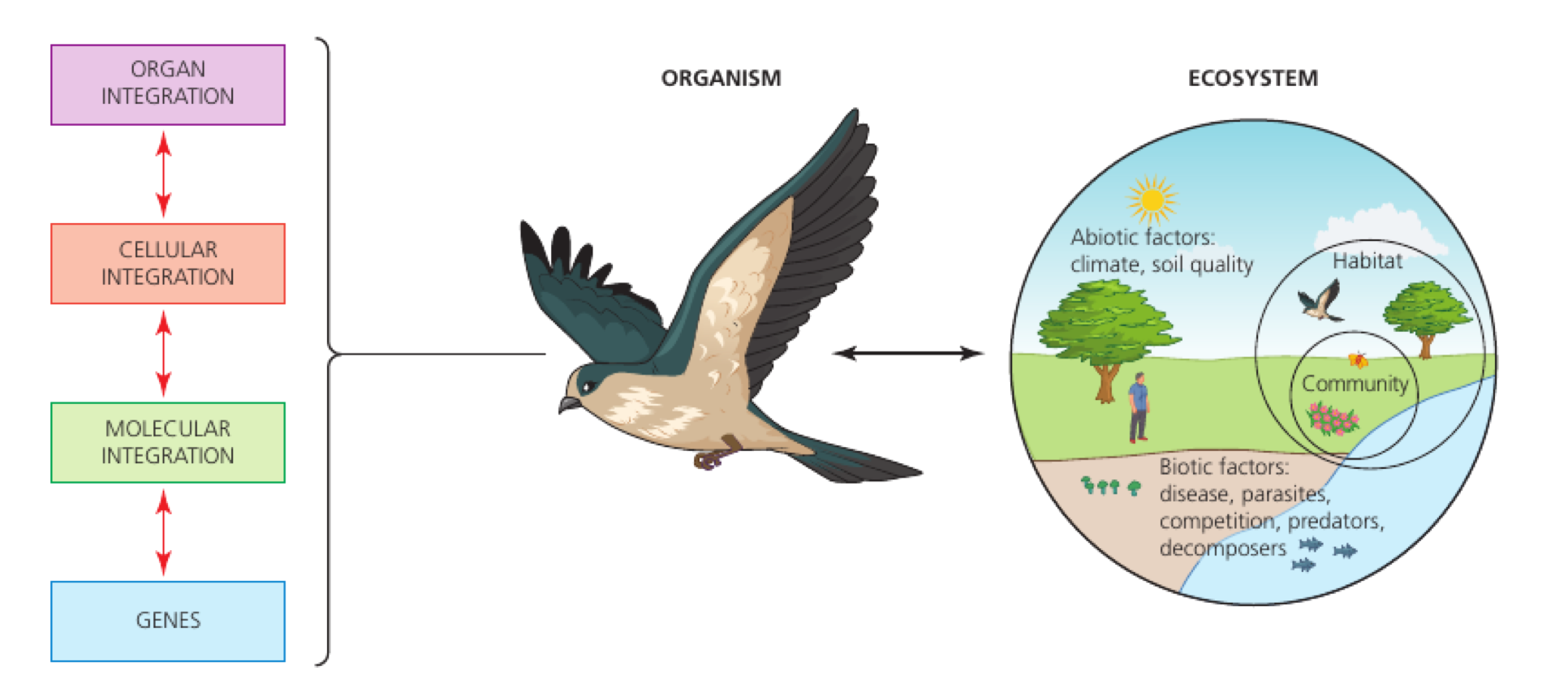
Integrated body systems coordinate neural, hormonal and plant responses through receptors, control centres and effectors, maintaining function across organisms and changing environmental conditions.

Syllabus
First assessment 2025
Topic
C3.1
Level
SL

Integration joins specialised parts into a working whole

Integration joins specialised parts into a working whole.

In a multicellular organism, tissues, organs and organ systems exchange information and materials so the organism can coordinate one response. Nervous impulses are fast; hormones travel in blood and can act on distant targets.

Name the parts; identify the signal or material crossing between them; then state the coordinated outcome.

During exercise, receptors and the brain increase heart and ventilation activity while blood delivers oxygen to working muscle.

Integration is more than a list of organs: the explanation must include communication and a shared function.

System integration

Assessment in practice

1–3 marks
How it is assessed

This objective is assessed through structured response, commonly using Identify / Explain.

Command terms

Identify / Explain

What earns marks

Mark schemes reward named structures, correct direction of information flow and explanations that keep the sequence causal.

Watch for

Naming a brain region without linking it to information integration or coordination.

Representative question

Question 1

[Maximum number: 3]

Using the diagram, explain the concept of emergent properties of biofilms.

Organisation builds new properties at each level

Organisation builds new properties at each level.

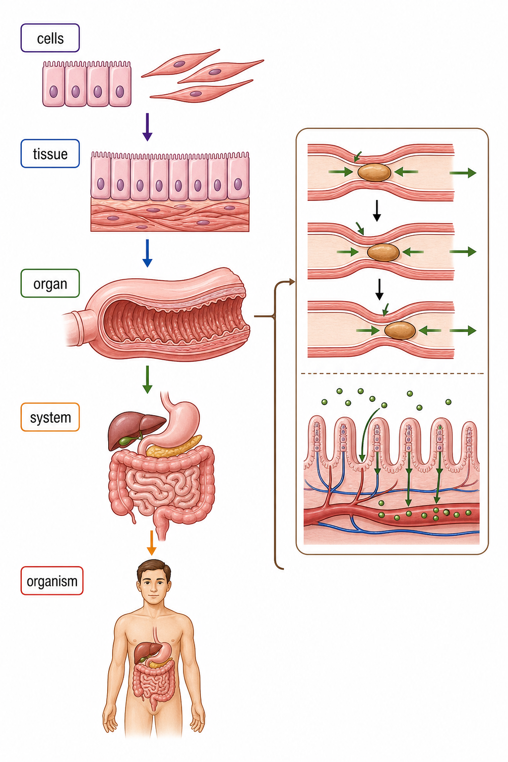
Cells of one type form tissues, different tissues form organs, and organs form systems. Interactions between parts can produce an emergent capability that no isolated cell performs alone.

cell → tissue → organ → system → organism; at each step ask what cooperation adds.

Cardiac muscle, connective tissue and blood vessels combine in a heart that pumps blood; no one tissue performs the whole job.

Hierarchy is not just size ranking; it is a functional relationship between levels.
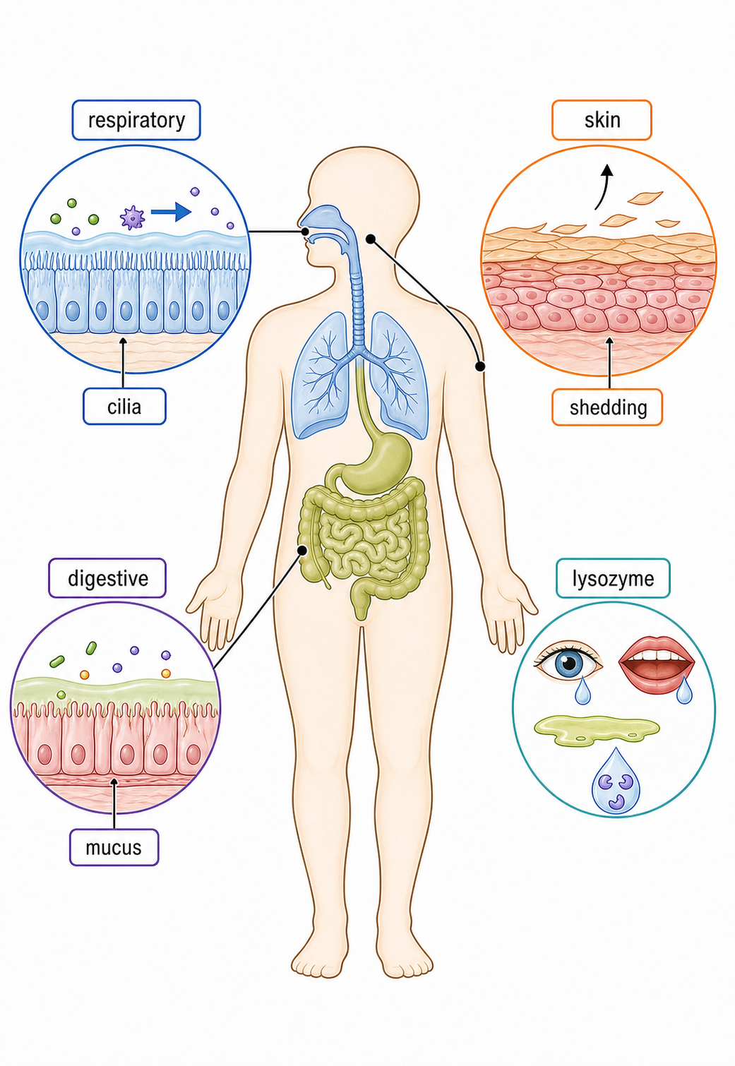
Hierarchy in multicellular organisms

Assessment in practice

1 marks
How it is assessed

This objective is assessed through multiple choice, commonly using Explain.

Command terms

Explain

What earns marks

Mark schemes reward named structures, correct direction of information flow and explanations that keep the sequence causal.

Representative question

Question 1

[Maximum number: 1]

Why do multicellular organisms have emergent properties?

A

They have more genes than unicellular organisms.

B

Properties of unicellular organisms are enhanced by having many cells.

C

All of their genes are expressed whereas unicellular organisms express only some.

D

They show properties that can only result from the interaction of many cells.

Organs integrate by sharing materials and control

Animal organs are integrated by nervous and hormonal messages and by blood transport of materials and energy between organs.

Integration route Message or material Typical pattern
Nervous signalling Electrical impulses and neurotransmitters Rapid, targeted, usually short-lived
Endocrine signalling Hormones carried in blood Slower, distributed to distant receptor-bearing cells, often longer-lived
Blood transport Oxygen, nutrients, heat and wastes Connects organ inputs and outputs continuously

During exercise, motor nerves stimulate skeletal muscles, epinephrine coordinates responses in several organs, and blood carries oxygen and glucose to muscle while transporting carbon dioxide away.

Blood is both a transport route for hormones and a carrier of non-message materials. Integration requires source, route, target and coordinated outcome—not just a list of organs.

Integration of organs

Assessment in practice

1–2 marks
How it is assessed

This objective is assessed through structured response, commonly using Identify / Explain / Draw.

Command terms

Identify / Explain / Draw / Deduce / Suggest / Outline / Describe

What earns marks

Mark schemes reward named structures, correct direction of information flow and explanations that keep the sequence causal.

Watch for

Giving a general body-system answer without the specific pathway step for integration of organs.

Representative question

Question 1

[Maximum number: 3]

Pancreatic secretions contain sodium hydrogen carbonate, making them basic.
Deduce the significance of the response by the pancreas to secretin.

The brain combines information before coordinating responses

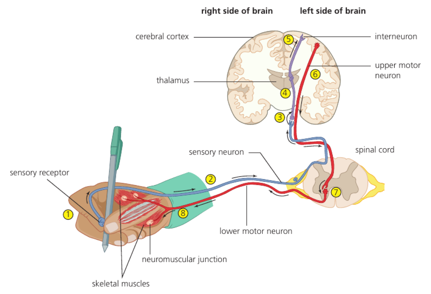
The brain is a central integration organ that combines information from several inputs before coordinating responses.

Current sensory inputs can be compared with stored information. Changes in neural connections support learning, while stored and retrievable information supports memory; both influence how later inputs are interpreted and acted on.

Trace input from several receptors → combined processing in the brain → comparison with learned or remembered information → coordinated output. Detailed slow-acting neurotransmitter mechanisms are not required.

A learner sees a traffic signal, hears an approaching vehicle and recalls the crossing rule; the brain combines these inputs and memory before coordinating whether to step forward or wait.

Integration is not simple forwarding of one sensory message. The objective concerns combining inputs plus learning and memory, not memorizing detailed functions of many brain regions.

Brain as information integration organ

Assessment in practice

1–3 marks
How it is assessed

This objective is assessed through essay response, commonly using Explain / Outline / Identify.

Command terms

Explain / Outline / Identify / Label / State / Discuss / Determine / Compare / Contrast

What earns marks

Mark schemes reward named structures, correct direction of information flow and explanations that keep the sequence causal.

Watch for

Naming a brain region without linking it to information integration or coordination.

Representative question

Question 1

[Maximum number: 6]

Explain two methods that scientists have used to determine the different functions of the brain.

The spinal cord can integrate a rapid response

The spinal cord can integrate a rapid response.

The spinal cord links body and brain, but grey matter can connect sensory input to motor output without waiting for conscious brain processing. White matter carries impulses along longer pathways.

receptor → sensory neurone → spinal integration → motor neurone → effector.

Touching a hot surface triggers a withdrawal through a spinal reflex; the brain receives information as the movement is already beginning.

A reflex is not brain-free information: the spinal cord handles the immediate response, while the brain can receive the signal afterwards.

Spinal cord

Assessment in practice

1 marks
How it is assessed

This objective is assessed through structured response, commonly using State.

Command terms

State

What earns marks

Mark schemes reward named structures, correct direction of information flow and explanations that keep the sequence causal.

Representative question

Question 1

[Maximum number: 1]

The spinal cord is involved in the coordination of involuntary actions such as the pain reflex.

Where are synapses between sensory neurons and interneurons found?

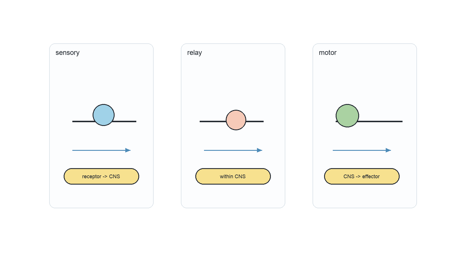
Sensory neurones carry transduced information to the CNS

Sensory neurones carry transduced information to the CNS.

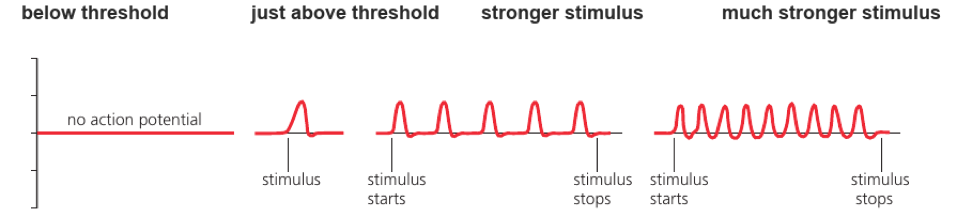
A receptor detects a stimulus and converts its energy into a change in membrane potential. If threshold is reached, an action potential travels along a sensory neurone to the CNS.

identify stimulus; receptor type; transduction; threshold; direction to CNS.

Salt ions entering taste-receptor channels depolarise the receptor cell, which then excites a sensory neurone carrying the signal to the brain.

A stronger stimulus is not encoded by a larger action potential; it can increase firing frequency or recruit receptors.

Input through sensory neurons

Assessment in practice

1–2 marks
How it is assessed

This objective is assessed through multiple choice, commonly using Identify / Outline.

Command terms

Identify / Outline

What earns marks

Mark schemes reward named structures, correct direction of information flow and explanations that keep the sequence causal.

Watch for

Giving a general body-system answer without the specific pathway step for sensory neuron input pathway.

Representative question

Question 1

[Maximum number: 1]

Between which structures do sensory neurons carry nerve impulses?

A

From receptors to muscles

B

From effectors to the central nervous system (CNS)

C

From the central nervous system (CNS) to receptors

D

From receptors to the central nervous system (CNS)

Motor neurones turn CNS decisions into muscle action

Motor neurones turn CNS decisions into muscle action.

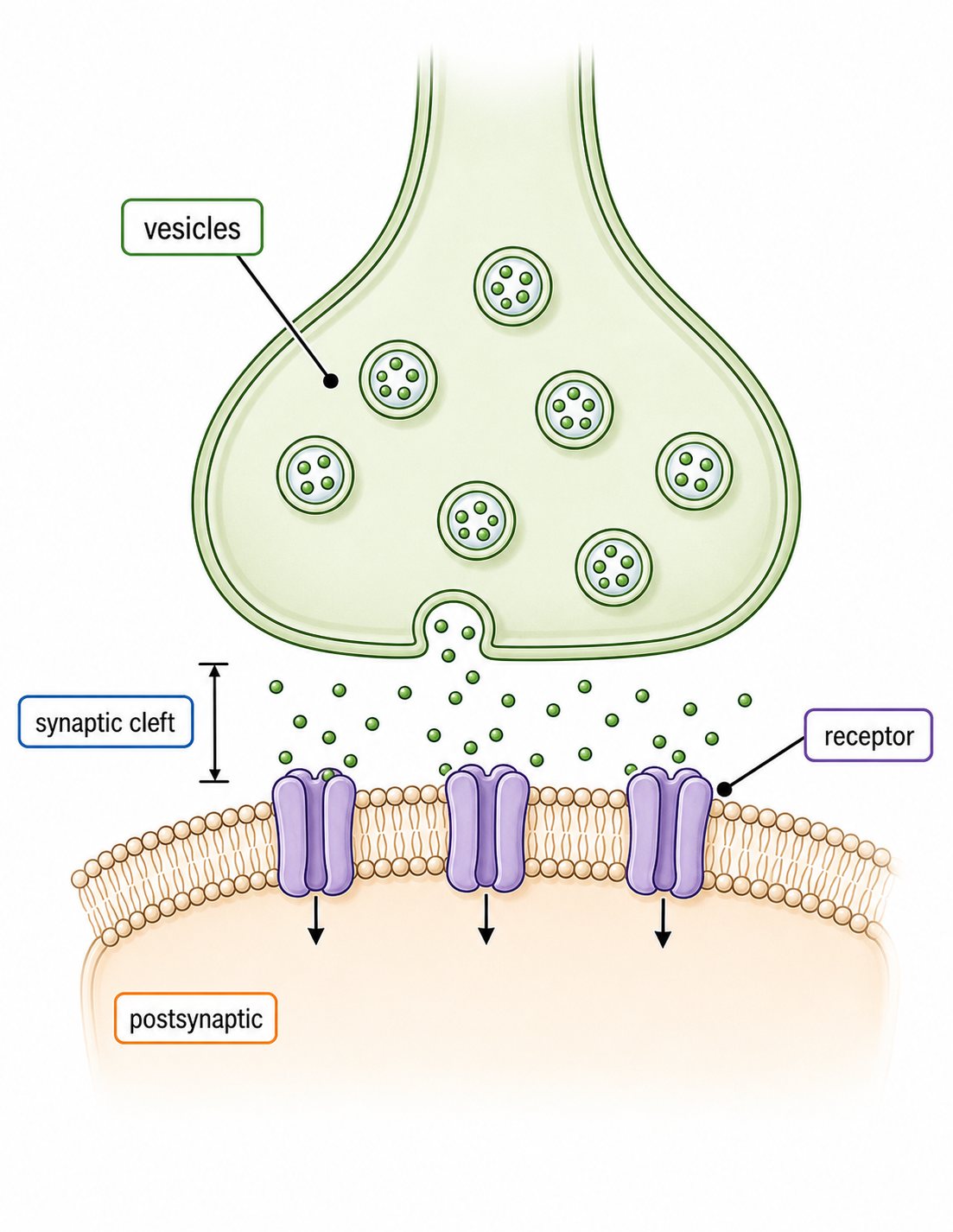
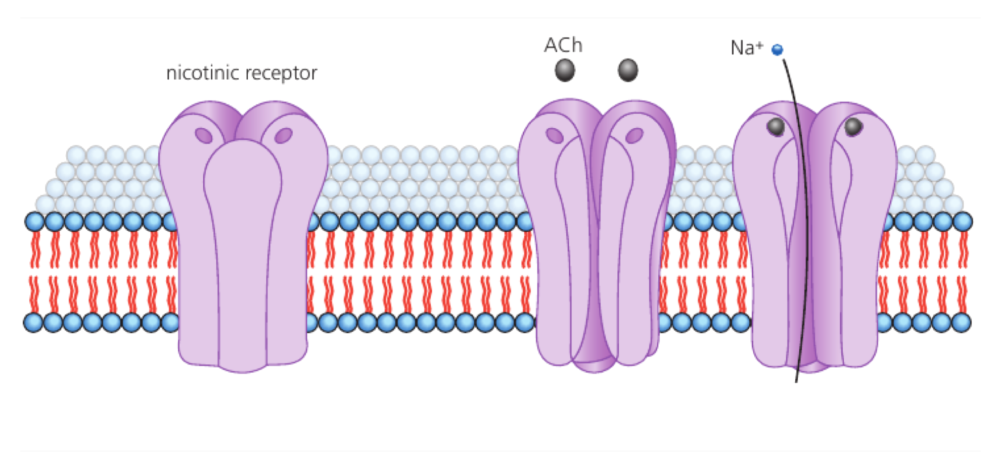
Motor neurones carry action potentials from the CNS to an effector. At a neuromuscular junction, acetylcholine depolarises the muscle membrane and starts the calcium-controlled contraction sequence.

CNS signal → motor axon → acetylcholine release → muscle action potential → calcium release.

An impulse at a motor end plate opens ion channels; the resulting muscle action potential travels along the fibre and triggers calcium release from the sarcoplasmic reticulum.

The motor neurone does not contract the muscle directly; transmitter and membrane events link the two cells.

Output through motor neurons

Assessment in practice

1 marks
How it is assessed

This objective is assessed through multiple choice, commonly using Describe / Identify.

Command terms

Describe / Identify

What earns marks

Mark schemes reward named structures, correct direction of information flow and explanations that keep the sequence causal.

Watch for

Giving a general body-system answer without the specific pathway step for motor neuron output pathway.

Representative question

Question 1

[Maximum number: 1]

What is the main role of nerves in human movement?

A

To cause muscles to stretch

B

To move joints

C

To transport pain signals that indicate muscle injuries

D

To stimulate muscle contraction

A nerve is a bundle of neurone axons

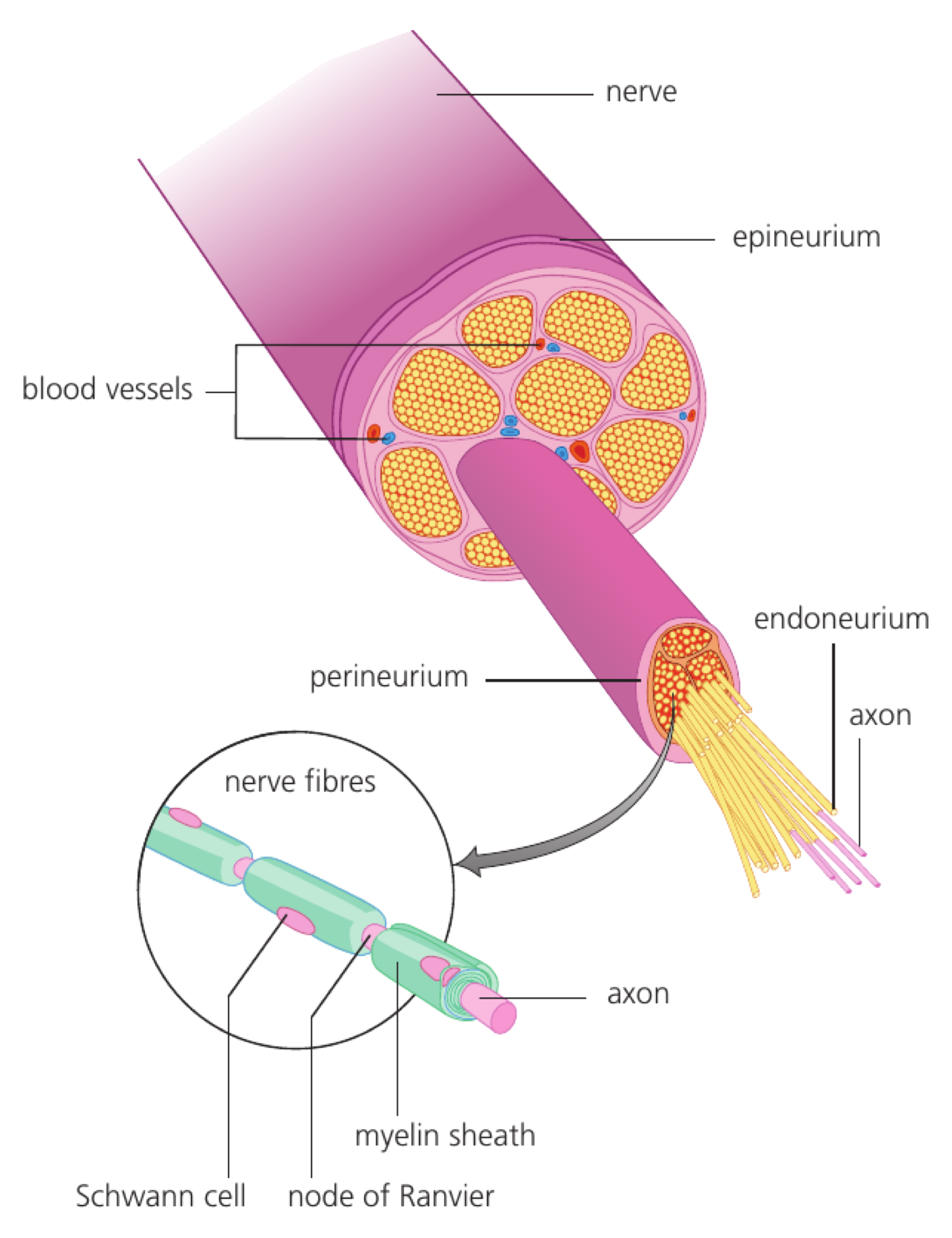
A peripheral nerve is a bundle of many nerve fibres—axons—from sensory and motor neurons, protected and organized by connective tissue sheaths.

In transverse section, each fibre appears cut across. Some have a visible myelin sheath around the axon and others are unmyelinated; groups of fibres are surrounded by protective connective tissue and supplied by blood vessels.
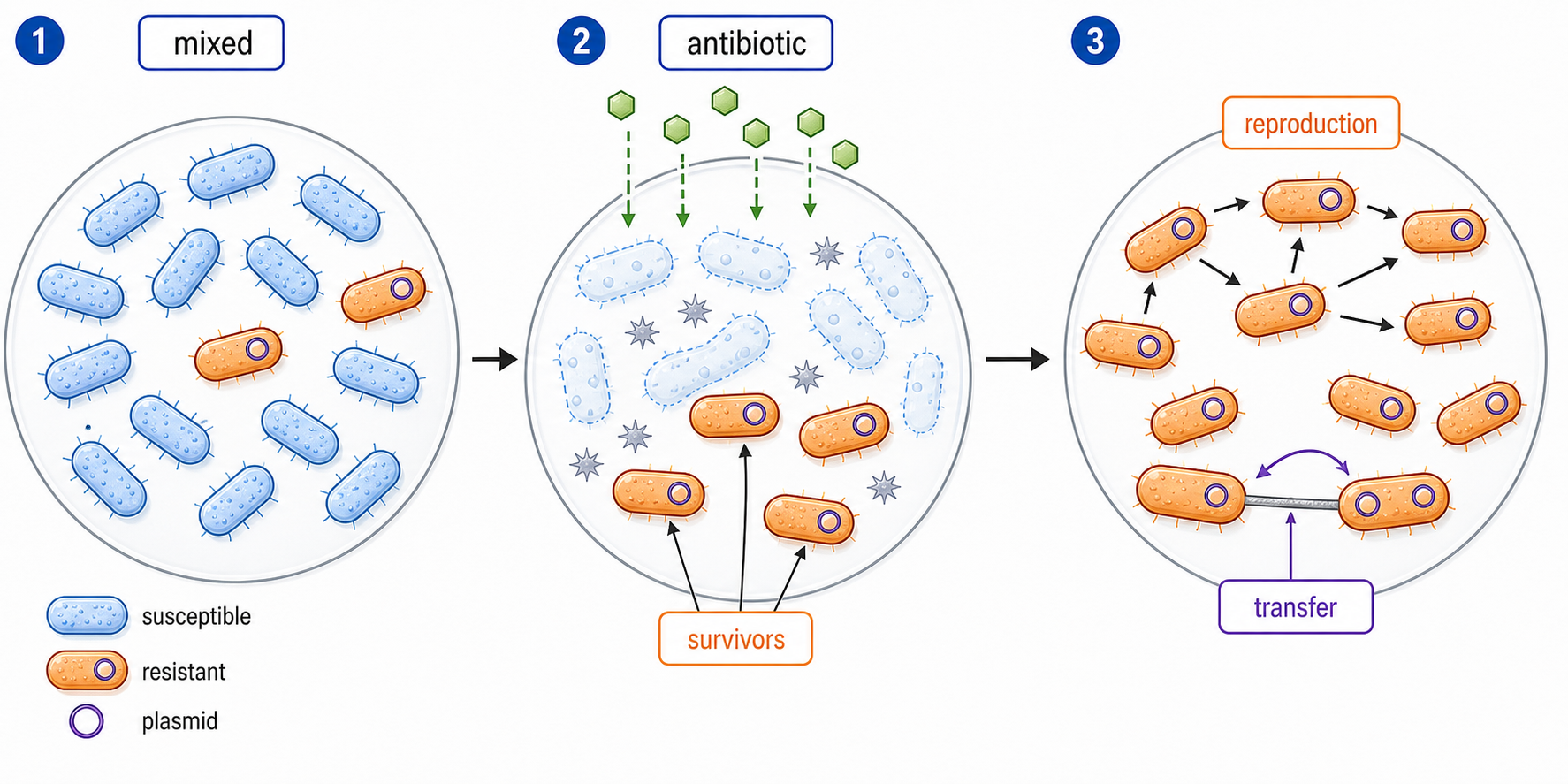
Identify outer protective sheath, bundles of fibres, myelinated fibres and unmyelinated fibres. Sensory axons carry input toward the CNS; motor axons carry output toward effectors.

A mixed nerve to a limb can contain sensory fibres carrying touch information toward the spinal cord and motor fibres carrying impulses back to skeletal muscle.

A nerve is not one giant neuron. Myelinated and unmyelinated fibres remain separate signal pathways inside the same protective bundle.

Nerves as bundles

Assessment in practice

1 marks
How it is assessed

This objective is assessed through structured response, commonly using State.

Command terms

State

What earns marks

Mark schemes reward named structures, correct direction of information flow and explanations that keep the sequence causal.

Representative question

Question 1

[Maximum number: 1]

State the role of the vagus nerve.

A reflex arc trades deliberation for speed

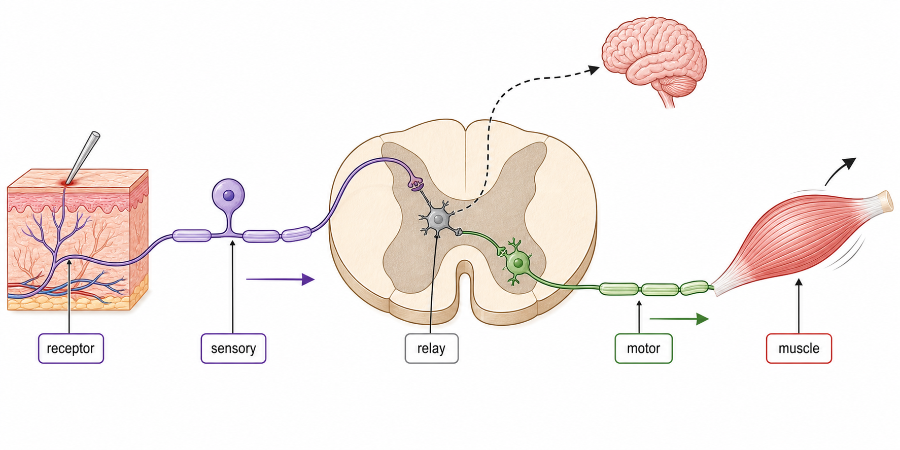
A pain withdrawal reflex is an involuntary response integrated in the spinal cord, with skeletal muscle as the effector.

A free sensory nerve ending in the hand detects a damaging stimulus. The sensory neuron enters the spinal cord and synapses with one interneuron in grey matter, which synapses with a motor neuron; the motor neuron stimulates a skeletal muscle to withdraw the hand.

Free nerve ending → sensory neuron → single interneuron in spinal grey matter → motor neuron → skeletal-muscle contraction. The short spinal route starts withdrawal before conscious pain processing is complete.

Touching a hot object activates the pain receptor; flexor muscle contraction withdraws the hand while impulses also travel to the brain and pain becomes conscious.

The reflex does not bypass the CNS—the spinal cord is part of the CNS. The brain can be informed in parallel but is not required to initiate this immediate response.

Pain reflex arcs

Assessment in practice

1–3 marks
How it is assessed

This objective is assessed through experimental design, commonly using Define / Label / Explain.

Command terms

Define / Label / Explain / Draw / Identify / Outline / Describe / Annotate

What earns marks

Mark schemes reward named structures, correct direction of information flow and explanations that keep the sequence causal.

Watch for

Putting the brain before the spinal relay in a rapid pain reflex.

Representative question

Question 1

[Maximum number: 4]

Draw a labelled diagram of a reflex arc for a pain withdrawal reflex.

The cerebellum coordinates movement and balance

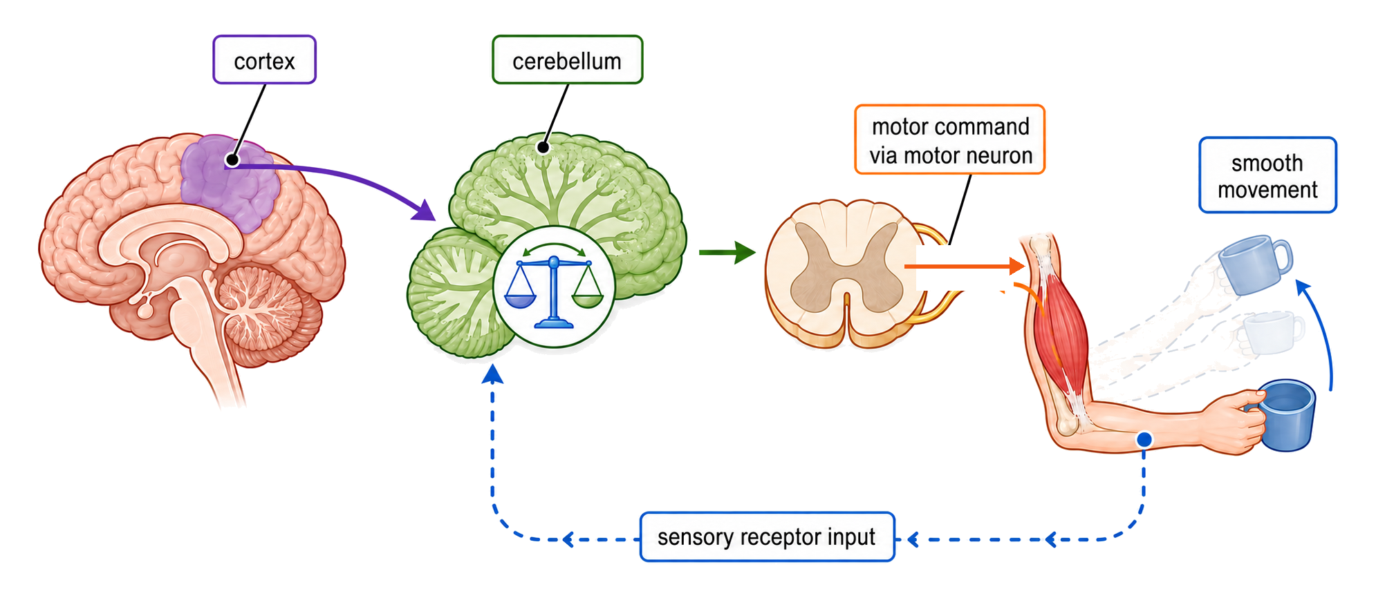
The cerebellum coordinates movement and balance.

The cerebellum compares intended movement with sensory feedback and adjusts motor output. It helps timing, posture, balance and smooth coordination rather than choosing the goal of a movement.

separate movement planning from coordination; link sensory feedback to corrective output.

If balance shifts while walking, cerebellar processing helps alter muscle activity before the person falls.
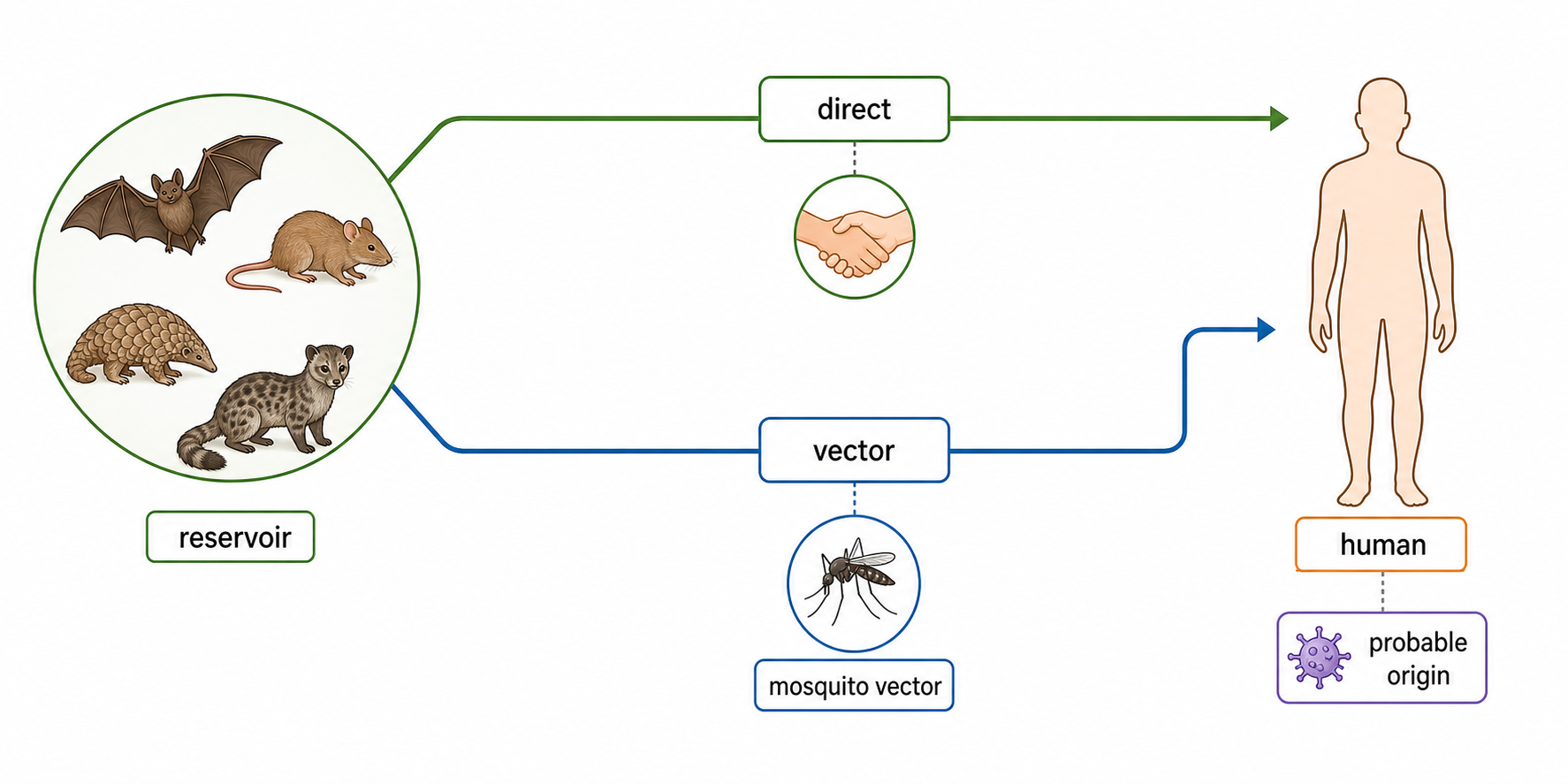
The cerebellum does not initiate every voluntary action; it refines the accuracy and timing of movement.

Cerebellum role

Assessment in practice

1 marks
How it is assessed

This objective is assessed through structured response, commonly using State / Identify / Describe.

Command terms

State / Identify / Describe

What earns marks

Mark schemes reward named structures, correct direction of information flow and explanations that keep the sequence causal.

Watch for

Naming a brain region without linking it to information integration or coordination.

Representative question

Question 1

[Maximum number: 2]

Describe the use of fMRI to identify the role of the cerebellum.

Melatonin links darkness to the body clock

Melatonin links darkness to the body clock.

Melatonin released by the pineal gland rises in darkness and helps synchronise circadian timing, including sleep propensity. Light reaching the retina can suppress its release.

light input → clock pathway → pineal melatonin → timing effect.

A bright screen late at night can delay the melatonin rise, shifting the timing signal even if the person feels tired.

Melatonin is a timing hormone, not an instant anaesthetic; sleep also depends on behaviour and other signals.
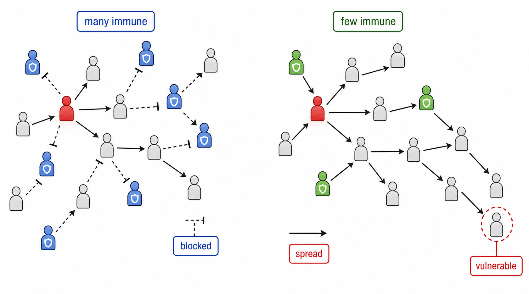
Melatonin and sleep

Assessment in practice

1 marks
How it is assessed

This objective is assessed through structured response, commonly using Explain / Identify / Describe.
Command terms

Explain / Identify / Describe / State / Deduce / Outline

What earns marks

Mark schemes reward named structures, correct direction of information flow and explanations that keep the sequence causal.

Watch for

Describing melatonin as a general energy hormone instead of a sleep-timing signal.

Representative question

Question 1

[Maximum number: 2]

Outline the role of melatonin in humans.

Epinephrine prepares several organs for immediate demand

Epinephrine prepares several organs for immediate demand.

Epinephrine from the adrenal medulla binds receptors in target tissues during acute stress. It raises cardiac output, widens airways and mobilises glucose so muscles can respond quickly.

hormone source; receptor-bearing target; response in heart, airways and liver; adaptive purpose.

A sudden threat increases epinephrine, accelerating heart rate and glycogen breakdown while reducing investment in digestion.

The hormone does not affect every cell equally: receptor type and tissue context determine the response.

Epinephrine (adrenaline)

Assessment in practice

1 marks
How it is assessed

This objective is assessed through structured response, commonly using State / Explain / Identify.

Command terms

State / Explain / Identify / Describe

What earns marks

Mark schemes reward named structures, correct direction of information flow and explanations that keep the sequence causal.

Watch for

Treating low oxygen as the only trigger and omitting carbon-dioxide-driven pH change.

Representative question

Question 1

[Maximum number: 2]

Describe two ways in which epinephrine in the body facilitates intense muscle contraction. [2]
1.
2.

The hypothalamus coordinates endocrine control through the pituitary

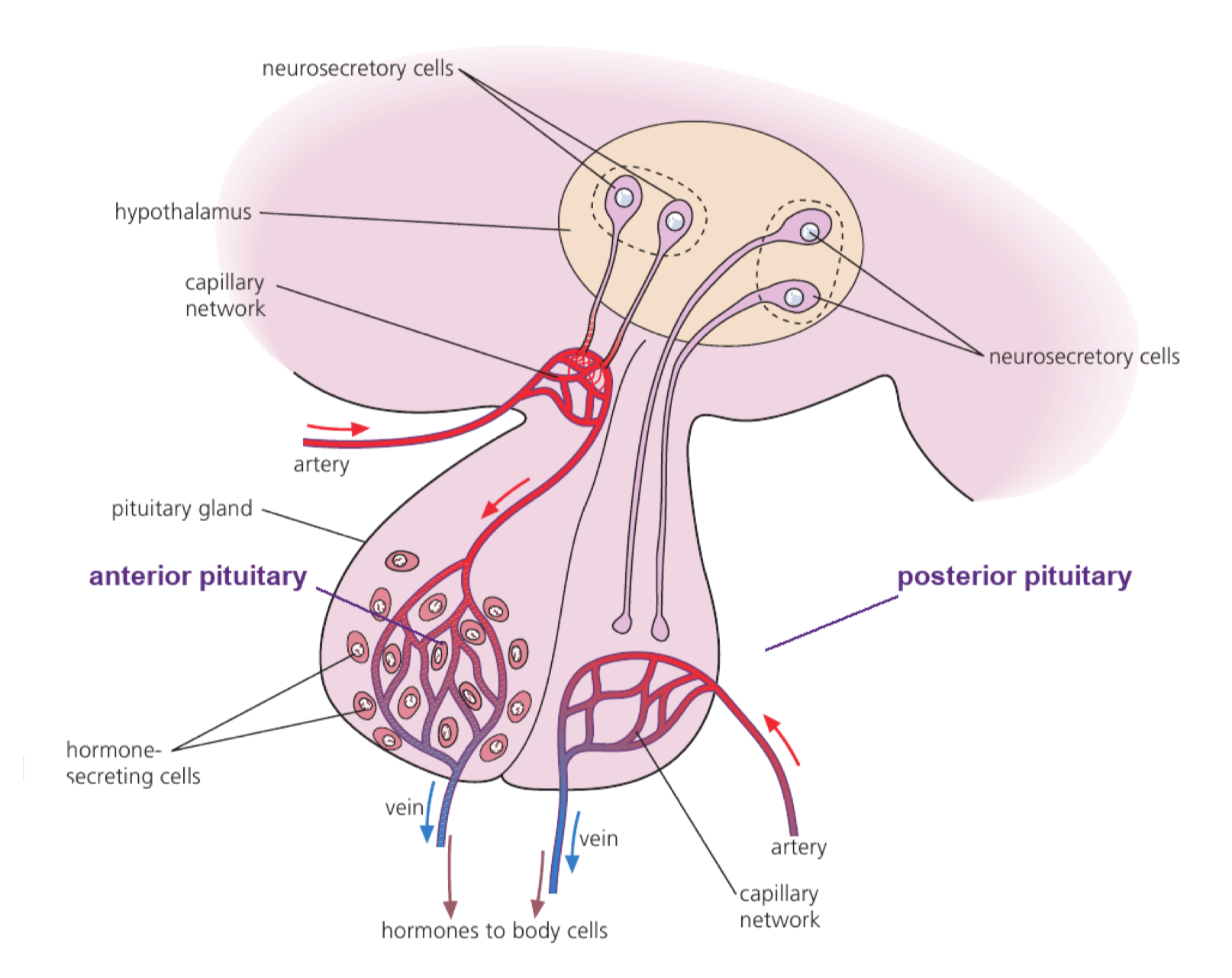
The hypothalamus coordinates endocrine control through the pituitary.

The hypothalamus links neural information to hormone release and regulates the pituitary with releasing or inhibiting signals. Pituitary hormones then act on distant glands or tissues.

stimulus or brain signal → hypothalamus → pituitary → target gland/tissue → feedback.

A hypothalamic signal can control pituitary FSH release, which then influences reproductive tissues.

The hypothalamus and pituitary are control centres, not the final target for every hormone they regulate.

Hypothalamus and pituitary control

Assessment in practice

1–2 marks
How it is assessed

This objective is assessed through structured response, commonly using Suggest / Describe / State.

Command terms

Suggest / Describe / State / Identify / Label / Explain / Outline

What earns marks

Mark schemes reward named structures, correct direction of information flow and explanations that keep the sequence causal.

Watch for

Confusing releasing hormones from the hypothalamus with hormones secreted by the pituitary.

Representative question

Question 1

[Maximum number: 7]

Explain the roles of named hormones in the development and function of the sexual reproductive systems in males and females.

Heart rate is adjusted by feedback from the body

Heart rate is adjusted by negative feedback after baroreceptors and chemoreceptors send sensory information to the medulla.

Baroreceptors in the carotid sinus and aortic arch monitor arterial pressure. Chemoreceptors in carotid and aortic bodies and the brainstem monitor blood pH and oxygen/carbon-dioxide concentrations.

The medulla integrates this input and changes autonomic nerve impulses to the heart. This alters heart rate and the strength of contraction, changing stroke volume so circulation opposes the detected disturbance.

If arterial pressure falls, reduced baroreceptor firing causes medullary output that increases heart rate and contraction strength, helping restore cardiac output and pressure.

Heart rate is only one component of cardiac output: cardiac output = heart rate × stroke volume. Receptors detect changes; the medulla coordinates the response.

Heart rate feedback control

Assessment in practice

1–4 marks
How it is assessed

This objective is assessed through structured response, commonly using Outline / Identify / State.

Command terms

Outline / Identify / State / Compare / Describe / Explain

What earns marks

Mark schemes reward named structures, correct direction of information flow and explanations that keep the sequence causal.

Watch for

Treating low oxygen as the only trigger and omitting carbon-dioxide-driven pH change.

Representative question

Question 1

[Maximum number: 7]

Explain the control mechanism of the heart rate.

Ventilation feedback matches breathing to CO₂ demand

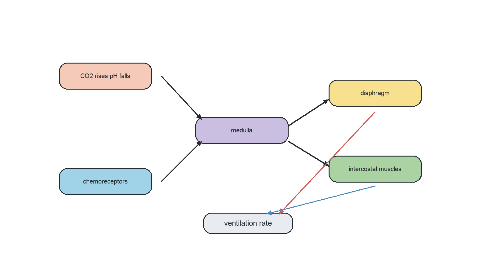
Ventilation rate is controlled by negative feedback from chemoreceptors that detect blood-pH changes linked mainly to carbon dioxide.

CO₂ dissolves and forms carbonic acid, increasing H⁺ and lowering pH. Chemoreceptors in the brainstem detect the change; the respiratory control centre sends nerve impulses to the diaphragm and intercostal muscles to alter breathing rate and depth.

Raised CO₂ → lower pH → brainstem chemoreceptor input → stronger or more frequent signals to diaphragm/intercostals → increased ventilation → more CO₂ removed → pH moves back toward normal.

During exercise, increased respiration produces extra CO₂. Ventilation rises, increasing gas exchange and limiting the fall in blood pH.

The lungs do not directly decide how to correct pH. Chemoreceptor detection and nervous output to breathing muscles coordinate the response.

Ventilation rate feedback control

Assessment in practice

1–3 marks
How it is assessed

This objective is assessed through structured response, commonly using Explain / State / Outline.

Command terms

Explain / State / Outline / Identify

What earns marks

Mark schemes reward named structures, correct direction of information flow and explanations that keep the sequence causal.

Watch for

Treating low oxygen as the only trigger and omitting carbon-dioxide-driven pH change.

Representative question

Question 1

[Maximum number: 8]

Explain the causes of a decreased blood pH and its effects on the ventilation rate in humans.

Peristalsis moves gut contents by coordinated smooth muscle

Control of movement through the digestive tract shifts between voluntary CNS actions at the endpoints and involuntary enteric control between them.

The central nervous system voluntarily initiates swallowing and participates in voluntary control of faecal egestion. Between these points, the enteric nervous system coordinates circular and longitudinal smooth-muscle contractions that move contents by peristalsis.

Voluntary initiation of swallowing → involuntary ENS-coordinated peristalsis through gut → voluntary component of egestion. Local stretch and chemical signals help the ENS coordinate contraction behind and relaxation ahead.

After a person chooses to swallow, an oesophageal peristaltic wave moves the bolus to the stomach without conscious control.

Peristalsis is not one simultaneous squeeze and is not consciously directed along the whole gut; the ENS coordinates the travelling pattern between voluntary endpoints.

Peristalsis control

Assessment in practice

1–3 marks
How it is assessed

This objective is assessed through structured response, commonly using Outline / Identify.

Command terms

Outline / Identify

What earns marks

Mark schemes reward named structures, correct direction of information flow and explanations that keep the sequence causal.

Watch for

Giving a general body-system answer without the specific pathway step for peristalsis control in the gut.

Representative question

Question 1

[Maximum number: 3]

Outline how food is moved from the stomach to the large intestine.

Body-System Integration

  • Nervous signals, hormones and blood transport integrate organs into coordinated systems; emergent functions arise from their interactions.
  • Sensory neurons carry receptor input to the CNS; motor neurons carry output to effectors. Mixed nerves contain both fibre types.
  • Reflex arcs provide rapid involuntary responses through sensory, relay and motor neurons. The spinal cord also links the brain and peripheral nerves.
  • Cerebral hemispheres support conscious processing; the cerebellum coordinates movement and balance; the medulla adjusts ventilation and heart activity.
  • The hypothalamus links nervous and endocrine control through the pituitary. Pineal melatonin helps time sleep; adrenal epinephrine supports acute stress responses.
  • Baroreceptors and chemoreceptors provide feedback about pressure, CO2, pH and O2.
  • The CNS controls voluntary swallowing and egestion, while the enteric nervous system coordinates gut peristalsis.

Objective notes

16 learning objectives
C3.1.1System integration• Integration lets interacting parts coordinate an overall biological function• Systems interact across molecular, cellular, organ, organism, and ecosystem levels1% of analysed papers 1 paper · 1 questionViewC3.1.2Hierarchy in multicellular organisms• Cells form tissues, organs, organ systems, and whole organisms• Emergent properties arise when subsystems interact, such as gut peristalsis and absorption1% of analysed papers 1 paper · 1 questionViewC3.1.3Integration of organs• Organs are integrated by nervous signals, hormones, and blood transport• Transport links nutrients, gases, wastes, hormones, and energy substrates between organs4% of analysed papers 6 papers · 7 questionsViewC3.1.4Brain as information integration organ• The brain integrates sensory input and coordinates complex responses• Cerebral hemispheres, cerebellum, hypothalamus, and medulla have distinct roles8% of analysed papers 11 papers · 14 questionsViewC3.1.5Spinal cord• The spinal cord links brain and peripheral nervous system• It integrates unconscious processes such as reflex arcs1% of analysed papers 1 paper · 1 questionViewC3.1.6Input through sensory neurons• Sensory neurons carry impulses from receptors to spinal cord and brain• Inputs may reach cerebral hemispheres for conscious perception3% of analysed papers 4 papers · 4 questionsViewC3.1.7Output through motor neurons• Motor neurons carry impulses from CNS to skeletal muscle effectors• Voluntary muscle contraction is coordinated through cerebral hemispheres1% of analysed papers 1 paper · 1 questionViewC3.1.8Nerves as bundles• Nerves are bundles of sensory and motor nerve fibres in connective tissue• Mixed nerves carry impulses both to and from the CNS1% of analysed papers 1 paper · 1 questionViewC3.1.9Pain reflex arcs• Pain reflexes are rapid involuntary withdrawal responses• Reflex arcs use receptors, sensory neurons, relay neurons, motor neurons, and skeletal muscle9% of analysed papers 12 papers · 16 questionsViewC3.1.10Cerebellum role• The cerebellum coordinates timing and force of skeletal muscle contractions• It maintains balance, posture, and smooth learned movements0% of analysed papers ViewC3.1.11Melatonin and sleep• The pineal gland secretes melatonin according to light-dark cycles• Melatonin modulates sleep timing as part of circadian rhythms4% of analysed papers 5 papers · 6 questionsViewC3.1.12Epinephrine (adrenaline)• Adrenal glands secrete epinephrine during stress or danger• It increases heart rate, ventilation, and respiratory substrate availability2% of analysed papers 3 papers · 3 questionsViewC3.1.13Hypothalamus and pituitary control• The hypothalamus links nervous inputs to endocrine control• Pituitary hormones regulate glands including thyroid, gonads, adrenals, and mammary glands3% of analysed papers 4 papers · 4 questionsViewC3.1.14Heart rate feedback control• Baroreceptors monitor blood pressure; chemoreceptors monitor CO₂, pH, and O₂• The medulla adjusts sinoatrial node activity by sympathetic and parasympathetic nerves7% of analysed papers 10 papers · 10 questionsViewC3.1.15Ventilation rate feedback control• Chemoreceptors detect CO₂-driven pH changes in blood and cerebrospinal fluid• The medulla alters diaphragm and intercostal muscle activity to change ventilation rate2% of analysed papers 3 papers · 3 questionsViewC3.1.16Peristalsis control• The CNS controls voluntary swallowing and egestion• The enteric nervous system coordinates involuntary peristalsis in gut smooth muscle1% of analysed papers 1 paper · 1 questionView