EduNinja

A-Level CAIE Biology AS2.3 ProteinsQuestion Bank

Question 1

[Maximum number: 5]

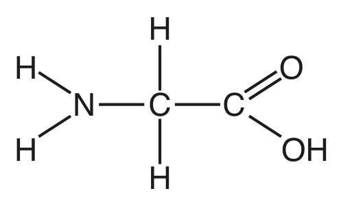
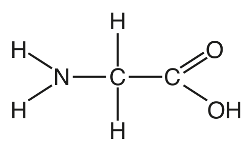
Fig. 1.1 shows the structure of the amino acid glycine.
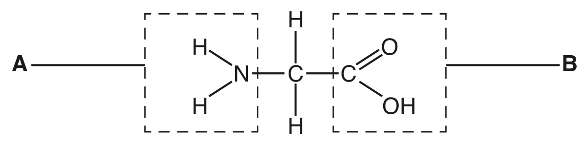
Fig. 1.1

Fig. 1.1

Question 1(a)

Question 1(a)(i)

(a)
(i)

Name the parts of the amino acid molecule labelled A and B in Fig. 1.1.

A

B

[ 2 ]

Question 1(a)(ii)

(ii)

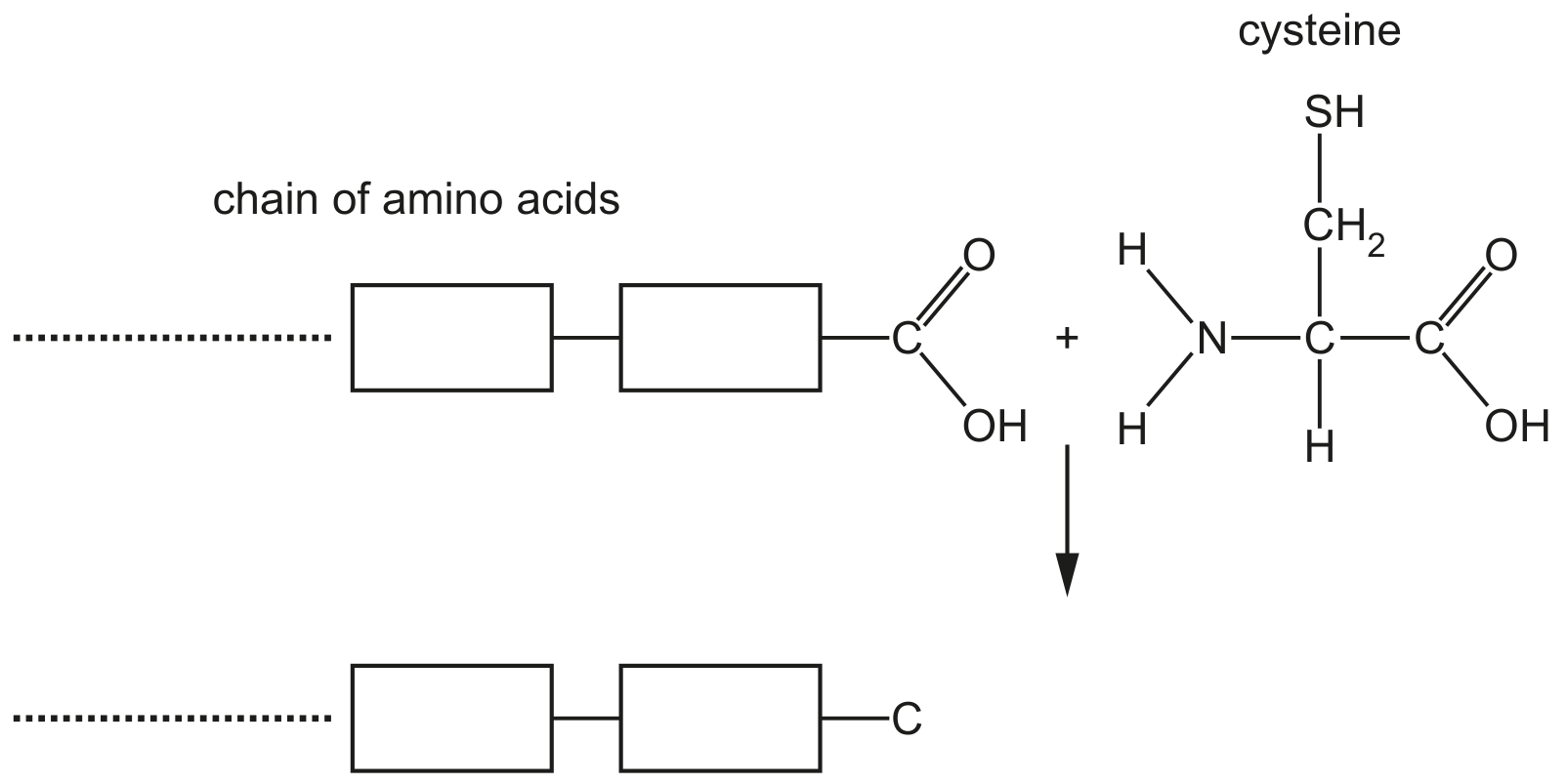
Amino acids are monomers used to build proteins.

Complete Fig. 1.2 by drawing a diagram to show the formation of a peptide bond between two molecules of glycine.

Question image
Question image
[ 3 ]

Question 1

[Maximum number: 5]

Fig. 1.1 is a diagram of a molecule of haemoglobin.

Fig. 1.1

Fig. 1.1

Question 1(b)

(a)

Haemoglobin is described as a globular protein. Explain why this protein is described as globular.

[ 2 ]

Question 1(d)

(b)

A single base change in the DNA of the gene H B B results in a change to the amino acid sequence of β\beta-globin. In the sequence, a single glutamic acid is replaced by valine.

Outline the effects of this change in the amino acid sequence of β\beta-globin on the structure and function of a haemoglobin molecule.

[ 3 ]

Question 1

[Maximum number: 3]

Fig. 1.1 is a diagram of an antibody molecule.

Fig. 1.1

Fig. 1.1

Question 1(a)

Question 1(a)(i)

(a)
(i)

Name the part labelled X.

[ 1 ]

Question 1(a)(ii)

(ii)

Name the bond labelled Y.

[ 1 ]

Question 1(a)(iii)

(iii)

The antibody molecule in Fig. 1.1 has quaternary structure.

Explain the meaning of the term quaternary structure as applied to proteins.

[ 1 ]

Question 1

[Maximum number: 2]

Fig. 1.1 shows the structures of four biological molecules A, B, C and D.

Fig. 1.1

Fig. 1.1

Question 1(a)

(a)

Give the letter, A to D, of the molecule in Fig. 1.1 which:

[ 3 ]

Question 1(a)(ii)

(i)

can form peptide bonds

Question 1(b)

(b)

Some of the molecules in Fig. 1.1 can form polymers.

[ 2 ]

Question 1(b)(iii)

(i)

Molecule D can form macromolecules with other similar monomers.

These macromolecules have three dimensional shapes held in place by interactions or bonds other than those between adjacent monomers.

Name two of these interactions or bonds.
1.
2.

[ 2 ]

Question 1

[Maximum number: 3]

The cell surface membrane has a fluid mosaic structure.

Question 1(d)

(a)

High temperature can damage cell membranes. One factor contributing to this damage is the denaturation of membrane proteins.

Describe how proteins become denatured at high temperature and explain how this could lead to damaging cell membranes.

[ 3 ]

Question 1

Question 1(a)

(a)

Antibody molecules are proteins that show primary structure, secondary structure, tertiary structure and quaternary structure.

Fig. 1.1 shows a ribbon diagram of an antibody molecule.

Fig. 1.1

Fig. 1.1

Describe how Fig. 1.1 shows the secondary structure and tertiary structure of the antibody molecule.
secondary structure
tertiary structure

[ 3 ]

Question 1

Question 1(b)

(a)

The perichondrium contains collagen fibres, composed of collagen molecules.

Collagen is an example of one type of biological molecule.
State the name of the type of biological molecule of which collagen is an example.

[ 1 ]

Question 1

Question 1(a)

(a)

During translation, a polypeptide is synthesised when amino acids are added to a growing chain of amino acids.

Fig. 1.1 shows part of a growing chain of amino acids and the amino acid cysteine.

Fig. 1.1

Fig. 1.1

[ 1 ]

Question 1(a)(ii)

(i)

State the name of the covalent bond that forms when cysteine is added to the growing chain of amino acids.

[ 1 ]

Question 1(b)

(b)

Fig. 1.2 is a ribbon diagram showing the three-dimensional structure of a protein from the bacterium Streptococcus.

Fig. 1.2

Fig. 1.2

[ 7 ]

Question 1(b)(i)

(i)

Describe the secondary structure of the protein shown in Fig. 1.2.

[ 2 ]

Question 1(b)(ii)

(ii)

Explain why the protein shown in Fig. 1.2 has tertiary structure, but not quaternary structure.

[ 2 ]

Question 1(b)(iii)

(iii)

An analysis of the amino acid composition of the protein in Fig. 1.2 showed that it does not contain any cysteine residues.

Explain how the three-dimensional structure of the protein shown in Fig. 1.2 is held in place.

[ 3 ]

Question 1

[Maximum number: 4]

Fig. 1.1 is a diagram showing a stage in protein synthesis.

Fig. 1.1

Fig. 1.1

Question 1(b)

(a)

Mutagenesis is a process that leads to a change in the amino acid sequences of proteins. Scientists carry out mutagenesis to investigate the importance of particular amino acids in protein structure and function.

Outline how changing one amino acid in the β\beta-globin polypeptide of haemoglobin may change the structure and function of a molecule of haemoglobin.

[ 4 ]

Question 1

[Maximum number: 5]

Antibodies are secreted by activated B-lymphocytes known as plasma cells.
Fig. 1.1 is a diagram showing the cellular processes involved in the production of a polypeptide of an antibody molecule (not drawn to scale).

Fig. 1.1

Fig. 1.1

Question 1(b)

(a)

Antibodies are glycoproteins.

State what is meant by the term glycoprotein.

[ 1 ]

Question 1(d)

(b)

Describe and explain how the structure of an antibody molecule is related to its functions.

[ 4 ]
0 selected