EduNinja

A-Level CAIE Biology AS7.1 Structure of Transport TissuesQuestion Bank

Question 1

[Maximum number: 2]

Fig. 1.1 shows the structure of the amino acid glycine.
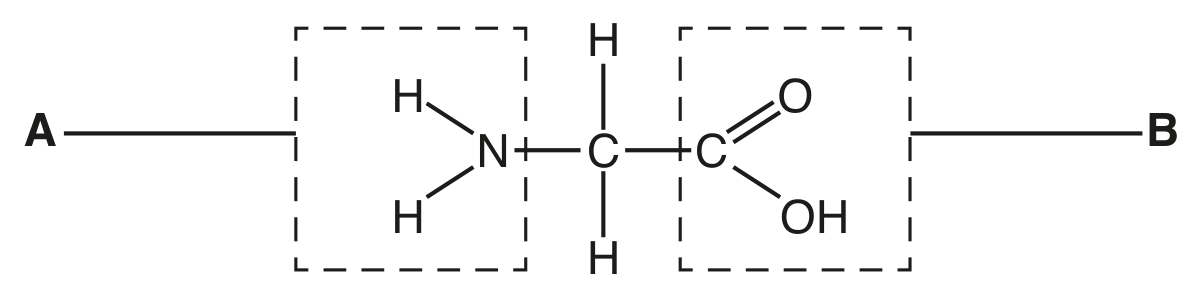
Fig. 1.1

Fig. 1.1

Question 1(c)

(a)

Sieve tube elements in plants have very few organelles such as mitochondria.

Explain how having very few organelles is an adaptation of the sieve tube element to its function.

[ 2 ]

Question 1

Question 1(a)

(a)

Fig. 1.1 shows a transverse section through a dicotyledonous leaf.

Fig. 1.1

Fig. 1.1

Name A, B and C.
A

B
C

[ 3 ]

Question 1

Question 1(a)

(a)

Fig. 1.1 shows a section through part of a dicotyledonous leaf of the tea plant Camellia sinensis.

Fig. 1.1

Fig. 1.1

On Fig. 1.1, use label lines and letters to label each of the following parts:
X - xylem tissue
P - palisade mesophyll tissue.

[ 2 ]

Question 1

Question 1(a)

(a)

Fig. 1.1 shows a section through part of a dicotyledonous leaf of the tea plant Camellia sinensis.

Fig. 1.1

Fig. 1.1

On Fig. 1.1, use label lines and letters to label each of the following parts:
X - xylem tissue
P - palisade mesophyll tissue.

[ 2 ]

Question 1

Question 1(a)

(a)

Fig. 1.1 is a diagram of a nephron.

Fig. 1.1

Fig. 1.1

Label Fig. 1.1 using:
- one labelling line and the letter A to identify a region that contains urine
- one labelling line and the letter B to identify a region that contains podocytes
- one labelling line and the letter C to identify a region of the nephron that is within the medulla of the kidney
- one labelling line and the letter D to identify the afferent arteriole.

[ 4 ]

Question 1

[Maximum number: 1]

Water and mineral ions are transported up the stem of a plant to the leaves within xylem vessels.
Some water and mineral ions can pass out of xylem vessel elements to supply parenchyma tissue in the stem.

Question 1(a)

(a)

Fig. 1.1 is a plan diagram of a section through a stem.

Fig. 1.1

Fig. 1.1

Identify one location where xylem tissue occurs in the stem by drawing a label line and the letter X on Fig. 1.1.

[ 1 ]

Question 1

[Maximum number: 3]

Fig. 1.1 and Fig. 1.2 are photomicrographs of sections through the leaves of two different plants.

Fig. 1.1
Fig. 1.2 is a photomicrograph of a section through a leaf of Himalayan cedar, Cedrus deodara.

Fig. 1.1 Fig. 1.2 is a photomicrograph of a section through a leaf of Himalayan cedar, Cedrus deodara.

Fig. 1.2
Fig. 1.1 and Fig. 1.2 are not shown at the same magnification.

Fig. 1.2 Fig. 1.1 and Fig. 1.2 are not shown at the same magnification.

Question 1(a)

(a)

Cells labelled A, B and C in Fig. 1.1 and Fig. 1.2 each form a different tissue.

Name each tissue formed.
tissue formed from A
tissue formed from B
tissue formed from C

[ 3 ]

Question 1

[Maximum number: 2]

Fig. 1.1 is a photomicrograph of a transverse section through the stem of creeping buttercup, Ranunculus repens.

Fig. 1.1

Fig. 1.1

Question 1(a)

(a)

Draw one label line on Fig. 1.1 to indicate one precise location where phloem sieve tubes occur.

[ 1 ]

Question 1(c)

(b)

A good quality plan diagram of Fig. 1.1 will show only the tissues in the stem in their correct location and in the correct proportions.

[ 1 ]

Question 1(c)(i)

(i)

State the name of the tissue that forms the outer layer of the stem section of R. repens in Fig. 1.1.

[ 1 ]

Question 1

[Maximum number: 7]

Fig. 1.1 is a photomicrograph of a low power image of part of the common sunflower, Helianthus annuus. Fig. 1.1 is a transverse section.

Fig. 1.1

Fig. 1.1

Question 1(a)

(a)

State, with a reason, whether Fig. 1.1 shows a section through the root or the stem of H. annuus.

[ 1 ]

Question 1(b)

(b)

Cell type A in Fig. 1.1 has a large central vacuole.

Suggest, with reasons, the role of the tissue formed by this type of cell.

[ 2 ]

Question 1(c)

(c)

Structure B in Fig. 1.1 contains phloem tissue and xylem tissue and other tissues that provide support.

[ 1 ]

Question 1(c)(i)

(i)

Name structure B.

[ 1 ]

Question 1(d)

(d)

When structure B is observed at a higher magnification, more detail of xylem vessel elements and phloem sieve tube elements can be seen.

Outline the differences in the structure of a xylem vessel element and a phloem sieve tube element.

[ 3 ]

Question 2

[Maximum number: 1]

Four students were asked to suggest a set of four labels to add to a plan diagram of a transverse section of a dicotyledonous leaf.

Which student, A, B, C or D, suggested a correct set of labels?

label 1

label 2

label 3

label 4

lower epidermis

phloem

spongy mesophyll

palisade mesophyll

phloem sieve tubes

upper epidermis

spongy mesophyll

xylem vessel elements

spongy mesophyll

endodermis

xylem vessel elements

palisade mesophyll

waxy cuticle

xylem

companion cells

spongy mesophyll

0 selected