EduNinja

A-Level CAIE Biology AS5.2 Chromosome Behaviour in MitosisQuestion Bank

Question 1

[Maximum number: 1]

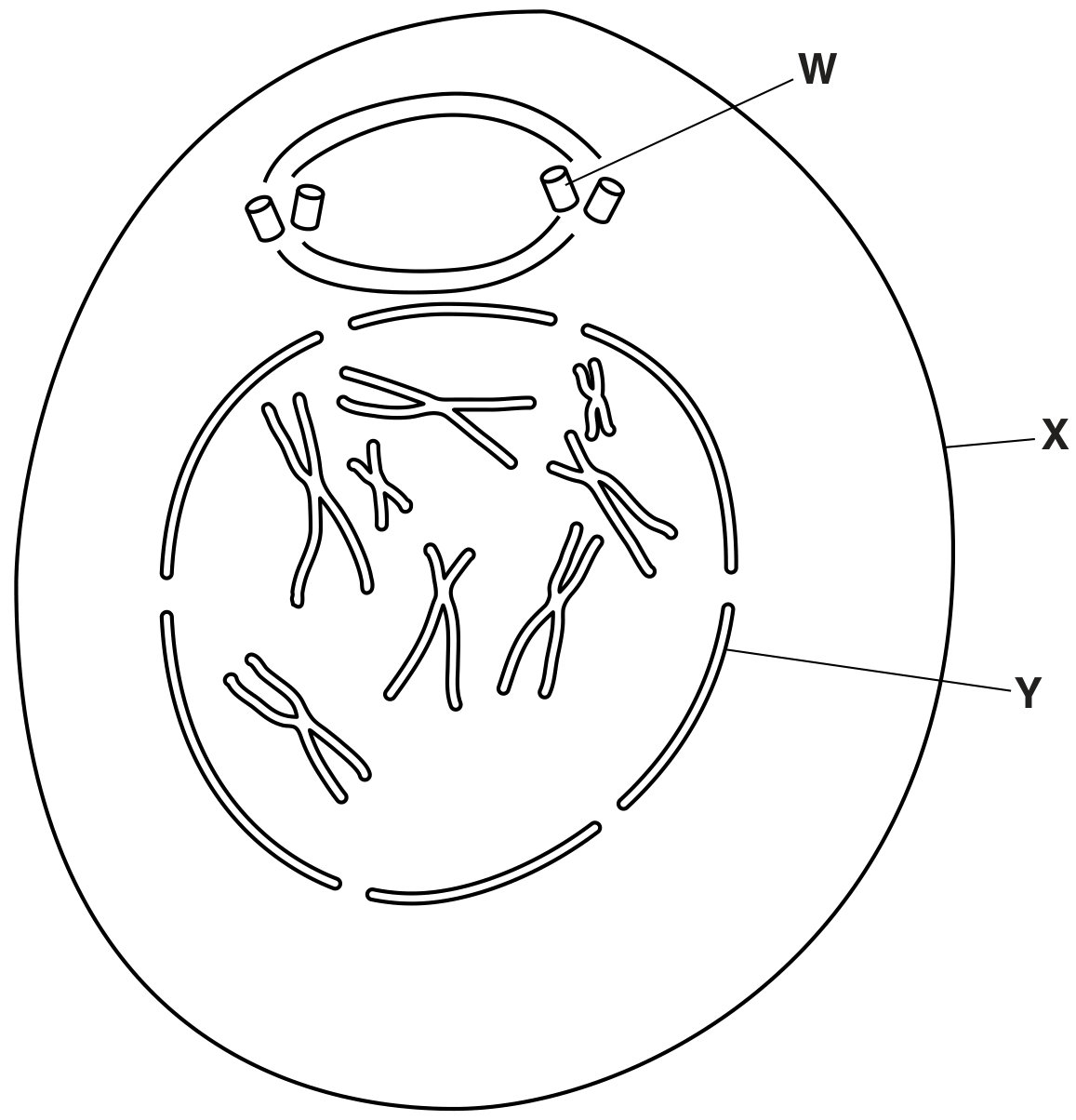
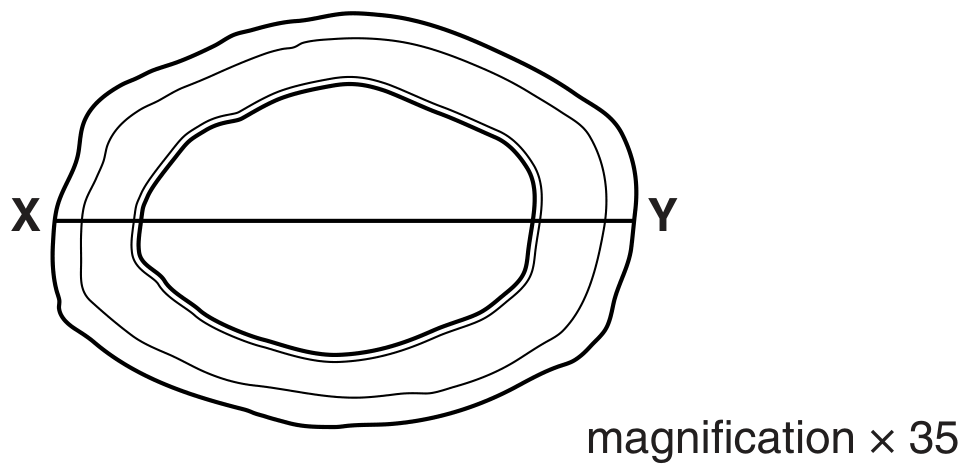
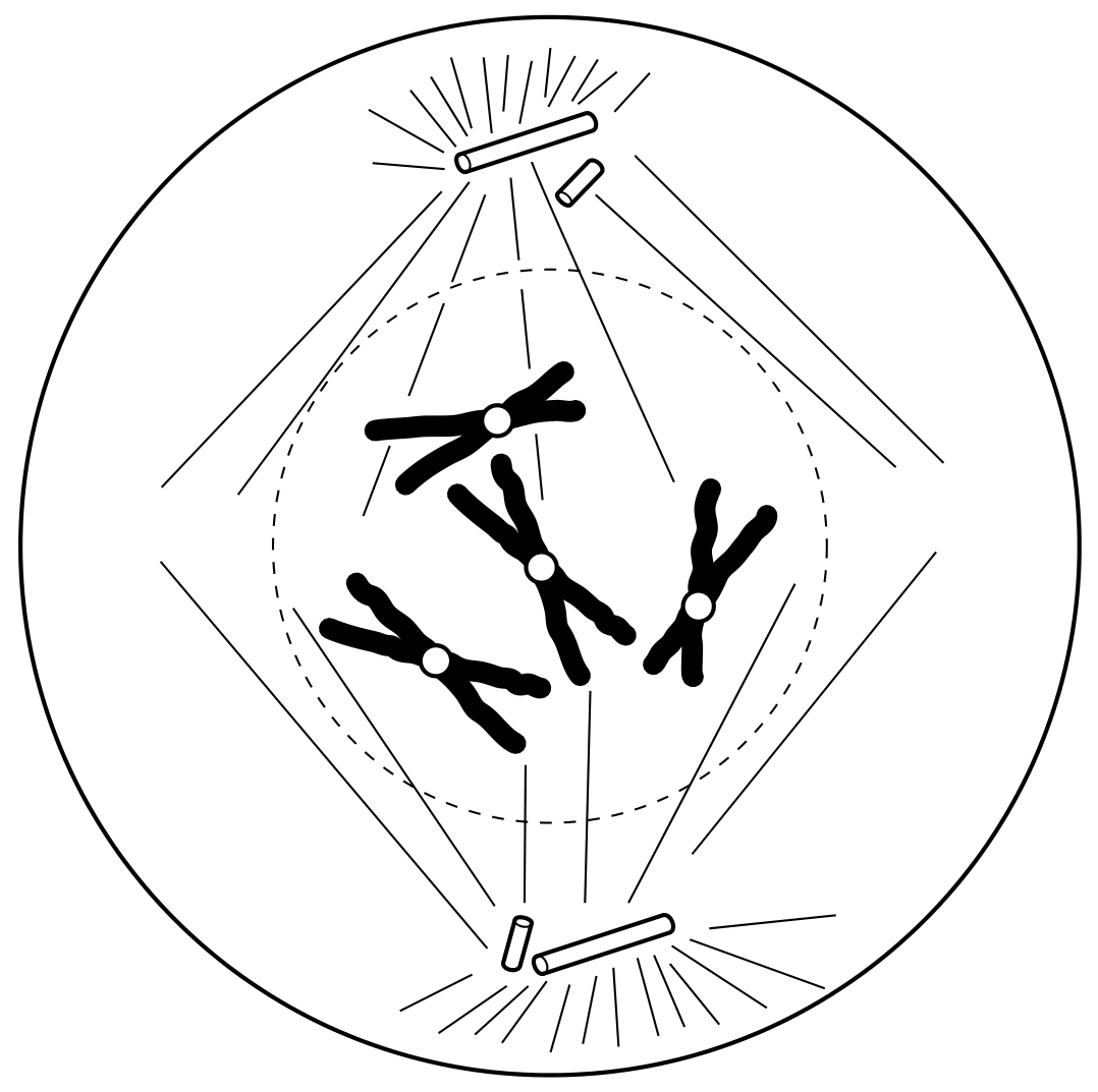
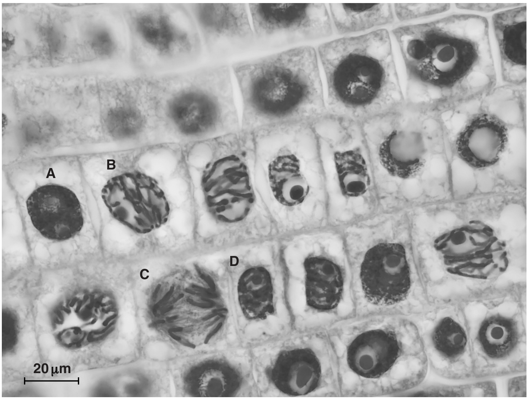
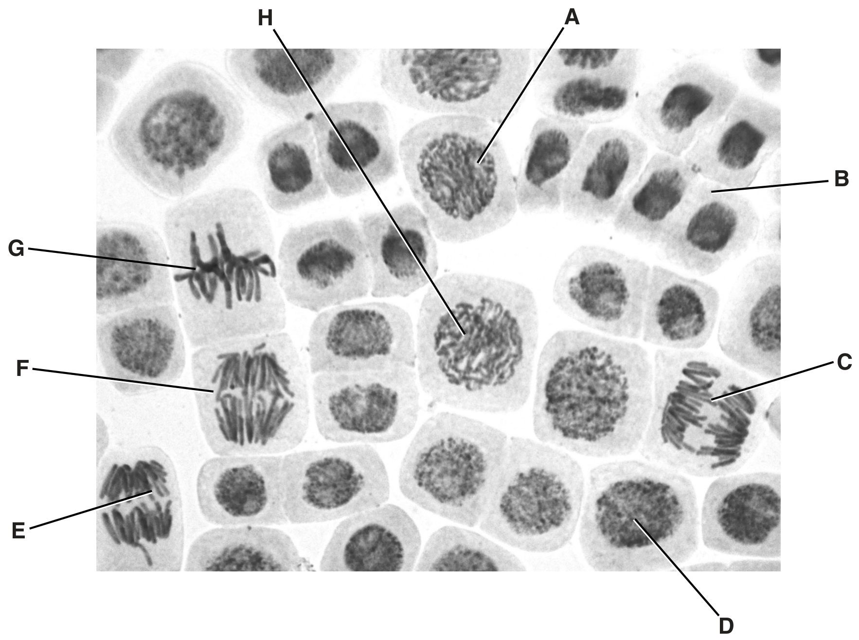
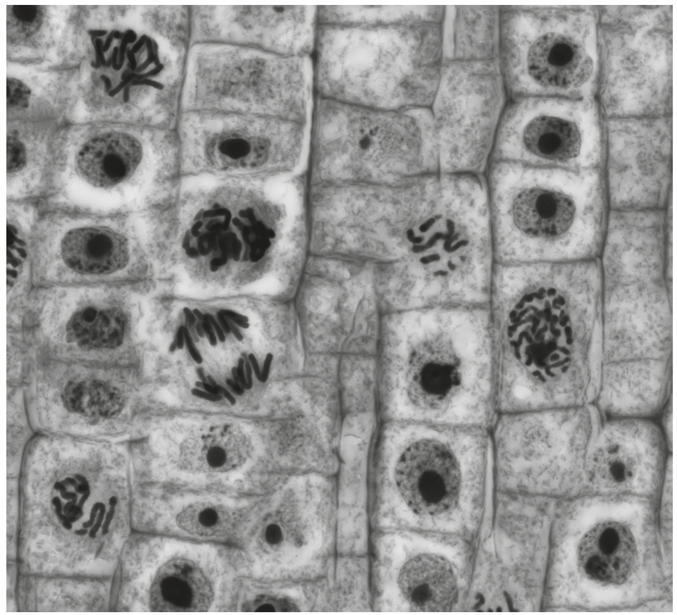
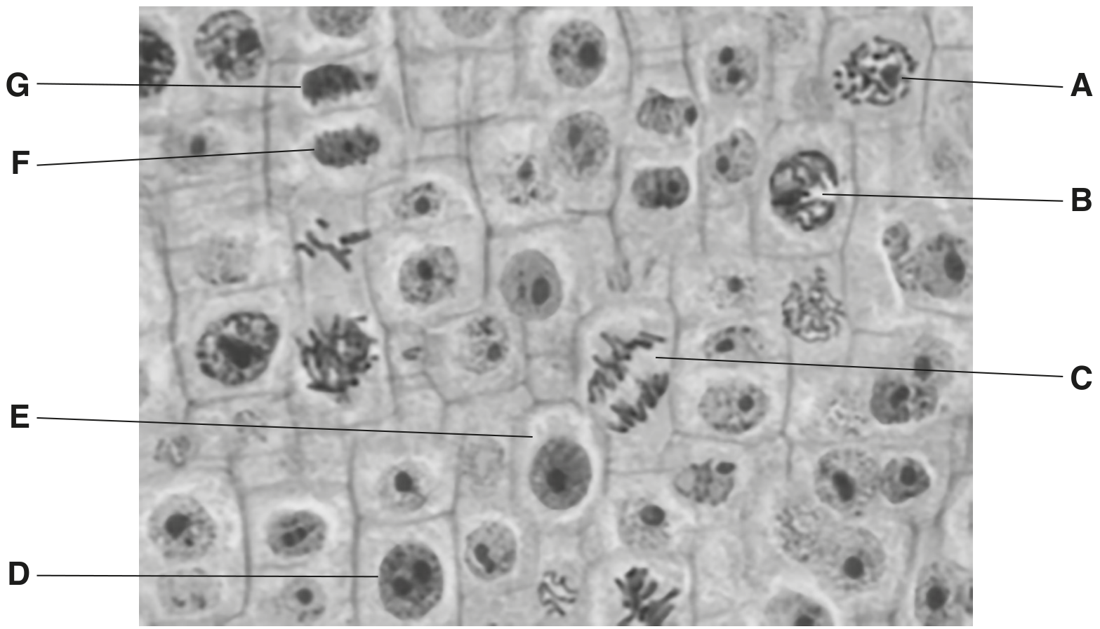
Fig. 1.1 shows four animal cells in different stages of the mitotic cell cycle.
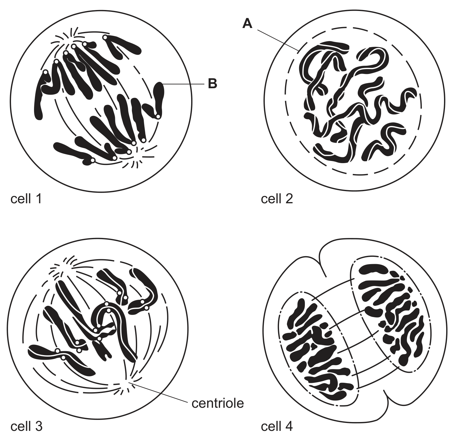
Fig 1.1

Fig 1.1

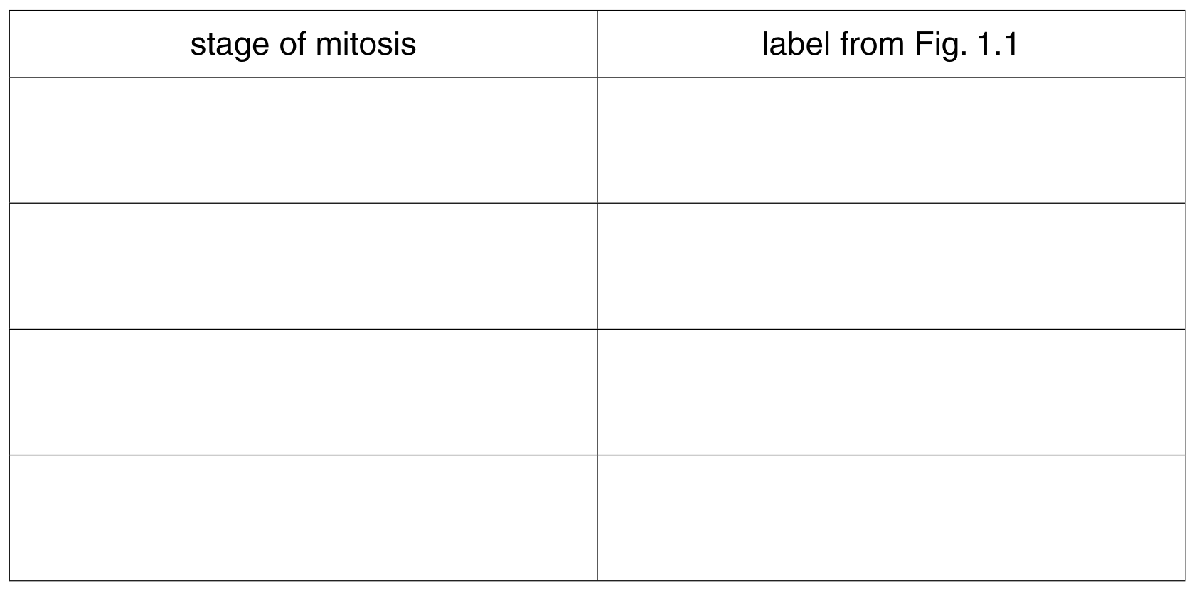
(a) Using the number given to each cell in Fig. 1.1, arrange the stages as they occur in the mitotic cell cycle.

Question 1(b)

Question 1(b)(i)

(a)
(i)

State what is occurring at A in cell 2 .
(ii) Label B is pointing to a region of the chromatid that contains repetitive nucleotide sequences.
State the name given to this region.

[ 1 ]

Question 1

Question 1(a)

(a)

Fig. 1.1 is a diagram of a human chromosome at a stage in mitosis.

Fig. 1.1

Fig. 1.1

[ 1 ]

Question 1(a)(ii)

(i)

State one stage during mitosis when the chromosome would appear as shown in Fig. 1.1.

[ 1 ]

Question 1

[Maximum number: 4]

Fig. 1.1 shows a cell of a female fruit fly, Drosophila melanogaster, during a stage of mitosis.

Fig. 1.1

Fig. 1.1

Question 1(a)

Question 1(a)(i)

(a)
(i)

Name the stage of mitosis shown in Fig. 1.1.

[ 1 ]

Question 1(b)

(b)

State what happens to structure X and to structure Y between the stage shown in Fig. 1.1 and the end of cell division.

[ 3 ]

Question 1

[Maximum number: 2]

Fig. 1.1 is a diagram of a transverse section through a vein.

Fig. 1.1

Fig. 1.1

Question 1(e)

(a)

Fig. 1.2 is a diagram of a cell in late prophase of mitosis.

Fig. 1.2

Fig. 1.2

Complete Fig. 1.3 to show the same cell in the anaphase stage of mitosis.

Fig. 1.3

Fig. 1.3

[ 2 ]

Question 1

[Maximum number: 5]

Fig. 1.1 is a photomicrograph of plant root cells near the growing tip. Some of the cells are undergoing mitosis.

Fig. 1.1

Fig. 1.1

Question 1(b)

(a)

State the letter, A to D, of the cell in Fig. 1.1 which is in:

[ 3 ]

Question 1(b)(i)

(i)

prophase

[ 1 ]

Question 1(b)(ii)

(ii)

anaphase.

[ 2 ]

Question 1(c)

(b)

Describe two events occurring in cell B.
1.
2.

[ 2 ]

Question 1

[Maximum number: 7]

A student investigated growth in the roots of broad bean, Vicia faba. The student cut sections of the root tip of this plant and viewed them with a light microscope.

Fig. 1.1 is a photomicrograph of one of the sections. The cell labelled D is in interphase.

Fig. 1.1

Fig. 1.1

Question 1(a)

(a)

Complete the table below by:
- naming the stages of mitosis in the correct sequence following interphase
- identifying one example from the cells labelled A to H that is in each stage of mitosis that you have named.

Table
[ 5 ]

Question 1(b)

(b)

In animal cells, centrioles are responsible for assembling microtubules to make the spindle at the beginning of mitosis.

Describe the role of the spindle during mitosis.

[ 2 ]

Question 1

[Maximum number: 1]

Fig. 1.1 is a photomicrograph of plant cells showing stages in cell division.

Fig. 1.1

Fig. 1.1

Question 1(a)

(a)

On Fig. 1.1, draw a circle around a cell in anaphase.

[ 1 ]

Question 1

[Maximum number: 5]

Fig. 1.1 is a photomicrograph of root tip meristem. Different stages of the cell cycle are visible. Some cells are in the same stage of the cell cycle and some are in the same stage of mitosis.

Fig. 1.1

Fig. 1.1

Question 1(b)

(a)

Name the stage of mitosis occurring in each of cells A, B and C.
cell A
cell B
cell C

[ 3 ]

Question 1(c)

(b)

Cells F and G are newly formed cells. Cytokinesis has occurred with the formation of a cell plate.

Describe the events that have occurred in the stage of mitosis immediately before cytokinesis.

[ 2 ]

Question 1

[Maximum number: 3]

The root apical meristem is a region of undifferentiated cells in the root tips of plants. Mitosis occurs in this region.

Question 1(a)

(a)

Fig. 1.1 is an image of the root tip of Allium as observed using a microscope with a low-power objective lens.

Fig. 1.1

Fig. 1.1

State which region, B, C, or D, should be chosen in order to observe the highest proportion of cells in stages of mitosis.

[ 1 ]

Question 1(b)

Question 1(b)(ii)

(b)
(i)

Describe the behaviour of the nuclear envelope during mitosis.

[ 2 ]

Question 1

[Maximum number: 6]

During interphase and mitosis of the cell cycle, the chromosomes within a cell go through a number of changes. Each chromosome is composed of DNA complexed with proteins.

Question 1(b)

(a)

When viewed through a microscope, a chromosome is most clearly visible during the metaphase stage of mitosis.

Complete Fig. 1.1 to produce a labelled diagram of the metaphase stage of mitosis in an animal cell with two chromosomes.

Fig. 1.1

Fig. 1.1

[ 3 ]

Question 1(c)

(b)

Outline the changes that occur to the structure and behaviour of a chromosome:
- from the start of the S phase to the end of interphase
- during prophase of mitosis.

[ 3 ]
0 selected