EduNinja

A-Level CAIE Biology AS9.1 The Gas Exchange SystemQuestion Bank

Question 1

[Maximum number: 4]

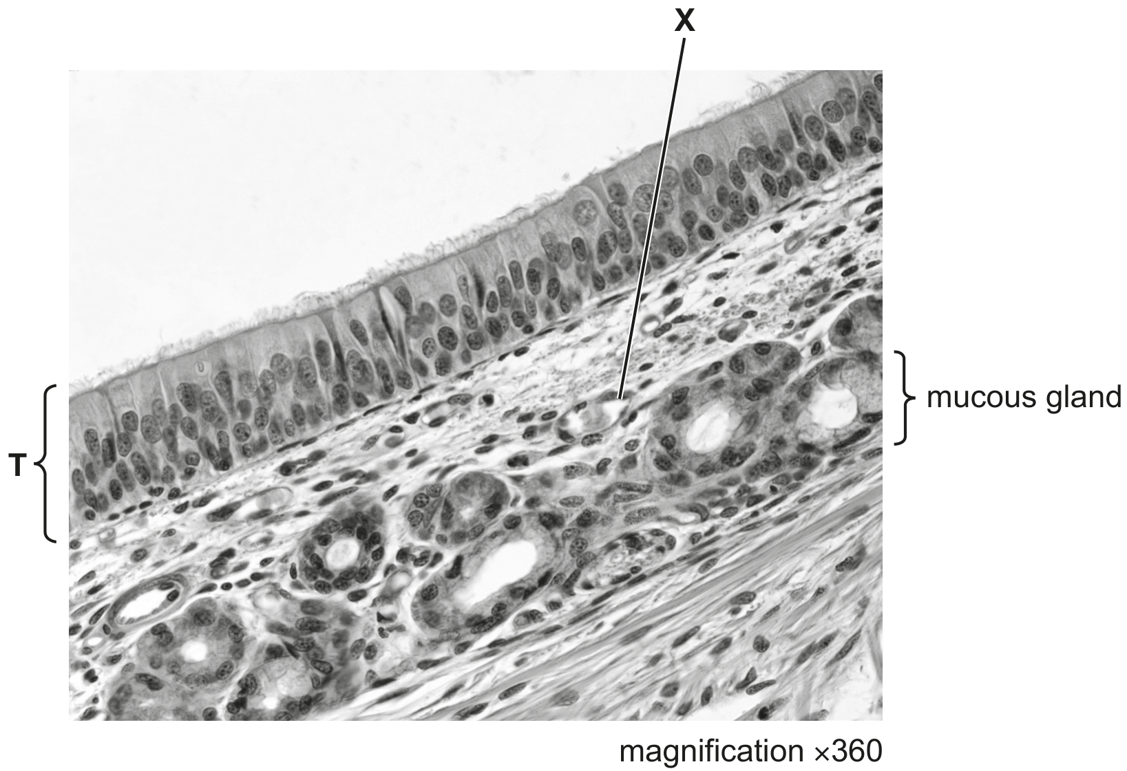
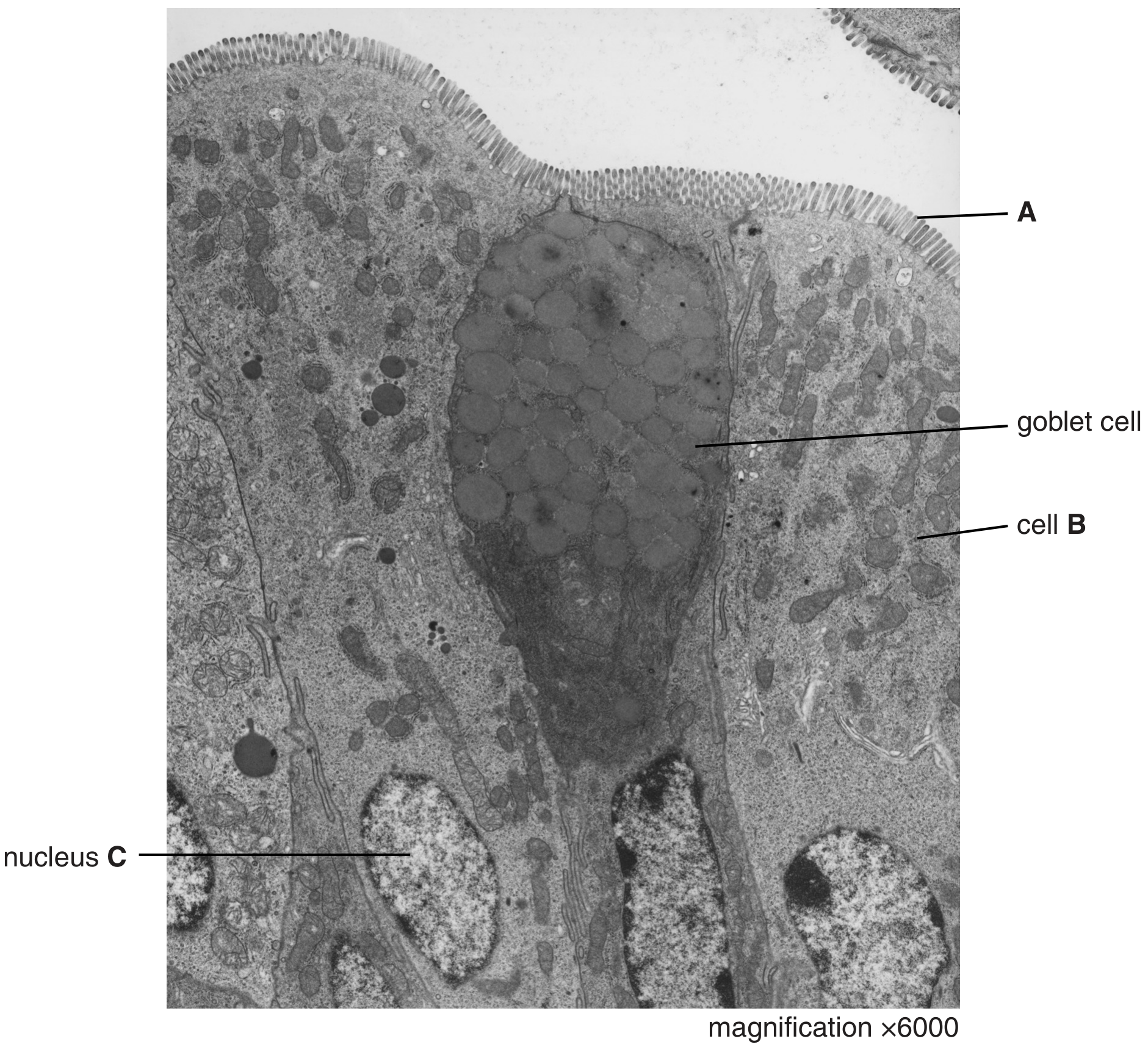
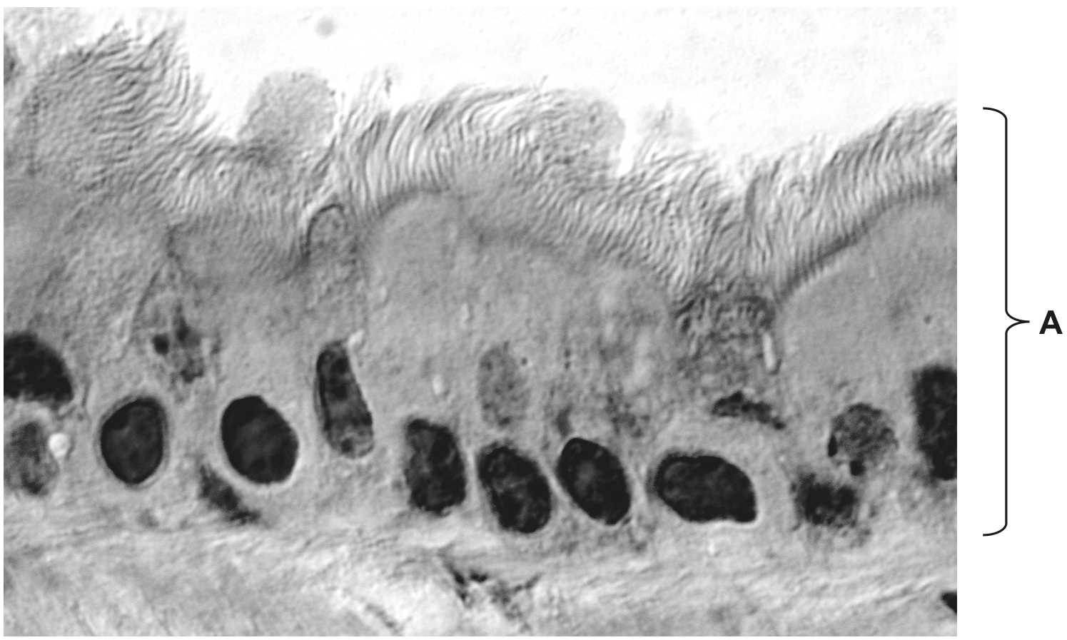
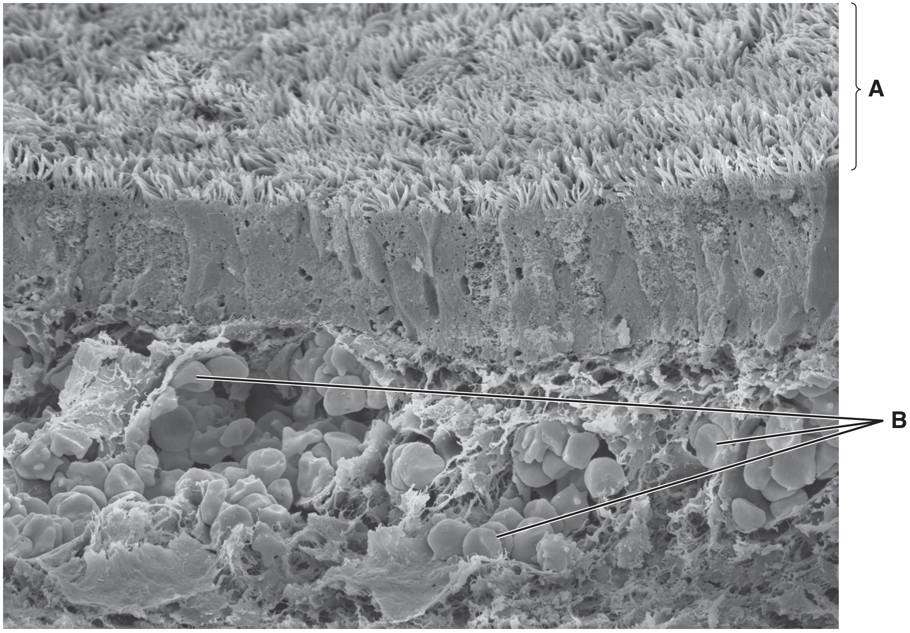
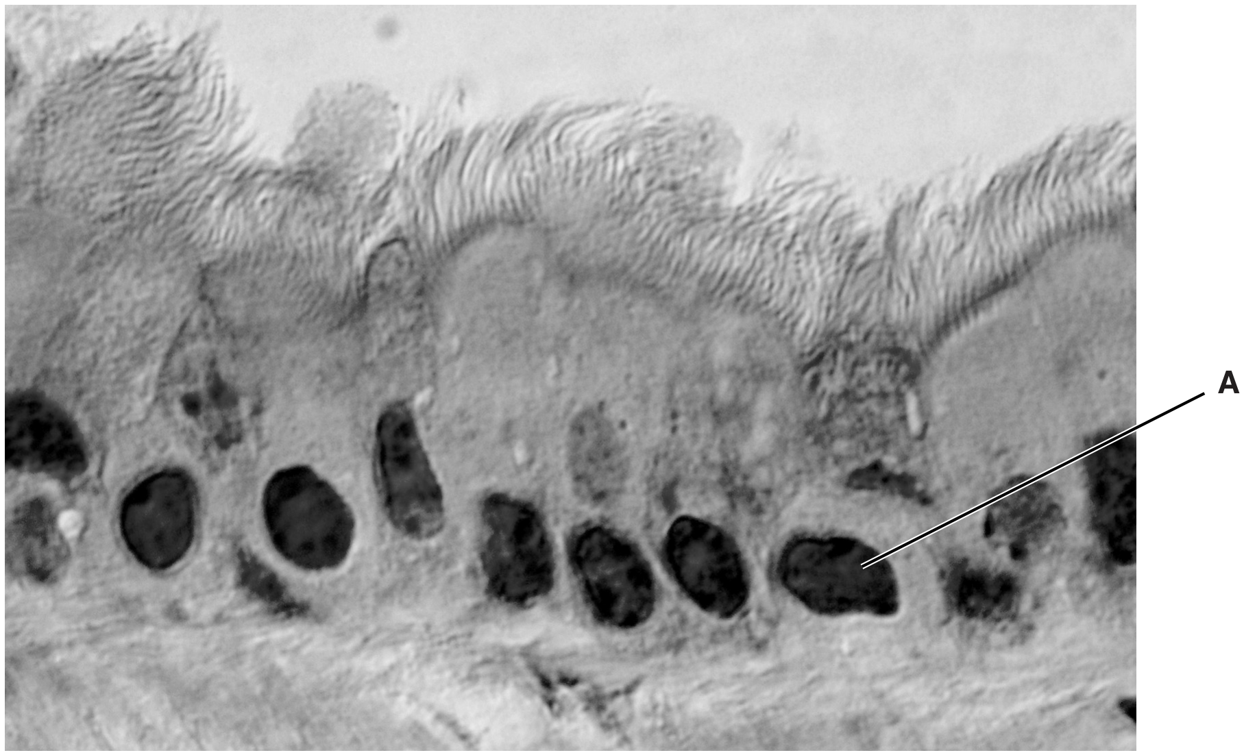
Fig. 1.1 is an electron micrograph of cells from the ciliated epithelium of the trachea.
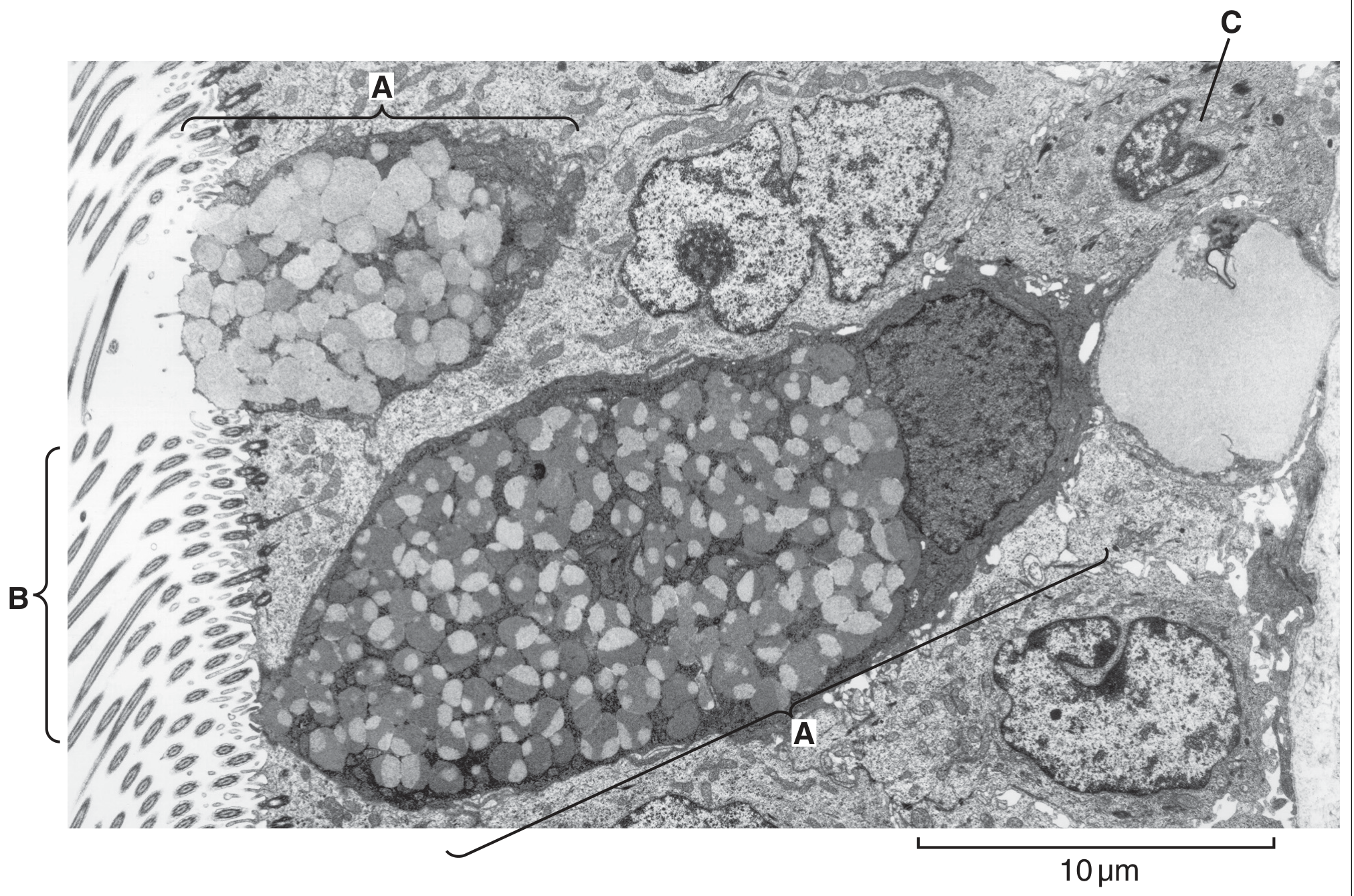
Fig. 1.1

Fig. 1.1

Question 1(b)

(a)

Explain how the cells labelled A and the structures labelled B in Fig. 1.1 protect the lining of the trachea.

[ 4 ]

Question 1

[Maximum number: 7]

The trachea of the gas exchange system branches into two airways, each of which enters a lung.

Question 1(a)

(a)

Name the airways that branch from the trachea to enter the lungs.

[ 1 ]

Question 1(c)

(b)

Fig. 1.1 is a photomicrograph of a section through part of the trachea.

Fig. 1.1

Fig. 1.1

[ 6 ]

Question 1(c)(i)

(i)

In Fig. 1.1, one of the tissues in the trachea is labelled T.

Describe the structural features of tissue T visible in Fig. 1.1.

[ 3 ]

Question 1(c)(ii)

(ii)

Identify structure X in Fig. 1.1 and outline the features that helped your identification. structure X=

[ 3 ]

Question 1

[Maximum number: 5]

Fig. 1.1 is a light micrograph of a section through part of the gas exchange system.
A, B and C are three different types of tissue.

Fig. 1.1

Fig. 1.1

Question 1(a)

(a)

The cell types in tissue A have different functions.

Describe how the cell types work together to maintain the health of the gas exchange system.

[ 3 ]

Question 1(b)

(b)

Suggest why the cells in tissue B have many mitochondria.

[ 1 ]

Question 1(c)

(c)

Name the parts of the gas exchange system where tissue C is distributed.

[ 1 ]

Question 1

[Maximum number: 5]

Fig. 1.1 is an electron micrograph of cells from the lining of the small intestine.

Fig. 1.1

Fig. 1.1

Question 1(d)

(a)

There are many goblet cells within the epithelium lining the trachea and the bronchi in the gas exchange system.

Describe the role of goblet cells in the gas exchange system.

[ 3 ]

Question 1(e)

(b)

State two ways in which the cells lining the alveoli in the lungs differ from cell B shown in Fig. 1.1.
1.

[ 2 ]

Question 1

[Maximum number: 6]

Fig. 1.1 and Fig. 1.2 are photomicrographs showing the distribution of tissues in the lungs.
Fig. 1.1 is a photomicrograph of a section through part of the lungs.
Fig. 1.2 is a high-power view of the area indicated on Fig. 1.1.

Fig. 1.1

Fig. 1.1

section enlarged in Fig. 1.2

Fig. 1.2

Fig. 1.2

Question 1(a)

(a)

State the names of the tissues A, B and D.

A

B
D

[ 3 ]

Question 1(b)

(b)

Describe the role of the glands labelled C in Fig. 1.1 in maintaining the health of the gas exchange system.

[ 3 ]

Question 1

[Maximum number: 2]

Name as precisely as you can the structure described in each of the following statements.

Question 1(d)

(a)

The epithelial cell that secretes mucus in the trachea.

[ 1 ]

Question 1(e)

(b)

The tissue that prevents the collapse of the trachea during inhalation.

[ 1 ]

Question 1

[Maximum number: 8]

Fig. 1.1 is a scanning electron micrograph of part of the wall of the bronchus of a healthy human.

Fig. 1.1

Fig. 1.1

Question 1(a)

Question 1(a)(i)

(a)
(i)

Name the structures labelled A.

[ 1 ]

Question 1(a)(ii)

(ii)

State the function of the cells labelled B.

[ 1 ]

Question 1(b)

(b)

Name two tissues found in the wall of the bronchus that are not visible in Fig. 1.1.
1.

[ 2 ]

Question 1(d)

(c)

Describe the appearance of a section through the wall of a bronchus in a person with chronic bronchitis.

[ 4 ]

Question 1

[Maximum number: 5]

Fig. 1.1 is a photomicrograph of epithelial cells in the bronchus.

Fig. 1.1

Fig. 1.1

Question 1(a)

Question 1(a)(i)

(a)
(i)

Write a letter X on Fig. 1.1 to show the lumen of the bronchus.

[ 1 ]

Question 1(a)(ii)

(ii)

Name the structure in Fig. 1.1 labelled A.

[ 1 ]

Question 1(a)(iii)

(iii)

State one feature of the cells, visible in Fig. 1.1, which indicates that these are not epithelial cells from the alveolus.

Question 1(b)

(b)

Epithelial cells are replaced when they are damaged.

[ 3 ]

Question 1(b)(ii)

(i)

The cells shown in Fig. 1.1 are from a non-smoker.

Smoking causes damage to the epithelial cells of the lungs.
Describe the appearance of the lining of the bronchus in a long-term smoker.

[ 3 ]

Question 1

Question 1(a)

(a)

Fig. 1.1 is an image of a transverse section through the trachea of the gas exchange system.

Fig. 1.1

Fig. 1.1

Write the names of structures X and Y on Fig. 1.1 in the boxes provided.

[ 2 ]

Question 1(c)

(b)

Some structures in the gas exchange system are listed in alphabetical order in Table 1.1.
- Write YES in the box provided if the structure contains smooth muscle.
- Write NO in the box provided if the structure does not contain smooth muscle.

Table 1.1

Table 1.1

[ 1 ]

Question 1

[Maximum number: 4]

Fig. 1.1 shows the human gas exchange system.

Fig. 1.1

Fig. 1.1

Question 1(a)

(a)

Name the structures labelled A, B, and C in Fig. 1.1.

A
B
C

[ 3 ]

Question 1(b)

(b)

Name a non-infectious disease that affects the human gas exchange system.

[ 1 ]
0 selected